
Coronary circulation diagram
Reading File
The coronary circulation consists of the right and left coronary arteries arising from the aortic root, supplying the myocardium, and draining via the coronary sinus into the right atrium.
Searching sources...
Finding Sources
Finding Sources
Reading File
Reading File
Reading File
I found two excellent diagrams. Let me fetch them both now.
Loading Image
Loading Image
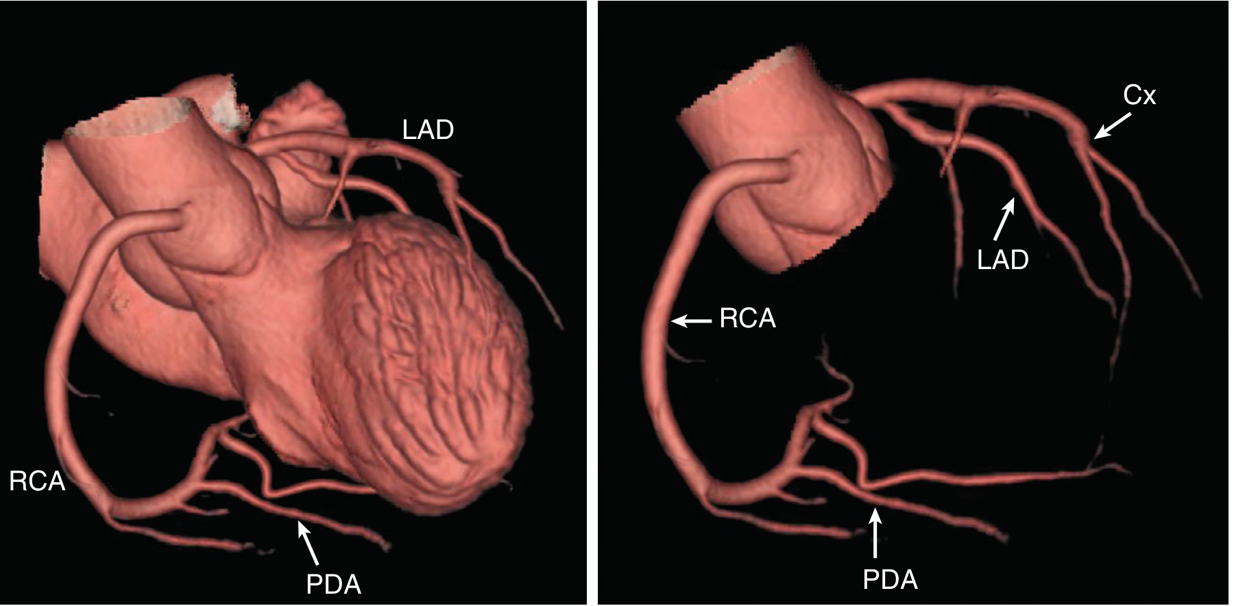
Here are two authoritative diagrams of the coronary circulation from the medical library:
Fig. 1 - Coronary Arteries (Illustrated) - Guyton and Hall Textbook of Medical Physiology
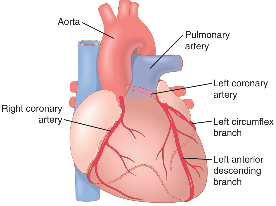
Fig. 2 - Normal Coronary Anatomy (CT Reconstruction) - Textbook of Clinical Echocardiography
Coronary Circulation - Key Anatomy
Arterial Supply
| Artery | Origin | Territory |
|---|---|---|
| Left Main (LM) | Left coronary sinus of Valsalva | Bifurcates into LAD and Cx |
| Left Anterior Descending (LAD) | Left main | Anterior septum, anterior LV wall, LV apex |
| Circumflex (Cx) | Left main | Lateral LV wall (obtuse marginal branches) |
| Right Coronary Artery (RCA) | Right coronary sinus of Valsalva | Right ventricle, posterior LV (in 80-90% of people) |
| Posterior Descending Artery (PDA) | RCA (right dominant, ~80%) or Cx (left dominant, ~20%) | Inferior septum, inferior LV free wall |
Venous Drainage
- ~75% of left ventricular coronary venous blood returns via the coronary sinus to the right atrium
- Right ventricular venous blood drains via anterior cardiac veins directly into the right atrium
- A small amount returns via thebesian veins directly into all heart chambers
Physiology Highlights (from Costanzo Physiology and Guyton & Hall)
- Flow is controlled almost entirely by local metabolites - primarily hypoxia and adenosine
- Resting coronary flow: ~225 mL/min (~4-5% of cardiac output)
- During strenuous exercise, coronary flow increases 3-4 fold
- Phasic flow pattern: Left coronary flow is reduced during systole due to myocardial compression, and the bulk of perfusion occurs in diastole - this is why tachycardia (shortened diastole) is dangerous in ischemic heart disease
- The right coronary artery maintains more continuous flow throughout the cardiac cycle since right ventricular wall tension is lower
This is a shared conversation. Sign in to Orris to start your own chat.