
Sick sinus syndrome
Reading File
Now let me search the medical library and PubMed in parallel.
Finding Sources
Searching PubMed
"sick sinus syndrome"[MeSH Terms]
Good results. Now let me read the key sections from Braunwald's and Tintinalli's in parallel.
Reading File
Reading File
Reading File
Let me get more content from Braunwald's block 8 (management section) and also fetch some of the ECG images.
Reading File
Finding Sources
Loading Image
Now let me read the specific management section from Braunwald's block 8 and the Ganong physiology section.
Reading File
Reading File
Reading File
Now let me also read the Braunwald's block 6 section on SSS clinical features and fetch the sinus node arrest ECG image.
Reading File
Loading Image
I now have comprehensive information to write a thorough answer. Let me also get the intrinsic vs extrinsic categorization from Braunwald's block 8.
Reading File
I now have enough comprehensive information from multiple authoritative textbooks. Let me compile the full answer.
Sick Sinus Syndrome (SSS)
Definition
Sick sinus syndrome (SSS), also called sinus node dysfunction (SND), is not a single arrhythmia but a collection of sinus nodal abnormalities that include:
- Persistent spontaneous sinus bradycardia inappropriate for the physiologic circumstance
- Sinus arrest or SA exit block
- Combinations of SA and AV conduction disturbances
- Bradycardia-tachycardia syndrome - alternating paroxysms of rapid atrial tachyarrhythmias (most often atrial fibrillation) and periods of slow atrial and ventricular rates
More than one of these patterns can occur in the same patient on different occasions, and their mechanisms are often causally interrelated.
- Braunwald's Heart Disease, p. 575
Epidemiology
- Leading diagnosis for permanent pacemaker implantation
- Predominantly a disease of the elderly (1 in 600 cardiac patients >65 years)
- Associated with cardiomyopathy, congestive heart failure, ischemic heart disease, and metabolic diseases
- Can also occur without identifiable cardiac abnormality ("idiopathic SND") - at any age, even in utero
- In children, most often associated with congenital or acquired heart disease, especially after corrective cardiac surgery (Mustard/Senning operations for transposition of great vessels; Fontan procedure)
- Course is frequently intermittent and unpredictable
Pathophysiology / Anatomic Basis
Structural Changes
- Total or subtotal destruction of the sinus node
- Areas of nodal-atrial discontinuity
- Inflammatory or degenerative changes in the surrounding nerves and ganglia
- Fibrosis and fatty infiltration of the sinus node
- Sclerodegenerative processes that can extend to involve the AV node, bundle of His, and its branches
- Occlusion of the sinus node artery
Intrinsic vs. Extrinsic Causes
Patients can be categorized as having intrinsic sinus node disease unrelated to autonomic abnormalities, or combinations of intrinsic and autonomic abnormalities. Excessive physical training can heighten vagal tone and produce syncope via sinus bradycardia or AV conduction abnormalities even in otherwise normal individuals.
Genetic Basis
Familial (idiopathic) SSS is linked to four genes:
| Gene | Channel/Protein | Mechanism |
|---|---|---|
| SCN5A | Nav1.5 sodium channel | Loss-of-function variants; nonfunctional or biophysically impaired channels; can produce mixed SSS + Brugada + cardiac conduction disease phenotype |
| HCN4 | "Funny current" (If) pacemaker channel | Loss-of-function; impaired automaticity; severe forms cause bradycardia, syncope, QT prolongation, TdP |
| ANK2 | Ankyrin-B | Scaffolding protein dysfunction |
| MYH6 | Alpha-myosin heavy chain | Structural protein |
SCN5A variants cause autosomal recessive SSS (compound heterozygotes); HCN4 variants can be lethal. Inheritance is autosomal dominant with reduced penetrance (or recessive with complete penetrance).
- Braunwald's Heart Disease, p. 574
ECG Findings
Sinus Bradycardia - rate <60 bpm, inappropriate for physiologic state.
Sinus Arrest / Sinoatrial (SA) Exit Block:
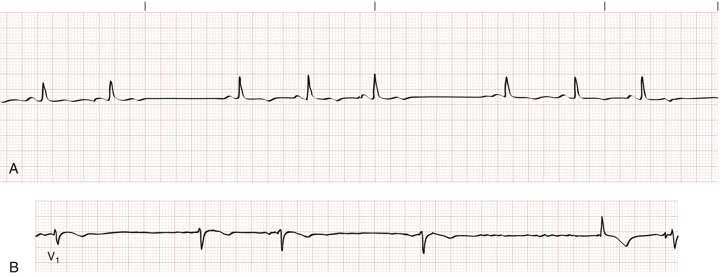
- Type I SA exit block: P-P interval shortens progressively (Wenckebach-like), then a pause shorter than twice the shortest cycle
- Type II SA exit block: Pauses equal exactly twice the basic P-P interval (abrupt block)
Bradycardia-Tachycardia Syndrome: An implanted loop recorder recording showing paroxysmal sinus node arrest with a pause of nearly 30 seconds is the classic finding:
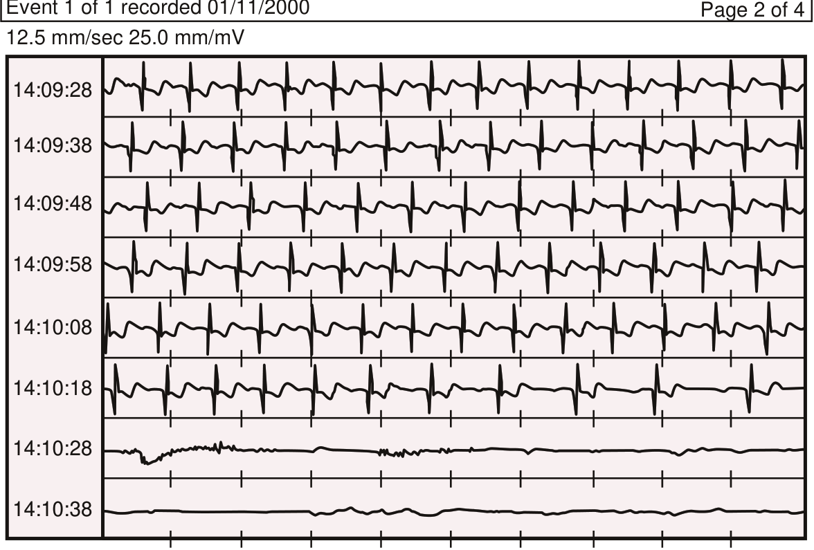
Chronotropic Incompetence (CI): Failure to reach 80-85% of the maximum age-predicted heart rate or inadequate heart rate reserve with exercise.
Clinical Manifestations
Symptoms arise from intermittent cerebral hypoperfusion due to the bradycardias, or from the tachycardias:
| Symptom | Cause |
|---|---|
| Dizziness, lightheadedness | Transient bradycardia |
| Syncope (Stokes-Adams attacks) | Sinus arrest / prolonged pause |
| Palpitations | Tachycardia phase |
| Fatigue, exercise intolerance | Chronotropic incompetence |
| Sudden cardiac death | Prolonged asystole or VT |
- Isolated sinus node dysfunction rarely causes syncope on its own; syncope in SSS is more likely due to a reentrant atrial tachycardia
- SSS is listed as a cause of syncope when sinus pauses exceed 3 seconds
Diagnosis
- 12-lead ECG: May show sinus bradycardia, long PP pauses, or SA block
- Holter monitoring: Most effective tool because of the episodic nature; captures sinus pauses, episodes of atrial tachycardias
- Implantable loop recorder: For infrequent syncope (see tracing above)
- Exercise stress testing: Reveals chronotropic incompetence
- Electrophysiologic study: Sinus node recovery time (SNRT), sinoatrial conduction time (SACT); abnormal responses in symptomatic patients with sinus pauses/SA exit block
- SSS is one of the causes in Box 11.2A of dysrhythmias potentially associated with syncope (alongside Mobitz type II, third-degree block, sinus pause >3 s)
Associations / Drug Causes
Many drugs can precipitate or worsen SSS:
- Beta-blockers
- Calcium channel blockers (especially non-dihydropyridines: verapamil, diltiazem)
- Digoxin
- Antiarrhythmics: quinidine, procainamide, flecainide
These are also conditions in which these drugs are contraindicated.
Management
Acute Setting (Emergency)
- Treat the specific rhythm present
- For bradycardia: atropine, temporary pacing
- For tachycardia (e.g., atrial fibrillation): nodal blocking agents (calcium channel blockers), with awareness that subsequent bradycardia may require temporary pacing
Long-Term Management
| Problem | Treatment |
|---|---|
| Symptomatic bradycardia | Permanent pacemaker implantation |
| Bradycardia-tachycardia syndrome | Pacemaker (for bradycardia) + antiarrhythmic drugs (for tachycardia) |
| Atrial fibrillation in SSS | Pacemaker to allow pharmacologic AF therapy safely |
| Tachycardias alone (without bradycardia) | Medications alone |
-
Preferred pacing mode: Atrial pacing (AAI or DDDR) - preserves AV synchrony; reduces AF and stroke risk compared to ventricular pacing (VVI)
-
Important caveat from Tintinalli's: after pacemaker placement, syncope and sudden death can still occur because the pacemaker prevents bradycardia but not tachycardia
-
Braunwald's Heart Disease, p. 576; ROSEN's Emergency Medicine, p. 1046; Ganong's Review of Medical Physiology, p. 568
SSS and Stroke Risk
A recent 2023 meta-analysis (PMID 37977871) found that sinus node dysfunction is associated with increased stroke risk, further supporting the importance of anticoagulation considerations alongside pacing decisions, particularly given the high co-prevalence of atrial fibrillation.
Special Situations
- Pediatric: Usually linked to prior cardiac surgery (Mustard, Senning, Fontan procedures); pacemaker placement does not eliminate sudden death risk
- Anesthesia / Perioperative: Patients with SSS require device interrogation pre-operatively and identification of rate-responsive programming; avoid drugs that suppress sinus node automaticity
- Aging: A 2025 review (PMID 40497650) highlights age-related mechanisms of sinus node dysfunction including progressive fibrosis, oxidative stress, and ion channel remodeling as active areas of research
Sources:
- Braunwald's Heart Disease: A Textbook of Cardiovascular Medicine (Chapters 63, 68, 69)
- ROSEN's Emergency Medicine (Chapter 65)
- Tintinalli's Emergency Medicine: A Comprehensive Study (Chapter 130)
- Ganong's Review of Medical Physiology, 26th Ed.
- Recent PubMed: Stroke risk meta-analysis | Tachy-brady syndrome review | Aging and SND
A 35-year-old patient presents after a stroke. Cardiac evaluation reveals a large and prominent pulmonary artery on chest X-ray. Which cardiac anomaly is most likely associated with these findings?... Ventricular septal defect... Mitral stenosis... Aortic coarctation... Patent ductus arteriosus... Atrial fibrillation...
Reading File
Finding Sources
Finding Sources
The textbook evidence is clear. Let me pull the specific Goldman-Cecil and Bailey & Love passages confirming the CXR findings of mitral stenosis.
Reading File
Reading File
Reading File
The answer is unambiguous and thoroughly supported. Here is the full explanation:
✅ Answer: Mitral Stenosis
Why Mitral Stenosis?
The two key findings in this question are:
- Stroke in a 35-year-old (young patient)
- Large and prominent pulmonary artery on chest X-ray
These two findings together point directly to mitral stenosis (MS), most commonly rheumatic in origin.
The Chest X-Ray in Mitral Stenosis
Goldman-Cecil Medicine describes the classic CXR constellation of mitral stenosis as:
"Left atrial enlargement, pulmonary venous hypertension, a small aortic knob, and enlargement of the main pulmonary artery."
Bailey & Love's Surgery reinforces this:
"There is a small aortic outline and a prominent pulmonary artery. The left atrium is enlarged (sometimes to an enormous degree) along with upper lobe diversion as a result of the raised pulmonary venous pressure. The right ventricle also appears enlarged."
The mechanism: MS obstructs flow from the left atrium into the left ventricle → left atrial pressure rises → pulmonary venous hypertension → pulmonary arterial hypertension → the main pulmonary artery dilates and becomes prominent on CXR.
- Goldman-Cecil Medicine, p. 448; Bailey & Love's Surgery, p. (Ch. 59)
Why Mitral Stenosis Causes Stroke
Bradley and Daroff's Neurology states:
"Most cases of mitral stenosis are due to rheumatic heart disease. Emboli occur in 9-14% of patients with mitral stenosis, with 60-75% causing stroke or TIA. Embolism may be the first manifestation of mitral stenosis, particularly if it is associated with AF."
The mechanism: MS causes left atrial enlargement and stasis → thrombus forms in the left atrium (especially the left atrial appendage) → cardioembolic stroke.
- Bradley and Daroff's Neurology in Clinical Practice
Why the Other Options Are Wrong
| Option | Why it doesn't fit |
|---|---|
| Ventricular septal defect (VSD) | CXR shows enlarged left atrium + left ventricle with shunt vascularity - not a prominent main pulmonary artery predominantly. Paradoxical embolism possible but not the classic pairing. |
| Aortic coarctation | CXR shows "figure-of-3 sign," rib notching, enlarged ascending aorta - not prominent pulmonary artery. Stroke risk exists via hypertension/bicuspid aortic valve, not embolism. |
| Patent ductus arteriosus (PDA) | CXR shows enlarged pulmonary vasculature and enlarged left heart, but it does not classically produce the prominent main pulmonary artery + stroke combination. |
| Atrial fibrillation | AF is a major cause of stroke, but it is not itself a "cardiac anomaly" that causes a prominent pulmonary artery on CXR. AF is actually a complication of mitral stenosis. |
Summary Diagram
Rheumatic Fever
      ↓
Mitral Valve Scarring / Thickening (Mitral Stenosis)
      ↓
Left Atrial Obstruction → Left Atrial Enlargement
      ↓                            ↓
Pulmonary Venous HTN         LA Thrombus → Embolism
      ↓                            ↓
Pulmonary Arterial HTN      STROKE / TIA
      ↓
Prominent Main Pulmonary Artery on CXR
The answer is Mitral Stenosis - the one cardiac anomaly that simultaneously explains a prominent pulmonary artery on CXR (via pulmonary hypertension from LA obstruction) and stroke risk (via left atrial thrombus and cardioembolism) in a young patient.
This is a shared conversation. Sign in to Orris to start your own chat.