
Write a long answer on topic poller screws for Ms orthopedics theory examination gold medal level
poller screws blocking screws intramedullary nailing orthopedics technique biomechanics
poller screw blocking screw intramedullary nail
PMID: 42098782
PMID: 38579780
poller screw blocking screw intramedullary nail technique metaphyseal fracture
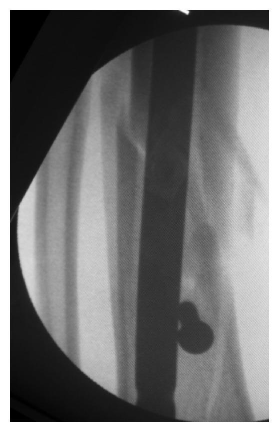
This diagnostic image is a fluoroscopic intraoperative radiograph of a human lower leg, likely the tibia, demonstrating the surgical management of a long bone fracture. The visual content shows a large-diameter, radiopaque intramedullary nail (IM nail) positioned centrally within the medullary canal to provide internal fixation and restore anatomical alignment. A notable feature is the presence of a 'Poller screw' (blocking screw), visible as a distinct, dark circular hardware component placed adjacent to the IM nail. This technique is used to guide the nail during insertion, narrowing the canal and preventing malalignment or translation in metaphyseal fractures. The cortical outlines of the bone are visible, showing restored longitudinal continuity despite the underlying fracture site. This image serves as an educational example of orthopedic trauma surgery, specifically illustrating the use of adjunctive blocking screws to optimize alignment during intramedullary nailing.
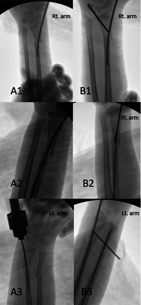
This composite of six diagnostic fluoroscopic images illustrates the surgical management of distal radius diaphyseal-metaphyseal junction (DRDMJ) fractures in pediatric patients. The left column (A1, A2, A3) demonstrates post-reduction failure using Elastic Stable Intramedullary Nailing (ESIN) alone. In these images, a single radiopaque intramedullary nail is visible within the radius, yet significant residual angular deformity and radial translation of the distal fragment persist at the fracture site. The right column (B1, B2, B3) demonstrates the 'poller K-wire' technique used to correct these deformities. A transverse 1.6 mm Kirschner wire (K-wire) is inserted as a blocking screw to act as a fulcrum. This poller wire guides the longitudinal ESIN, creating a lever effect that successfully reduces the translation and realigns the anatomical axis of the radius. The images represent a comparison of intraoperative fixation strategies in orthopedic trauma surgery, specifically focusing on achieving stable reduction in regions with wide metaphyses and narrow medullary canals.
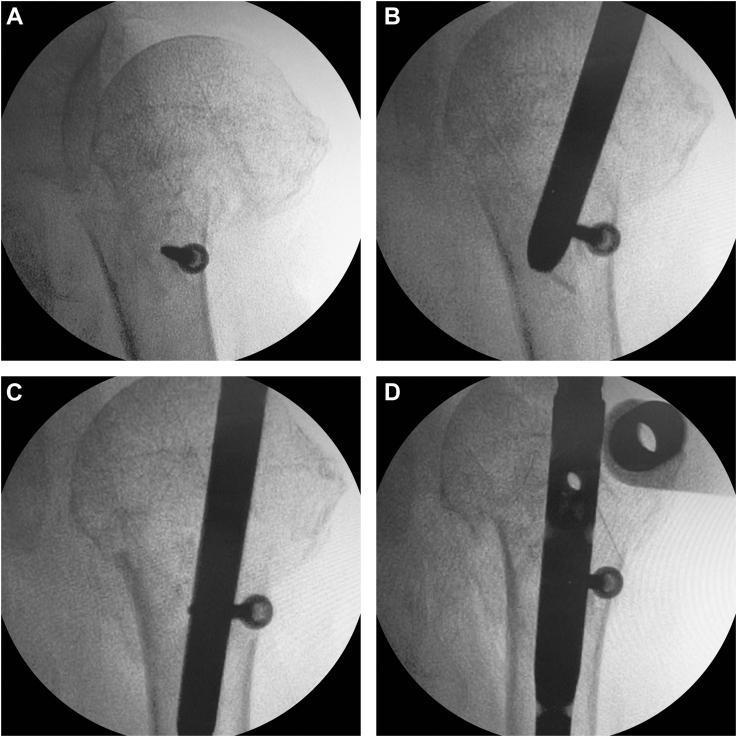
A series of four intraoperative fluoroscopic images (A-D) demonstrating the surgical technique of blocking screw augmentation for intramedullary nailing of a proximal humeral fracture. Panel A shows a single radiopaque cortical screw placed in the lateral aspect of the distal humeral shaft fragment to serve as a 'Poller' or blocking screw. Panel B illustrates the antegrade insertion of a radiodense intramedullary nail, which is guided medially by the presence of the blocking screw. Panel C shows the progression of the nail, demonstrating how the blocking screw facilitates lateral translation of the humeral shaft to improve fracture alignment and reduction. Panel D displays the final construct with the nail fully seated to an adequate depth through the humeral head, showing successful reduction of the surgical neck fracture. The technique illustrates the use of mechanical interference to narrow the effective medullary canal, preventing nail deviation and ensuring anatomical alignment in orthopedic trauma surgery.
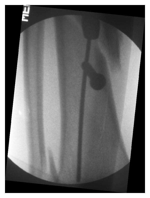
**Imaging Modality:** Intraoperative fluoroscopy (C-arm X-ray). **Anatomical Region:** Long bone diaphysis, likely the tibia or femur. **Observed Pathology/Procedures:** The image demonstrates an orthopedic surgical procedure involving intramedullary (IM) nailing for fracture fixation. A radiopaque intramedullary nail is visible traversing the medullary canal. **Characteristic Visual Features:** - **Poller (Blocking) Screw:** A single, large-diameter radiopaque screw is positioned perpendicular to the long axis of the bone, situated on the concave side of the deformity near the fracture site in the distal segment. - **Nail-Screw Interaction:** The Poller screw is strategically placed to narrow the medullary canal, acting as a physical guide to redirect the intramedullary nail. This contact ensures central alignment within the distal fragment and prevents malalignment (valgus/varus or prorecurvatum/recurvatum) during insertion. - **Hardware Positioning:** The distal tip of the IM nail is visible extending past the level of the blocking screw. **Key Diagnostic Features:** The primary feature is the utilization of a blocking screw (Poller screw technique) to achieve indirect reduction and stability of an intramedullary nail in a wide metadiaphyseal region or a distal fracture segment.
POLLER SCREWS (BLOCKING SCREWS) IN INTRAMEDULLARY NAILING
Definition and Etymology
Why Poller Screws Are Needed: The Problem They Solve
The Nail-Metaphysis Mismatch
- Angular malalignment (varus/valgus, procurvatum/recurvatum)
- Translational malalignment of fracture fragments
- Rotational instability
Why Standard Interlocking Screws Don't Fully Solve This
Historical Background and Evolution
| Year | Development |
|---|---|
| 1999 | Krettek et al. first described blocking screws for proximal/distal tibial fractures with small-diameter nails |
| Early 2000s | Technique applied to femoral fractures, subtrochanteric region |
| 2010s | Extended use in humeral nailing, pediatric fractures, limb lengthening |
| 2020s | "Epicentric" and "3rd/4th generation" poller screws for reduction + compression; use with motorized lengthening nails |
- 1st generation: Screw placed before nailing as a guide/redirect tool
- 2nd generation: Screw placed after nailing if malalignment detected
- 3rd generation: Screw placed to achieve direct reduction (epicentric technique)
- 4th generation: Novel configurations for complex deformity correction and limb lengthening
Biomechanical Principles
Mechanism of Action
-
Guiding (alignment): By narrowing the canal on the concave side of an anticipated deformity, the screw guides the reamer and nail into the center of the distal/proximal fragment rather than the cortex of least resistance.
-
Stabilizing (anti-toggle): After the nail is seated, the screw resting against the nail shaft reduces the bending moment arm - it decreases the lever arm available for the bone segment to pivot around the nail. This increases construct stiffness in the plane perpendicular to the screw.
-
Fracture site motion reduction: The 2026 meta-analysis (Yang et al., JOSS, PMID 42098782) demonstrated via finite element analysis that Poller screws reduced fracture site displacement by 54.88% under axial loading, and increased union rates significantly (OR = 2.48; 95% CI, 1.13-5.46; p = 0.020).
The "Pseudo-Canal" Concept
Cadaveric Evidence (Krettek's Original Study)
- Reduced translation of the proximal fragment by >50%
- Increased stiffness of the bone-nail construct significantly
- Was most effective when placed close to the fracture site on the concave side of deformity
The Golden Rule of Poller Screw Placement
"The screw is placed on the CONCAVE side of the anticipated deformity, in the SHORT (near) fragment."
Rationale
- Placing the screw on the concave side blocks the nail from being deflected by the cortex on the concave side (the side toward which the deformity bends).
- The screw acts as a "wall" that pushes the nail centrally or toward the convex side - restoring the anatomical axis.
Plane-Specific Rules
| Deformity | Screw Position |
|---|---|
| Valgus apex lateral (e.g. proximal tibia after nailing) | Screw on lateral side (concave = lateral) in proximal fragment |
| Varus deformity | Screw on medial side of the near fragment |
| Procurvatum (apex anterior) | Screw on anterior side of the near fragment |
| Recurvatum (apex posterior) | Screw on posterior side of the near fragment |
Indications
1. Proximal Tibial Fractures (Classic Indication)
- Valgus deformity (from reamer creep medial-to-lateral, patella pushing start point medially)
- Procurvatum (apex anterior, due to extensor mechanism pull)
2. Distal Tibial Fractures
3. Subtrochanteric Femoral Fractures
- Flexion (iliopsoas)
- Abduction (hip abductors)
- External rotation (short external rotators)
4. Distal Femoral Fractures
5. Humeral Shaft and Proximal Humeral Fractures
6. Pediatric Fractures (Poller K-wire)
7. Limb Lengthening and Deformity Correction
8. Malunion Correction / Closed Nailing of Malunions
Timing of Placement
| Timing | Purpose | Notes |
|---|---|---|
| Before reaming/nailing (Pre-emptive/1st generation) | Guide the reamer and nail into desired path | Ideal - prevents deformity from occurring |
| After reaming, before nail insertion | Correct trajectory that went wrong during reaming | Most common scenario |
| After nail insertion (2nd generation/salvage) | Reduce toggle if malalignment detected on fluoroscopy | Nail must be partially withdrawn, screw placed, nail re-advanced |
- Reamer creep already creates a biased canal path - even if the nail is directed correctly, it will follow the reamed path
- Placing the screw first redirects the reaming path, not just the nail
Surgical Technique
Step-by-Step
-
Preoperative planning: Assess the fracture pattern and predict the likely deformity (based on fracture location and level). Identify which plane of deformity to address.
-
Positioning and standard nailing approach: Begin with standard nail entry point and approach.
-
Reduction and guide wire insertion: Reduce the fracture (closed or with percutaneous aids). Pass the guide wire.
-
Assess trajectory under fluoroscopy: Check AP and lateral views. If the guide wire is heading toward a cortex rather than centrally, a Poller screw is indicated.
-
Identify the screw position:
- Concave side of the deformity
- In the short (near) fragment
- Close to the fracture site (maximizes the mechanical advantage)
- On the side toward which the fracture is displacing
-
Screw insertion:
- Use a standard 3.5 mm or 4.5 mm cortical screw (or a K-wire for children)
- Pass the screw through both cortices of the metaphyseal fragment
- The screw should occupy the canal space immediately adjacent to where the nail will run
- The screw tip must be adjacent to (not in the path of) the nail's intended track
-
Re-ream and advance the nail: The reamer and nail are now forced away from the blocking screw, following the narrowed corridor into proper alignment.
-
Fluoroscopic confirmation: Verify alignment on AP and lateral views.
-
Complete interlocking: Finish with standard proximal and distal interlocking screws.
-
Decision to keep or remove the Poller screw: The screw may be left in place permanently (adding to construct stability) or removed depending on surgeon preference and stability achieved. Most surgeons retain them.
Technical Tip for Accurate Placement (Hannah et al., 2014 - PMID 24629702)
Fluoroscopic Images
Specific Scenarios and Rules of Thumb
Proximal Tibial Nail - Classic Deformity
- Valgus + procurvatum = classic pattern
- Lateral blocking screw for valgus
- Anterior blocking screw for procurvatum
- Both can be placed simultaneously for combined deformity
Distal Femur Nailing (Retrograde)
- Valgus with short distal fragment: medial blocking screw (concave side = medial) in the distal fragment
- For short distal segments, bilateral blocking screws create a pseudo-canal
Subtrochanteric Femur Nailing
- Flexion deformity of proximal fragment: anterior blocking screw on the anterior cortex of the proximal fragment forces the nail posteriorly
- Can be combined with a bone hook or Schanz pin in the proximal fragment for provisional reduction
Advantages
- Technically simple - no special instrumentation required beyond standard screws
- Minimally invasive - can be placed percutaneously under fluoroscopy
- No added hardware cost - uses screws already in any implant set
- Improves stability - reduces metaphyseal toggle without adding another implant type
- Reversible - can be removed if causing soft tissue problems
- Versatile - applicable to any long bone, any age group
- No increase in infection risk - confirmed in the 2026 meta-analysis
- Reduces need for secondary surgery - same meta-analysis showed fewer secondary procedures in the Poller screw group
Disadvantages and Complications
- Increased operative time - documented in multiple studies
- Fluoroscopy exposure - additional time under C-arm
- Risk of inadvertent nail channel blockage - if screw is placed in the exact path of the intended nail
- Screw breakage - if too large a screw is placed and the nail contacts it repeatedly
- Technical error - placing on the wrong (convex) side worsens deformity
- Stress riser - at the screw hole if the screw is removed early
- Cannot prevent rotational malalignment - Poller screws address angular/translational but not torsional deformity
Evidence Base
2026 Meta-Analysis (Yang et al., JOSS - PMID 42098782)
- 5 RCTs/trials, 413 participants
- Union rate: significantly higher with Poller screws (OR = 2.48, p = 0.020)
- Secondary surgery: significantly decreased
- Infection: no difference
- Malalignment: no significant difference (both groups acceptable)
- Finite element analysis: 54.88% reduction in fracture site displacement
- Conclusion: Poller screws should be considered the standard adjunct in extra-isthmic lower limb fractures treated with IM nailing
Krettek's Original Cadaveric Study (1999)
Zhou et al. (2024, EFORT Open Reviews - PMID 38579780)
Comparison with Other Anti-Deformity Techniques in Metaphyseal Nailing
| Technique | Advantage | Disadvantage |
|---|---|---|
| Poller screw | Simple, no special hardware | Cannot correct rotation |
| Suprapatellar nailing | Better start point geometry | Different patient positioning, learning curve |
| Percutaneous reduction clamps | Temporary, good control | Require assistant, skin complications |
| Provisional unicortical plate | Rigid hold | Additional incision, infection risk |
| Femoral distractor | Excellent traction | Bulky, limits fluoroscopy |
| External fixator-assisted | Strong reduction | Time-consuming |
Common Examination Points
- Who coined the term and when? - Krettek et al., 1999
- Etymology? - Named after harbor bollards (metal posts that block/redirect traffic)
- Where is the screw placed? - Concave side of deformity, in the short/near fragment, close to fracture site
- Classic deformity in proximal tibial nailing? - Valgus + procurvatum (apex anterior)
- What does a Poller screw create in the metaphysis? - A pseudo-canal / synthetic isthmus
- What does a Poller screw NOT correct? - Rotational malalignment
- Current meta-analysis finding? - OR 2.48 for union, 54.88% reduction in displacement (2026)
- When is the screw ideally placed? - Before reaming (pre-emptive) for maximum effect
- Bilateral Poller screws? - Create a pseudo-canal for periarticular/very wide metaphyseal fractures
- Pediatric equivalent? - Poller K-wire (used with ESIN)
Summary
On the concave side - in the short fragment - close to the fracture - before reaming
Concise this into a 5 marks answer
Poller Screws (Blocking Screws) — 5 Marks Answer
Definition
- Term coined by Krettek et al. (1999) - named after harbor bollards that redirect traffic.
The Core Problem
Golden Rule of Placement
Screw is placed on the CONCAVE side of the anticipated deformity, in the SHORT (near) fragment, close to the fracture site - ideally BEFORE reaming.
Indications
- Proximal tibia - valgus + procurvatum (classic, most common)
- Distal tibia - wide canal, varus/valgus toggle
- Subtrochanteric femur - proximal fragment flexion/abduction
- Distal femur retrograde nailing - short distal segment
- Limb lengthening with motorized nails
Key Points
| Feature | Detail |
|---|---|
| Placement side | Concave side of deformity |
| Fragment | Short/near fragment |
| Bilateral screws | Create a pseudo-canal (periarticular fractures) |
| Does NOT correct | Rotational malalignment |
| Evidence (2026 meta-analysis) | Union rate OR = 2.48, fracture displacement ↓ by 54.88% |