Blood supply of brain
Reading File
Finding Sources
Finding Sources
Reading File
Reading File
Reading File
Reading File
Reading File
Reading File
Loading Image
Loading Image
Loading Image
Loading Image
Here is a complete, textbook-based account of the blood supply of the brain.
Blood Supply of the Brain
Overview
The brain receives its entire arterial supply from two pairs of vessels - the internal carotid arteries (anterior circulation) and the vertebral arteries (posterior circulation). These join at the base of the brain to form an anastomotic ring called the Circle of Willis (cerebral arterial circle). Venous drainage is almost entirely via the internal jugular veins.
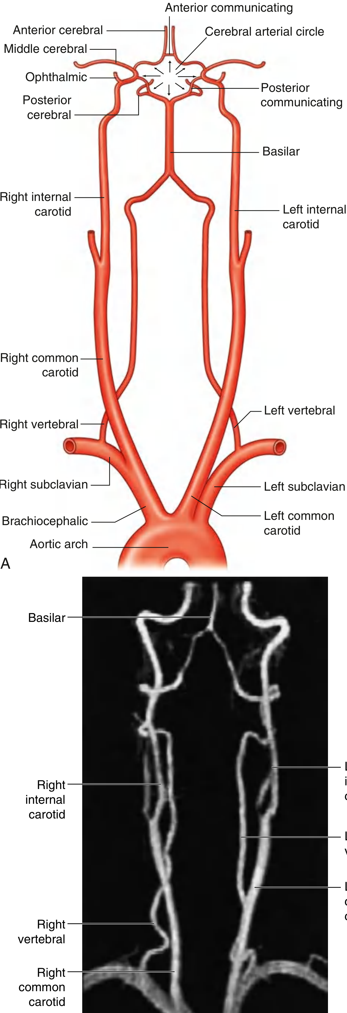
Fig. 8.39 - Arterial Supply to the Brain (Gray's Anatomy for Students)
1. Anterior Circulation: Internal Carotid Arteries (ICA)
- The two ICAs arise as terminal branches of the common carotid arteries in the neck.
- They ascend and enter the skull through the carotid canals.
- On entering the cranial cavity, each ICA gives off four main branches:
| Branch | Territory supplied |
|---|---|
| Ophthalmic artery | Orbit, retina |
| Posterior communicating artery (PComm) | Connects ICA to posterior cerebral artery (part of Circle of Willis) |
| Middle cerebral artery (MCA) | Most of the lateral surface of the cerebral hemisphere; basal ganglia and internal capsule via lenticulostriate branches |
| Anterior cerebral artery (ACA) | Medial wall of the cerebral hemisphere; superior surface of the corpus callosum |
2. Posterior Circulation: Vertebral and Basilar Arteries
Vertebral Arteries
- Arise from the first part of each subclavian artery in the lower neck.
- Pass superiorly through the foramina transversaria of the upper six cervical vertebrae.
- Enter the cranial cavity through the foramen magnum.
- Each vertebral artery gives off three branches before fusing with its partner to form the basilar artery:
- Posterior inferior cerebellar artery (PICA) - supplies posterior and inferior cerebellum and lateral medulla
- Posterior spinal artery - descends on posterior surface of spinal cord
- Anterior spinal artery (paired contributions unite into one) - descends in the anterior median fissure of the spinal cord
Basilar Artery
- Formed by the fusion of both vertebral arteries just inferior to the pons.
- Travels rostrally along the anterior aspect of the pons.
- Branches (caudal to rostral):
- Anterior inferior cerebellar arteries (AICA) - anterior and inferior cerebellum; facial nerve region
- Pontine arteries (small perforators)
- Superior cerebellar arteries (SCA) - superior cerebellum
- Terminates by bifurcating into the two posterior cerebral arteries (PCA)
Posterior Cerebral Arteries (PCA)
- Supply the medial wall of the occipital lobe and the inferior surface of the temporal lobe.
- Connected to the ICAs via the posterior communicating arteries (PComm).
3. The Circle of Willis (Cerebral Arterial Circle)
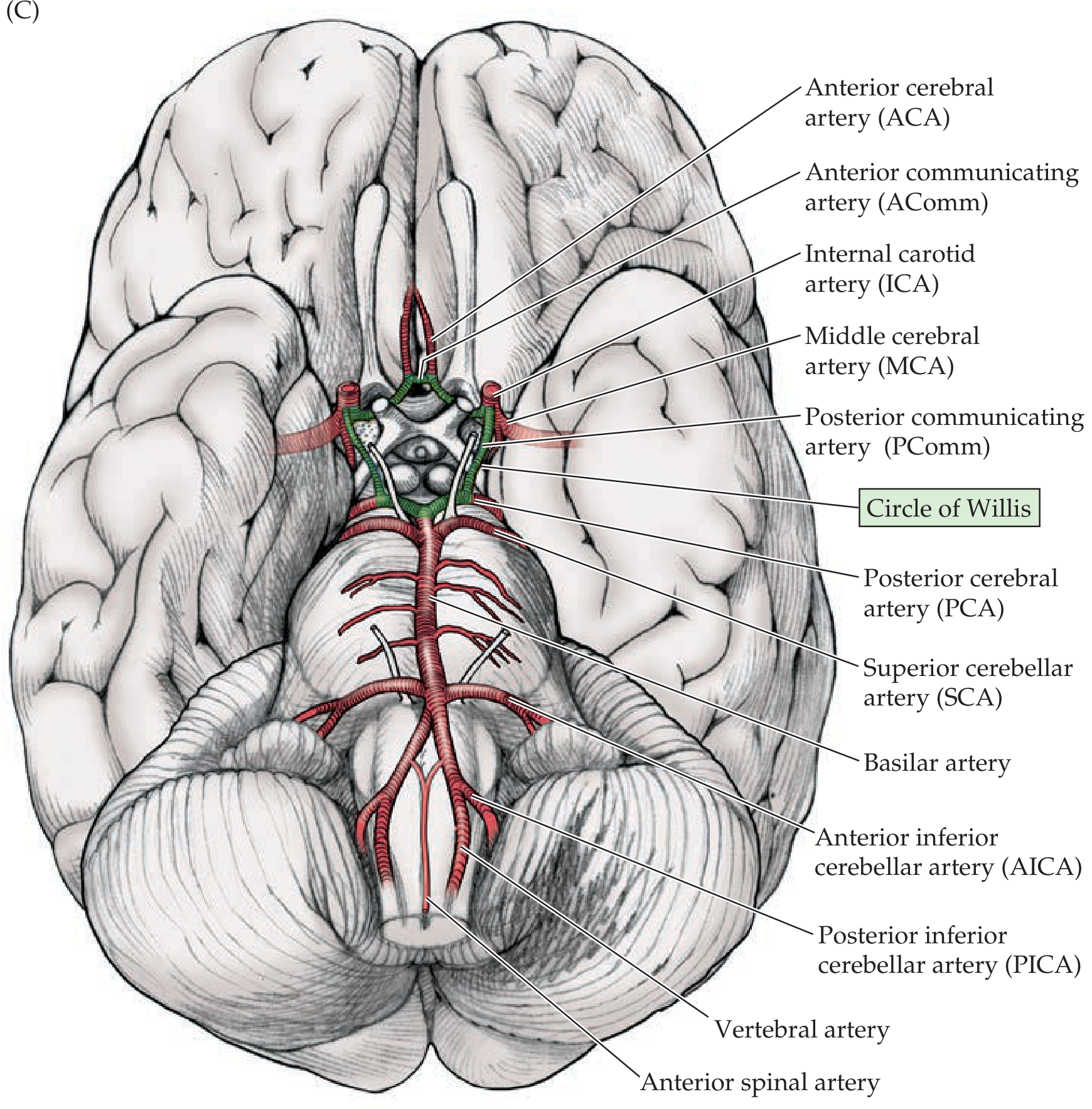
Circle of Willis showing all components - Neuroanatomy through Clinical Cases
The Circle of Willis is an anastomotic ring at the base of the brain formed by:
| Component | Connects |
|---|---|
| Anterior communicating artery (AComm) | Left and right anterior cerebral arteries to each other |
| Left and right ACAs | - |
| Left and right ICAs | - |
| Left and right posterior communicating arteries (PComm) | ICA to PCA on each side |
| Left and right PCAs | Terminal branches of basilar |
This ring provides an important collateral pathway - if one vessel is occluded, blood can theoretically be rerouted through the circle. However, in practice, the communicating arteries are often small and may not provide sufficient collateral flow.
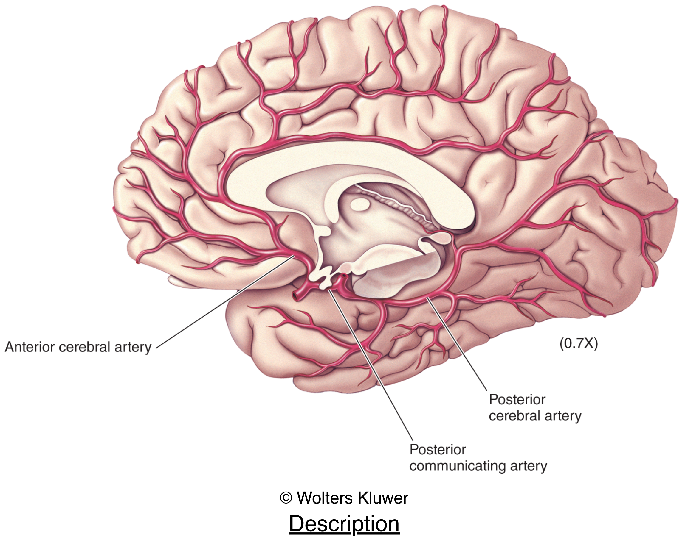
4. Cortical Territories - Which Artery Supplies What
Lateral surface: Primarily the MCA (most of the frontal, parietal, and temporal lobes on the convexity)
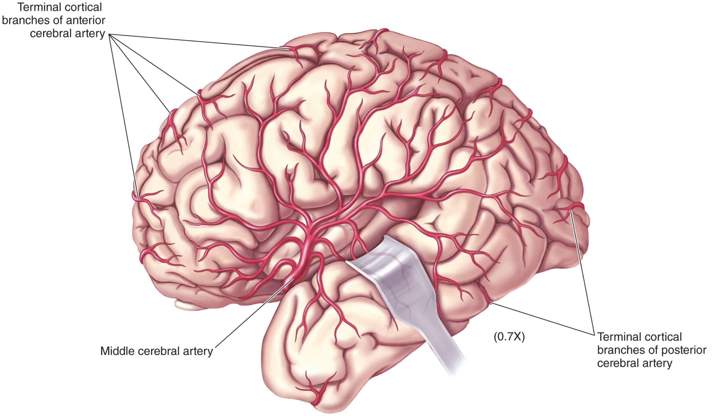
Medial surface: Primarily the ACA (medial frontal and parietal lobes); the PCA supplies the medial occipital lobe and inferior temporal lobe
Deep structures (basal ganglia, internal capsule): Lenticulostriate branches of the MCA (hence MCA strokes classically cause contralateral hemiplegia)
Brainstem and cerebellum: PICA, AICA, SCA, and pontine perforators from the vertebrobasilar system
5. Venous Drainage
- Venous blood drains from the brain into the dural venous sinuses (superior sagittal sinus, transverse sinus, sigmoid sinus, etc.).
- From there, it drains into the internal jugular veins bilaterally, which carry it back to the heart.
Clinical Correlate: Stroke
Interruption of blood flow causes a cerebrovascular accident (CVA):
- MCA occlusion (most common): contralateral hemiplegia/hemisensory loss, aphasia (dominant side), neglect (non-dominant side)
- ACA occlusion: contralateral leg weakness > arm weakness; personality changes
- PCA occlusion: contralateral homonymous hemianopia, memory deficits
- PICA/lateral medullary occlusion (Wallenberg syndrome): ipsilateral facial sensory loss, contralateral body sensory loss, dysphagia, vertigo, Horner's syndrome
- Basilar artery occlusion: locked-in syndrome (devastating - spares consciousness but paralyzes everything below the eyes)
Ischemic stroke can be treated with IV thrombolytics within 3-4.5 hours of symptom onset, or mechanical thrombectomy in suitable cases.
Sources: Gray's Anatomy for Students, p. 1017-1020; Neuroanatomy through Clinical Cases 3rd Edition, p. 70-71; Neuroscience: Exploring the Brain, 5th Edition, p. 705-708
This is a shared conversation. Sign in to Orris to start your own chat.