
Discuss the term alcoholic, chronic alcohol consumer a
chronic alcohol use disorder definition pathophysiology organ damage 2024
https://www.ncbi.nlm.nih.gov/books/NBK436003
"alcohol use disorder" AND "organ damage"
alcoholic liver disease cirrhosis fatty liver pathology
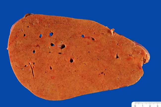
Imaging modality: gross pathology photograph of the liver (macroscopic specimen) illustrating diffuse hepatic steatosis characteristic of alcoholic fatty liver disease. The liver appears enlarged and homogenously pale yellow to orange, with a greasy, glistening cut surface due to lipid accumulation within hepatocytes. This macroscopic appearance reflects macrovesicular steatosis, which can progress to steatohepatitis and cirrhosis with continued alcohol exposure. The specimen is from a human liver; the background is a blue operational field used for pathology photography. Notable features include a relatively smooth capsule, uniform parenchymal coloration, and absence of discrete focal lesions in this view. The pathophysiology involves increased fatty acid influx, impaired beta-oxidation, and altered lipid export, with progression influenced by genetic and metabolic factors. Clinically, this pattern correlates with alcohol use disorder and elevated liver enzymes; correlation with history, serology, and imaging is essential. This gross finding establishes a groundwork for histologic confirmation of steatosis, inflammation, and fibrosis. The image is valuable for medical education, teaching hepatology, gastroenterology, and pathology about the spectrum from simple steatosis to steatohepatitis and cirrhosis, and for comparative studies of disease progression. Researchers can use this to illustrate macroscopic phenotypes in alcohol-related liver disease. This image supports diagnosis, education, and research efforts.
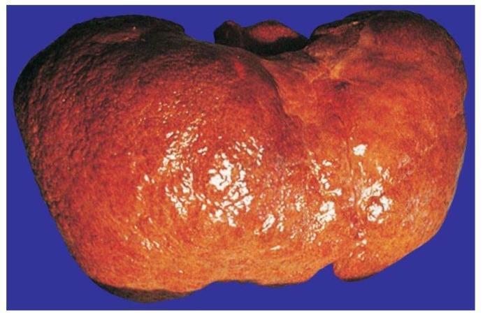
This clinical photograph displays a macroscopic view of a human liver specimen exhibiting advanced alcoholic cirrhosis. The organ demonstrates a characteristic micronodular surface, where the normally smooth capsule is replaced by a diffuse, granular texture. These numerous small, rounded protrusions represent regenerative nodules of hepatocytes surrounded by dense fibrotic septa. The liver color is predominantly reddish-brown with yellowish-tan areas, likely reflecting steatosis (fatty change) and variation in tissue density. Bright, fragmented light reflexes on the surface highlight the irregular, cobweb-like topography and uneven capsule tension typical of end-stage chronic liver disease. The specimen illustrates the pathological progression from chronic alcohol-induced injury to extensive fibrosis and architectural distortion, which are key diagnostic features of cirrhosis in hepatology and pathology education.
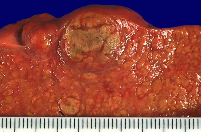
Gross pathology photograph of the liver surface from a case of alcoholic liver disease. The image shows diffuse micronodularity of the parenchyma with a coarse, irregular texture and reddish-brown coloration. Nodularity is predominantly micronodular (<3 mm) due to widespread fibrosis, with occasional larger regenerative nodules that may reflect hepatocyte regeneration during abstinence. The hepatic surface is nodular and uneven, indicating bridging fibrosis and loss of normal lobular architecture. A centimeter-scale ruler is present for size reference, confirming small nodules and expanded fibrous septa. This macroscopic pattern is characteristic of cirrhosis in advanced alcoholic liver disease, where irreversible architectural remodeling yields a nodular, firm liver with altered contour. Pathological significance includes portal hypertension risk, impaired synthetic function, and increased hepatocellular carcinoma risk. Clinically, this appearance correlates with long-standing alcohol exposure, potential ascites, and abnormal liver function tests. The image serves as an educational reference for hepatology, pathology, and medical education, illustrating end-stage liver disease, nodular regeneration, and fibrosis. Differential considerations include cirrhosis from other etiologies (viral hepatitis, biliary disease, nonalcoholic fatty liver disease) and variation in nodularity with disease stage and abstinence. This photograph aids gross-pathology correlation with histology and radiology in teaching cases and supports case-based learning.
The Alcoholic / Chronic Alcohol Consumer
1. Terminology and Definition
- Mild: 2-3 criteria
- Moderate: 4-5 criteria
- Severe: ≥6 criteria
2. Epidemiology
- In the US, 67.4% of adults report past-year alcohol use; 23.5% report binge drinking and 6.3% heavy alcohol use in the past month.
- An estimated 29.5 million Americans aged 12+ have AUD (2022 NSDUH data).
- Globally, harmful alcohol use causes approximately 5.9% of all deaths annually and accounts for 5.1% of the global disease burden.
- AUD is now the leading cause of liver transplantation in the United States, surpassing hepatitis C.
3. Pathophysiology of Ethanol
Metabolism
- Alcohol dehydrogenase (ADH) → acetaldehyde
- Microsomal ethanol oxidizing system (MEOS/CYP2E1) - induced with chronic use
- Catalase (minor)
- Altered redox state (excess NADH): impairs gluconeogenesis → hypoglycemia; promotes fatty acid synthesis → steatosis
- Acetaldehyde toxicity: protein adduct formation, DNA damage, mitochondrial injury, apoptosis
- CYP2E1 induction: generates reactive oxygen species (ROS) → oxidative stress and lipid peroxidation
- Gut permeability: increased intestinal permeability allows bacterial endotoxins (LPS) into portal blood → Kupffer cell activation → TNF-α, IL-1, IL-6, TGF-β1 → hepatic inflammation and fibrosis
4. Organ-by-Organ Effects of Chronic Alcohol Use
4.1 Liver (Most Common Site of Chronic Injury)
| Stage | Features | Reversibility |
|---|---|---|
| Fatty liver (Steatosis) | Develops in ~90% of heavy drinkers; hepatomegaly, mildly elevated LFTs | Reversible with abstinence |
| Alcoholic Steatohepatitis (ASH) | Steatosis + inflammation, Mallory-Denk bodies, neutrophilic infiltrate | Partially reversible |
| Alcoholic hepatitis | Acute-on-chronic liver injury, jaundice, fever, leukocytosis; high short-term mortality | Partially reversible |
| Cirrhosis | Irreversible fibrosis, nodular regeneration, portal hypertension | Irreversible |
- Cirrhosis accounts for ~50% of all cirrhosis deaths and 40-45% of all liver disease deaths.
- Only 30-35% of heavy long-term drinkers develop alcoholic hepatitis, and <20% develop cirrhosis - genetic and environmental factors play a role.
- Women are more susceptible at lower doses and shorter durations than men.
- Complications of cirrhosis: portal hypertension, esophageal varices (risk of massive hemorrhage), ascites, hepatic encephalopathy, hepatocellular carcinoma (HCC).
4.2 Nervous System
- Up-regulation of NMDA glutamate receptors and voltage-sensitive Ca²⁺ channels during chronic use. When alcohol is withdrawn, these channels are overactive → withdrawal seizures.
- Down-regulation of GABA-mediated responses explains why benzodiazepines (GABA enhancers) treat withdrawal effectively.
- Alcohol activates the mesolimbic dopamine reward circuit (nucleus accumbens) and modulates serotonin, opioid, and endocannabinoid systems → craving and dependence.
- Mild: tremors, anxiety, diaphoresis, tachycardia
- Severe: seizures, toxic psychosis, delirium tremens (confusion, hyperthermia, hypertension, hallucinations, death if untreated)
- Wernicke encephalopathy: classic triad of ophthalmoplegia, ataxia, and confusion (only ~10% present with all three). Reversible with prompt parenteral thiamine.
- Korsakoff psychosis: if Wernicke goes untreated or undertreated, patients develop dense anterograde and retrograde amnesia with confabulation. Less likely to be fully reversible. MRI shows mammillary body and thalamic atrophy.
- All patients with altered consciousness + history of alcohol use should receive IV thiamine immediately.
- Cerebellar degeneration (gait ataxia)
- Cerebral cortical atrophy and alcohol-related dementia
- Optic neuropathy with bilateral visual blurring
- Increased risk of stroke
4.3 Cardiovascular System
- Dilated cardiomyopathy ("alcoholic cardiomyopathy"): chronic heavy alcohol causes membrane disruption, mitochondrial dysfunction, sarcoplasmic reticulum dysfunction, intracellular phospholipid/fatty acid accumulation, and upregulation of voltage-gated Ca²⁺ channels → ventricular hypertrophy and dilatation. Prognosis is worse than idiopathic DCM if drinking continues; cessation can improve cardiac size and function.
- "Holiday heart" syndrome: acute atrial fibrillation triggered even by a single binge episode.
- Hypertension: alcohol use and withdrawal worsen hypertension.
- Accelerated coronary artery disease.
- Beri-Beri heart: thiamine deficiency leads to high-output cardiac failure with oedema.
- Munich beer heart syndrome: dilated cardiomyopathy described in heavy beer drinkers (partly from cobalt used as a beer additive historically).
4.4 Gastrointestinal Tract (beyond the liver)
- Gastritis: inflammation and increased susceptibility to gastric ulceration and GI bleeding.
- Esophageal varices: from portal hypertension secondary to cirrhosis - risk of fatal hemorrhage.
- Mallory-Weiss tears: from repeated vomiting.
- Pancreatitis: chronic alcohol use is the most common cause of chronic pancreatitis in the Western world. Alcohol directly injures pancreatic acinar cells, alters epithelial permeability, and promotes protein plug and calcium carbonate stone formation.
- Small intestinal injury: diarrhea, malabsorption of water-soluble vitamins (especially B vitamins).
- Malnutrition: ethanol provides "empty calories" (7 kcal/g) but displaces nutritious food intake.
4.5 Hematological Effects
- Bone marrow suppression: leukopenia, thrombocytopenia, anemia.
- Macrocytosis (MCV >100 fL): due to direct toxic effect on erythropoiesis AND folate/B12 deficiency. Useful as a screening marker.
- Coagulopathy: reduced hepatic synthesis of clotting factors (especially if cirrhosis is present).
- Thrombocythemia may occur in the context of underlying liver disease.
4.6 Endocrine and Metabolic Effects
- Hypoglycemia: impaired hepatic gluconeogenesis (excess NADH shifts substrate away from glucose).
- Ketosis: excess lipolytic hormones (cortisol, growth hormone) during withdrawal.
- Hypothyroidism: alcohol interferes with thyroid hormone metabolism.
- Osteoporosis: impaired bone formation and increased fracture risk.
- Gynecomastia and testicular atrophy: from alcohol-induced alteration of steroid hormone metabolism, compounded by liver disease.
- Hypogonadism: reduced testosterone.
- Fluid and electrolyte disturbances: hypokalemia (from vomiting, diarrhea, secondary hyperaldosteronism), hypomagnesemia, hypophosphatemia.
4.7 Immune System
- Immune function is suppressed in the lung: reduced alveolar macrophage function, impaired neutrophil chemotaxis, reduced T-cell function → predisposes to pneumonia and tuberculosis.
- Immune function is pathologically enhanced in the liver: Kupffer cell and hepatic stellate cell hyperactivation, excess cytokine production.
- Overall, chronic alcoholism increases infection susceptibility and worsens pneumonia outcomes.
4.8 Oncologic Effects
- Oral cavity, pharynx, larynx, esophagus
- Liver (secondary to cirrhosis → HCC)
- Colorectum
- Breast (even at low-to-moderate intake)
- Stomach, pancreas
4.9 Fetal Alcohol Spectrum Disorder (FASD)
- Intrauterine growth retardation
- Microcephaly
- Poor coordination
- Midfacial hypoplasia (flat face, short palpebral fissures, smooth philtrum)
- Minor joint anomalies
- Congenital heart defects
- Intellectual disability
5. Laboratory Markers of Chronic Alcohol Use
| Marker | Significance | Window of Detection |
|---|---|---|
| Blood alcohol level (BAL) | Acute intoxication | Hours |
| GGT (gamma-glutamyl transferase) | Induced by chronic use; sensitive but not specific | Days-weeks |
| MCV (mean corpuscular volume) | Macrocytosis from direct toxicity + folate deficiency | Weeks-months |
| CDT (carbohydrate-deficient transferrin) | Moderately specific; detects heavy drinking | ~2 weeks |
| Serum ethyl glucuronide (EtG) | Direct metabolite | ~5 days |
| Phosphatidylethanol (PEth) | Sensitive biomarker in blood | ~3-4 weeks |
| AST:ALT ratio >2:1 | Suggestive of alcoholic hepatitis | Days |
6. Withdrawal Management
- CIWA-Ar scale (Clinical Institute Withdrawal Assessment) guides severity monitoring.
- Benzodiazepines are the cornerstone of treatment (GABA-A agonism substitutes for alcohol).
- Thiamine (B1) IV should be given to all patients before glucose (to prevent precipitation of Wernicke encephalopathy).
- Severe cases (delirium tremens): ICU admission, IV diazepam or lorazepam, monitoring of electrolytes.
7. Treatment of AUD
- Naltrexone (oral or long-acting injectable): mu-opioid receptor antagonist - reduces craving and reward. Recent review confirms opioid circuit involvement.
- Acamprosate: NMDA/GABA modulator - reduces protracted withdrawal symptoms and craving. Best for abstinence maintenance.
- Disulfiram: ALDH inhibitor - causes aversive acetaldehyde accumulation if alcohol consumed (flushing, nausea, tachycardia, hypotension).
- Baclofen: GABA-B agonist - has some evidence in patients with severe liver disease.
- Cognitive behavioral therapy (CBT), motivational interviewing, 12-step programs (Alcoholics Anonymous), SMART Recovery.
8. Summary
- Acetaldehyde toxicity and oxidative stress
- Gut permeability and endotoxemia driving hepatic inflammation
- Nutritional deficiencies (especially thiamine, folate, B12)
- Neurotransmitter dysregulation (GABA, NMDA, dopamine) causing dependence and withdrawal
- Immune dysregulation and oncogenic mechanisms
- Goldman-Cecil Medicine (Chapter 138 - Alcohol-Associated Liver Disease)
- Katzung's Basic and Clinical Pharmacology, 16th Edition
- Robbins & Kumar Basic Pathology
- Kaplan & Sadock's Comprehensive Textbook of Psychiatry
- Parikh's Textbook of Medical Jurisprudence, Forensic Medicine and Toxicology
- StatPearls - AUD Screening and Management
- AUD Neurobiology and Therapeutics, PMC 2022