
Explain alpha gamma co activation from standard textbook of physiology for 5 markers
Alpha-Gamma Co-activation
Definition
- Approximately 31% of all motor nerve fibers to a muscle are small type-A gamma efferent fibers.
- Both extrafusal (skeletal) muscle fibers (via α-motor neurons) and intrafusal muscle fibers of the muscle spindle (via γ-motor neurons) contract at the same time.
Mechanism
- A voluntary motor command from the cerebral cortex (or any higher brain area) travels down to the spinal cord.
- It activates α-motor neurons - causing contraction of the extrafusal skeletal muscle fibers.
- Simultaneously, the same descending signal activates γ-motor neurons - causing the contractile polar ends of the intrafusal fibers within the muscle spindle to shorten.
- This shortening of the intrafusal fibers stretches the central nuclear bag/chain region, keeping the Ia afferent endings under tension throughout the contraction.
Purpose / Significance (the "why")
1. Prevents "Unloading" of the Spindle
2. Maintains Continuous Reflex Control
3. Preserves the Damping Function
4. Joint Stabilization
Control of γ-Motor Neurons in Co-activation
- Bulboreticular facilitatory region of the brain stem (most direct)
- Cerebellum (fine-tuning)
- Basal ganglia
- Cerebral cortex
Summary Table
| Feature | Detail |
|---|---|
| Definition | Simultaneous activation of α- and γ-motor neurons by descending motor commands |
| Effect on extrafusal fibers | Direct contraction (via α-motor neurons) |
| Effect on intrafusal fibers | Shortening of polar ends, keeping spindle taut (via γ-motor neurons) |
| Key benefit | Prevents spindle unloading; maintains Ia afferent feedback during voluntary movement |
| Controls smoothness | Preserves damping function at all muscle lengths |
| Higher control | Bulboreticular area, cerebellum, basal ganglia, cerebral cortex |
- Guyton and Hall Textbook of Medical Physiology - "Role of the Muscle Spindle in Voluntary Motor Activity"
- Ganong's Review of Medical Physiology, 26th Edition - "Effects of γ-Motor Neuron Discharge"
Give answer of alpha gamma co activation for 3 marker proper answer format
Alpha-Gamma Co-activation
- ~31% of all motor fibers to a muscle are small type-A gamma efferent fibers
- Both extrafusal fibers (via α) and intrafusal fibers of the muscle spindle (via γ) contract together
Taste pathway flowchart with diagram
taste pathway gustatory pathway brain diagram
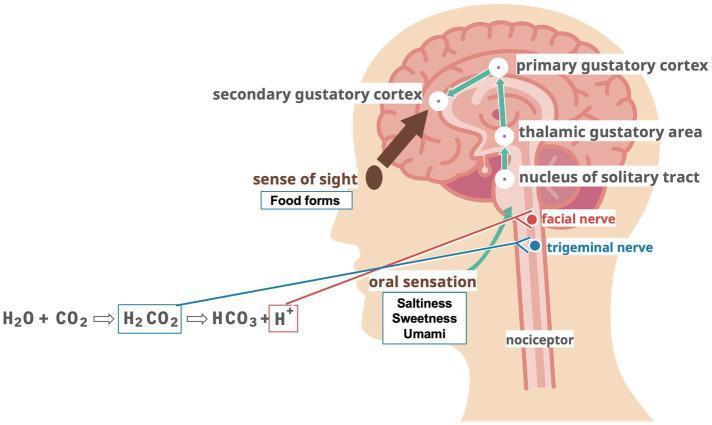
This medical illustration depicts the neuroanatomical pathway of taste and sensory integration in the human head and brain. The diagram illustrates 'oral sensation' (saltiness, sweetness, umami) and carbonic acid stimulation (chemical reaction: H2O + CO2 → H2CO2 → HCO3 + H+) being transmitted via the facial nerve and trigeminal nerve to the nucleus of the solitary tract in the brainstem. The ascending gustatory pathway continues through the thalamic gustatory area to the primary gustatory cortex, then to the secondary gustatory cortex. The illustration also incorporates the 'sense of sight' (food forms) as a contributing input to the secondary gustatory cortex, highlighting the multisensory nature of flavor perception. Nociceptors are also indicated along the spinal tract. This pathophysiology diagram is used to explain the hypothesized mechanisms of action for taste, vision, and chemical stimulation in the context of nutrition and swallowing function (dysphagia management).
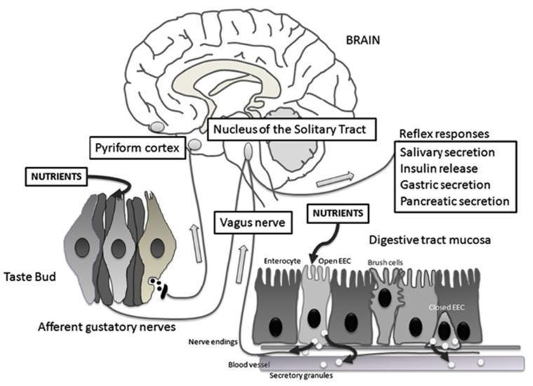
A medical pathophysiology diagram illustrating the gut-brain axis and taste circuitry. The visual depicts two parallel pathways for nutrient sensing: the oral gustatory system and the gastrointestinal system. In the oral pathway, nutrients stimulate a taste bud, activating afferent gustatory nerves that project to the pyriform cortex and the nucleus of the solitary tract (NTS) in the brainstem. In the gastrointestinal pathway, the digestive tract mucosa is shown with enterocytes, brush cells, and enteroendocrine cells (EECs). 'Open' EECs respond to luminal nutrients by releasing secretory granules (containing hormones like CCK and GLP-1) into the interstitial space or bloodstream, stimulating vagus nerve endings. These visceral signals also terminate at the NTS. The brain integrates these inputs to trigger physiological reflex responses, explicitly listed as salivary secretion, insulin release, gastric secretion, and pancreatic secretion. This diagram highlights the neuroendocrine coordination between peripheral sensory organs and central homeostatic regulation.
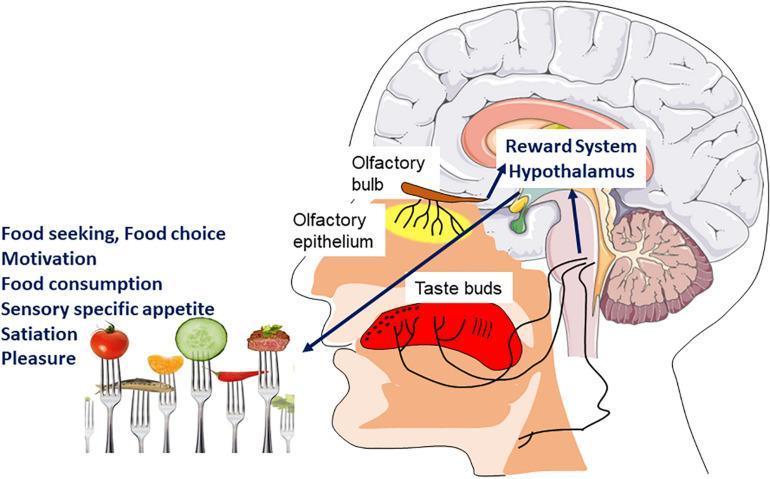
A medical anatomical diagram illustrating the neurobiological pathways of food perception and the hedonic sensory signal. The illustration uses a sagittal view of the human head and brain to trace olfactory and gustatory inputs. The olfactory pathway is shown starting at the olfactory epithelium in the nasal cavity, projecting to the olfactory bulb. The gustatory pathway is depicted via taste buds on the tongue. Both chemosensory signals converge and project toward integrated central brain regions labeled as the Reward System and Hypothalamus. The diagram connects visual food stimuli (shown on forks) to complex physiological and psychological outcomes including food seeking, motivation, food consumption, sensory-specific appetite, satiation, and pleasure. This flowchart-style diagram serves as an educational tool for understanding the integration of sensory inputs into the homeostatic and hedonic control of feeding behavior.
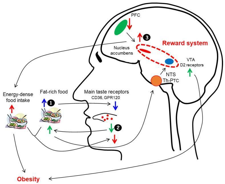
This medical schematic diagram illustrates the complex neurobiological pathophysiology and feedback loops linking fat taste perception, the brain's reward system, and obesity. The illustration highlights three central hypotheses or mechanisms. First (1), high intake of fat-rich foods leads to the downregulation of lingual taste receptors CD36 and GPR120 via negative feedback. Second (2), decreased fat taste sensitivity leads to increased consumption of high-fat foods to achieve hedonic satisfaction. Third (3), overactivation of the brain's reward system—comprising the Nucleus Accumbens and the Ventral Tegmental Area (VTA) with D2 receptors—stimulates energy-dense food intake while inhibiting the Prefrontal Cortex (PFC) and further decreasing fat taste sensitivity. The diagram also maps the ascending gustatory pathway from the tongue through the Nucleus Tractus Solitarius (NTS) to the ventroposteromedial nucleus of the Thalamus (Th) and Primary Taste Cortex (PTC). Arrows indicate directed influence, showing how these interlocking systems promote the development of obesity through a self-reinforcing cycle of dysregulated eating behavior and neural reward signaling.
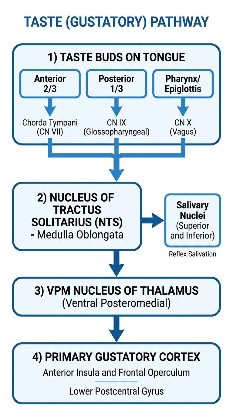
A clean vertical flowchart diagram of the taste (gustatory) pathway, showing the following steps in labeled boxes connected by arrows: 1) Taste Buds on Tongue - Anterior 2/3 via Chorda Tympani (CN VII), Posterior 1/3 via CN IX (Glossopharyngeal), Pharynx/Epiglottis via CN X (Vagus); all three arrows merge into 2) Nucleus of Tractus Solitarius (NTS) - Medulla Oblongata; then arrow to 3) VPM Nucleus of Thalamus (Ventral Posteromedial); then arrow to 4) Primary Gustatory Cortex - Anterior Insula and Frontal Operculum / Lower Postcentral Gyrus. Also show a branch from NTS going to Salivary Nuclei (Superior and Inferior) for reflex salivation. Use clean medical illustration style with blue and white color scheme, clear labels, professional anatomy diagram look.
Taste (Gustatory) Pathway
Flowchart
Textbook Diagram (Guyton & Hall)
Detailed Pathway (Step by Step)
TASTE BUDS
(~5000 buds on tongue, soft palate, epiglottis, pharynx)
        |
        |── Anterior 2/3 of tongue
        |       ↓
        |   Lingual Nerve → Chorda Tympani → CN VII (Facial Nerve)
        |       Cell body: Geniculate Ganglion
        |
        |── Posterior 1/3 of tongue
        |       ↓
        |   CN IX (Glossopharyngeal Nerve)
        |       Cell body: Petrosal (Inferior) Ganglion
        |
        |── Pharynx / Epiglottis / Base of tongue
                ↓
            CN X (Vagus Nerve)
                Cell body: Nodose (Inferior) Ganglion
                        |
                        ▼
        ╔═══════════════════════════════════╗
        ║  NUCLEUS OF TRACTUS SOLITARIUS   ║
        ║      (NTS) — Medulla Oblongata   ║
        ║   (1st order neuron synapses)    ║
        ╚═══════════════════════════════════╝
                |                   |
                |                   ↓
                |         Superior & Inferior
                |         Salivary Nuclei
                |         (Reflex: salivation)
                ↓
        2nd Order Neuron ascends via
        IPSILATERAL MEDIAL LEMNISCUS
                        |
                        ▼
        ╔═══════════════════════════════════╗
        ║  VPM NUCLEUS OF THALAMUS         ║
        ║  (Ventroposteromedial Nucleus)   ║
        ║  — slightly medial to facial     ║
        ║    representation                ║
        ╚═══════════════════════════════════╝
                        |
                        ▼
                3rd Order Neuron
                        |
                        ▼
        ╔═══════════════════════════════════╗
        ║  PRIMARY GUSTATORY CORTEX        ║
        ║  • Anterior Insula               ║
        ║  • Frontal Operculum             ║
        ║  • Lower Postcentral Gyrus       ║
        ║    (curling into Sylvian fissure)║
        ╚═══════════════════════════════════╝
Key Points to Remember
| Feature | Detail |
|---|---|
| Nerve for anterior 2/3 tongue | Chorda tympani branch of CN VII |
| Nerve for posterior 1/3 tongue | CN IX (Glossopharyngeal) |
| Nerve for pharynx/epiglottis | CN X (Vagus) |
| 1st relay | NTS (Nucleus Tractus Solitarius) - Medulla |
| 2nd relay | VPM nucleus of Thalamus |
| Cortical area | Anterior insula + Frontal operculum (ipsilateral) |
| Pathway laterality | Ipsilateral (does NOT cross) |
| Reflex branch from NTS | Superior & Inferior salivary nuclei → salivation |
Important Notes
- The taste pathway is predominantly ipsilateral - unlike most other sensory pathways, it does not decussate prominently
- The cortical taste area is rostral to the face area of the postcentral gyrus, lying in the parietal operculum/insula
- Taste buds survive only ~10 days and are continually replaced; if the sensory nerve is cut, taste buds degenerate
- Saliva acts as a solvent for tastants, which must dissolve before stimulating taste receptors
- From the NTS, taste signals also reach the hypothalamus and amygdala for emotional/behavioral responses to taste