
staphylococcus aureus with mostly pertaining to ent
Staphylococcus aureus gram stain microscopy clusters
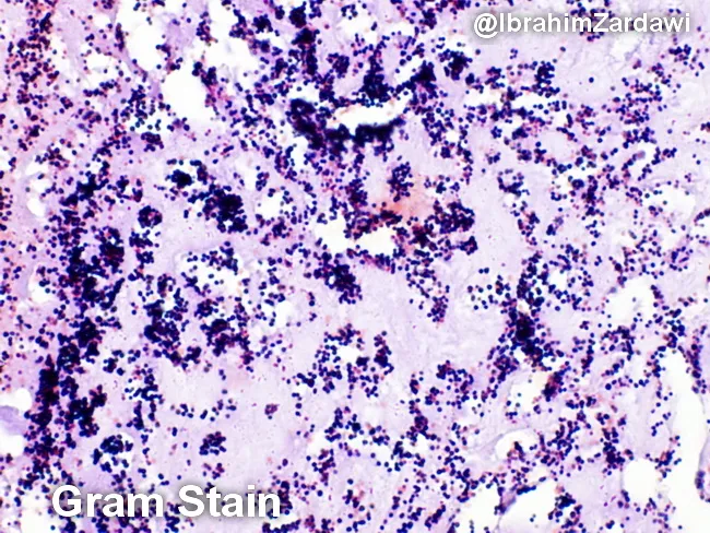
Imaging modality: Light microscopy; Gram staining of a histologic section of a cardiac valve vegetation from suspected infective endocarditis. The specimen shows a dense, friable vegetative mass within valve tissue characterized by a fibrinous, platelet-rich matrix in which numerous Gram-positive cocci are adherent. On the Gram stain, the cocci appear as purple spherical cells arranged in clusters and sometimes in short chains, consistent with Gram-positive organisms. The background demonstrates inflammatory cells and debris typical of endocardial infection. The histologic architecture indicates vegetative lesions with microbial colonization and a biofilm component. These findings correlate with infective endocarditis due to cocci such as Staphylococcus aureus or viridans-group streptococci in clinical practice, though Gram stain alone cannot specify species. The image is a micrograph provided with permission by Dr. Ibrahim Zardawi; image shows classic histopathology of bacterial endocarditis. The diagnostic significance lies in confirming bacterial endocarditis and guiding immediate management including empiric antibiotic therapy targeting Gram-positive cocci while awaiting culture results. This image is valuable for pathology education, microbiology correlation, and endocarditis research. Potential clinical use cases include training in Gram stain interpretation, distinguishing bacterial endocarditis from sterile vegetations, and supporting diagnosis when blood cultures are pending.
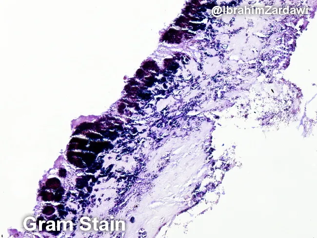
This histology image is a light-micrograph of a Gram-stained valvular vegetation from a case of infective endocarditis. The specimen is cardiac valvular tissue; the vegetation comprises a fibrin-rich, platelet-laden thrombus with embedded bacterial colonies along the endocardial surface. Gram staining highlights Gram-positive cocci as purple to deep purple clusters within the thrombus, indicating organisms such as Streptococcus spp., Staphylococcus spp., or Enterococcus. The bacterial colonies are adherent to the valvar surface within a dense inflammatory milieu, with neutrophilic infiltrate and tissue necrosis limited to microdomains. The histologic architecture demonstrates vegetations typical of endocarditis: a friable, topographically irregular lesion on the valve with microabscess-like foci and bacterial encasement within fibrin. The presence of Gram-positive bacteria on histology supports a bacterial endocarditis diagnosis and guides antimicrobial therapy pending culture confirmation. Clinically, such histologic confirmation is critical for distinguishing infectious from non-infectious valve pathology, for clarifying the etiologic category of pathogens (oral flora-related viridans Streptococcus, Staphylococcus aureus, enterococci), and for correlating with blood culture, echocardiographic findings, and patient risk factors. This image is valuable for educational purposes, illustrating Gram stain morphology in endocardial vegetations and reinforcing the need for targeted antibiotics and possible surgical consultation.
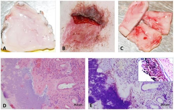
This composite image provides a comparative study of Staphylococcus aureus infection in an abrasion model, utilizing clinical photographs and histopathology. Panels A-C show macroscopic views: A displays an uninfected abrasion control with smooth, uniform pinkish-white tissue; B demonstrates an S. aureus-infected lesion characterized by a dark brown-red scab, serous exudate, and significant inflammation; C illustrates the ventral side of the infected site with visible hypervascularization. Panels D and E show corresponding microscopic findings at 24 hours post-infection. Panel D is an H&E stained section (50.0 µm scale) showing dense infiltration of inflammatory cells within the dermis and disrupted tissue architecture. Panel E features a Gram stain revealing large, deep purple clusters of Gram-positive cocci embedded within the extracellular matrix, with a higher-magnification inset detailing the bacterial morphology. This sequence serves as an educational resource for identifying macroscopic signs of acute pyogenic infection and correlating them with the presence of bacteria and host immune response.
Staphylococcus aureus in ENT - A Comprehensive Overview
Microbiology Basics (ENT-Relevant)
| Factor | Mechanism | ENT Implication |
|---|---|---|
| Coagulase | Clots plasma around organism | Abscess formation (peritonsillar, retropharyngeal, deep neck) |
| Hyaluronidase | Degrades connective tissue | Facilitates spread in head-neck spaces |
| Exfoliative toxin | Skin layer separation | Impetigo of external ear/nose |
| Enterotoxins (superantigens) | Non-MHC-restricted T-cell activation | Chronic rhinosinusitis with nasal polyps |
| Panton-Valentine Leukocidin (PVL) | Pore-forming toxin - destroys leukocytes | Recurrent furunculosis; rare necrotizing infection |
| Biofilm | Extracellular slime matrix | Chronic sinusitis, chronic otitis media, middle ear disease |
Nasal Carriage - The ENT Reservoir
- Nasal carriage is the reservoir for recurrent furunculosis of the nasal vestibule
- Carriers spread infection to surgical sites
- Pre-operative MRSA decolonization (mupirocin nasal ointment) targets this reservoir
ENT Conditions Caused by S. aureus
1. External Ear
- The most common bacteria are Pseudomonas aeruginosa (first) and S. aureus (second), followed by S. epidermidis. Polymicrobial infection is common.
- Management: Ototopical antibiotics. Acetic acid drops are active against both methicillin-resistant and sensitive S. aureus (MRSA and MSSA) as well as quinolone-resistant strains.
- If initial ototopical therapy fails, consider: self-instrumentation trauma, malignant otitis externa, contact dermatitis, or failure of adherence.
- Infection of a hair follicle, predominantly S. aureus. Presents with severe, exquisite localized pain in the cartilaginous EAC.
- Systemic antibiotics (flucloxacillin) are required for cellulitis surrounding furunculosis.
- If associated with abscess, incision and drainage.
- Although Pseudomonas aeruginosa is the overwhelming causative agent (>95%), rare cases due to S. aureus and S. epidermidis are documented.
- Ciprofloxacin is the drug of choice for malignant OE regardless of the organism (covers Pseudomonas primarily).
- ENT key point: When MRSA is the causative agent, ciprofloxacin-resistant MRSA has been reported - in vitro susceptibility testing is necessary.
2. Middle Ear & Mastoid
- S. aureus is a less common but recognized cause of AOM, particularly in neonates and in complicated/refractory cases. The dominant pathogens are S. pneumoniae and H. influenzae.
- Antibiotic controversy: Most cases resolve spontaneously. If antibiotics are used, amoxicillin is first line; co-amoxiclav or cephalosporins for treatment failure.
- S. aureus-caused AOM is more likely to progress to complications.
- S. aureus is a significant causative agent.
- Conservative management (IV antibiotics without surgery) can be attempted first; about one-third settle within 24-48 hours.
- Subperiosteal abscess formation - a hallmark of S. aureus virulence via coagulase activity - mandates surgical drainage.
- S. aureus can form biofilms on the mucosal surfaces of the middle ear, just as it does on sinuses and tonsils. Biofilm organisms:
- Show reduced metabolic activity, dividing infrequently
- Are resistant to antibiotic penetration through the slime matrix
- Communicate via quorum sensing, regulating virulence gene expression
- Can shed planktonic organisms, causing acute exacerbations on a background of chronic disease
3. Nose & Paranasal Sinuses
- Infection of hair follicles in the nasal vestibule - almost always S. aureus. Presents as painful, tender swelling at the nasal tip/columella.
- Treatment: topical mupirocin for mild cases; flucloxacillin orally for spreading cellulitis.
- Critical danger: The facial "danger triangle" - thrombosis of veins in this area can propagate to the cavernous sinus via the ophthalmic veins. All cases of nasal furunculosis with systemic features require IV antibiotics.
- S. aureus is a recognized but less common causative agent compared to S. pneumoniae, H. influenzae, and M. catarrhalis.
- It becomes more important in:
- Hospital-acquired sinusitis (nasotracheal intubation patients)
- Immunocompromised patients
- Complications (orbital/intracranial extension)
- S. aureus colonizes the nasal/sinus mucosa and produces staphylococcal enterotoxins that act as superantigens - bypassing normal MHC-II antigen presentation to non-specifically activate large numbers of T-cells
- This drives a polyclonal Th2-skewed inflammatory response with:
- Elevated total and specific IgE against staphylococcal toxins in polyp tissue
- Eosinophilic infiltration
- IL-5, IL-13 overproduction
- S. aureus colonization and IgE formation against enterotoxins is significantly increased in patients with nasal polyposis compared to controls
- Staphylococcal exotoxins have been detected directly in nasal polyp tissue
- Intracellular S. aureus has been associated with worse prognosis in CRS
4. Throat and Deep Neck Spaces
- S. aureus contributes to tonsillar biofilm formation. Biofilms are well recognized on tonsillar mucosal surfaces.
- In peritonsillar abscess, the organism is often found in mixed cultures alongside streptococci and anaerobes.
- The coagulase-mediated abscess formation is a key virulence mechanism.
- S. aureus is a major causative organism, especially in children (lymphadenitis suppuration).
- Management: IV antibiotics (flucloxacillin for MSSA, vancomycin for MRSA) + surgical drainage when fluctuant.
- Flucloxacillin is the antibiotic of choice for staphylococcal cellulitis in the head and neck.
- Penicillin V/amoxicillin covers streptococcal cellulitis/erysipelas, but not staphylococci.
- If MRSA is suspected (health-care-associated, previous MRSA history, failed flucloxacillin): vancomycin IV or doxycycline/trimethoprim orally for mild cases.
MRSA - The ENT Challenge
| Feature | Detail |
|---|---|
| Emerged | 1960s; now community and hospital |
| Mechanism | mecA gene cassette - alters cell wall penicillin-binding proteins |
| Resistance | All beta-lactams including flucloxacillin |
| Treatment (invasive) | Vancomycin IV (gold standard) |
| Treatment (mild/topical) | Mupirocin topically; TMP-SMX or doxycycline orally |
| Dual therapy | May add rifampicin, fusidic acid, clindamycin, gentamicin for biofilm-associated infection |
| VRSA | Extremely rare (case reports only); vancomycin remains effective |
| PVL-positive MRSA | Recurrent furunculosis; specific infection control measures from public health |
- Anterior nares mupirocin ointment (2%, 5 days pre-op)
- Chlorhexidine body wash
- Reduces surgical site infection risk in head and neck procedures
Biofilm in ENT - The Unifying Concept
- Tonsils
- Sinus mucosa
- Middle ear mucosa
- Surgical implants (cochlear implants, ossicular prostheses, tympanostomy tubes)
- Reversible planktonic attachment to surface
- Irreversible attachment with extracellular slime matrix
- Quorum sensing - coordinated gene regulation
- Shedding of planktonic bacteria - causing acute-on-chronic flares
- Antibiotics penetrate poorly; reduced metabolic activity renders bactericidal drugs less effective
Treatment Summary for ENT
| Condition | Organism | Drug of Choice |
|---|---|---|
| Nasal furunculosis (mild) | MSSA | Topical mupirocin |
| Nasal furunculosis / EAC furunculosis (systemic) | MSSA | Flucloxacillin PO/IV |
| Cellulitis of pinna/face | MSSA | Flucloxacillin PO/IV |
| AOM (S. aureus) | MSSA | Amoxicillin-clavulanate |
| Deep neck abscess / PTA | MSSA | Flucloxacillin IV (± metronidazole for anaerobes) |
| Any invasive MRSA infection | MRSA | Vancomycin IV |
| Malignant OE | Pseudomonas (rarely MRSA) | Ciprofloxacin (check sensitivities if MRSA) |
| Biofilm-associated (refractory) | MSSA/MRSA | Add rifampicin, fusidic acid, or clindamycin |
Quick-Reference Key Points (Scott-Brown's)
- S. aureus = coagulase positive, major human pathogen, anterior nares reservoir
- 20% permanent nasal carriers, 60% intermittent
- Flucloxacillin = drug of choice for staphylococcal cellulitis in ENT
- Vancomycin = drug of choice for invasive MRSA
- Superantigen mechanism links S. aureus enterotoxins to nasal polyposis
- Biofilms form on tonsils, sinus mucosa, and middle ear - explain chronic/recurrent ENT disease
- PVL-positive strains: recurrent furunculosis, require special infection control
- MRSA resistance via mecA gene; VRSA remains extremely rare