
Paediatric airway in detail for long question answer according to standard ent textbooks and in simplified manner, do not miss any of the important points or information
paediatric airway anatomy differences adult child larynx
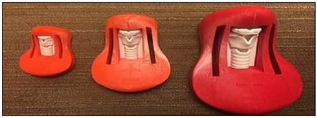
This clinical photograph displays three 3D-printed anatomical models used as cricothyroidotomy trainers, representing infant, child, and adult scales. Arranged from left to right in ascending size, the trainers consist of a molded red or orange-red plastic base housing a white internal anatomical insert. Each insert depicts the larynx and upper trachea, featuring landmarks such as the thyroid cartilage, cricoid cartilage, and tracheal rings. The models highlight developmental differences in airway anatomy, with the adult model on the right showing the most pronounced laryngeal prominence and larger cricothyroid membrane space. These specimens are used in surgical skills labs for emergency airway management training, specifically for practicing the identification of anterior neck landmarks and the performance of surgical cricothyroidotomy across different patient demographics. The models demonstrate the application of 3D printing in creating low-cost, age-specific medical simulation tools for emergency medicine and surgical education.
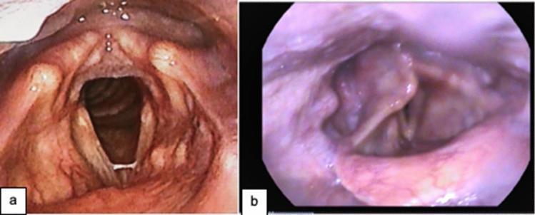
This comparative figure features two laryngeal endoscopic images (a and b) illustrating post-intubation sequelae in adult patients. Image 'a' displays the larynx of a 56-year-old male with thinned vocal folds and a visible fibrous band located mid-right vocal fold; however, the airway remains patent with symmetrical adduction and abduction preserved. Image 'b' shows a 58-year-old male with significant laryngeal structural irregularities, including bilateral vocal fold palsy, right arytenoid prolapse, and posterior glottic stenosis. The vocal folds are shown in maximal abduction, revealing a severely narrowed glottic and subglottic airway due to stenosis. These diagnostic images demonstrate typical late-stage complications of prolonged mechanical ventilation and endotracheal intubation, such as those observed in COVID-19 survivors. The focus is on laryngeal anatomy, vocal fold pathology, and airway patency, serving as an educational resource for speech-language pathology, otolaryngology, and intensive care medicine regarding the detection and classification of laryngeal trauma and stenosis.
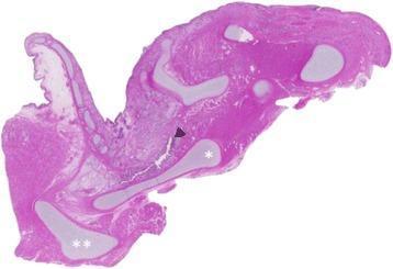
This diagnostic image is a low-magnification (1.25x) histological sagittal section of a human larynx stained with Hematoxylin and Eosin (H&E). The slide illustrates key upper airway anatomy, including the epiglottis, thyroid cartilage (marked with a single asterisk '*'), and the hyoid bone (marked with double asterisks '**'). The pathology demonstrated is tracheal agenesis, a rare congenital anomaly. The laryngeal lumen is seen terminating blindly, indicated by a black arrowhead (◀), rather than communicating with the trachea, which is absent from its expected anatomical position to the right of the larynx in this section. The cartilaginous structures appear as lighter, well-defined eosinophilic regions within the surrounding muscular and connective tissue. This image is clinically significant for neonatology and pediatric pathology as it provides visual evidence of Floyd Type I tracheal agenesis, highlighting the total disruption of the continuity between the larynx and the distal respiratory system.
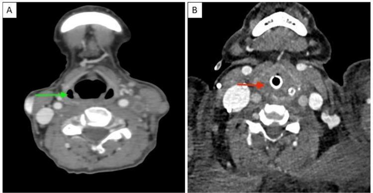
This Comparison Chart features two side-by-side axial contrast-enhanced computed tomography (CECT) images of the neck, illustrating the differences between normal anatomy and pathological upper airway edema. Image A (left) displays normal anatomy at the level of the larynx and hypopharynx. A green arrow indicates the bilateral, air-filled pyriform sinuses, which appear as clear, crescentic radiolucent areas. Image B (right) demonstrates a pathological state characterized by significant laryngeal and hypopharyngeal soft tissue edema. A red arrow points to the complete effacement and obliteration of the pyriform sinuses due to tissue swelling. Additionally, Image B shows an endotracheal tube in situ within the tracheal lumen, identifiable as a hyperdense circular ring with a central air column. The surrounding subcutaneous tissues in Image B exhibit increased density and reticulation consistent with subcutaneous edema. This comparison is clinically relevant for diagnosing airway compromise and identifying radiological signs of severe inflammation or angioedema in the cervical region.
laryngomalacia stridor infant endoscopy
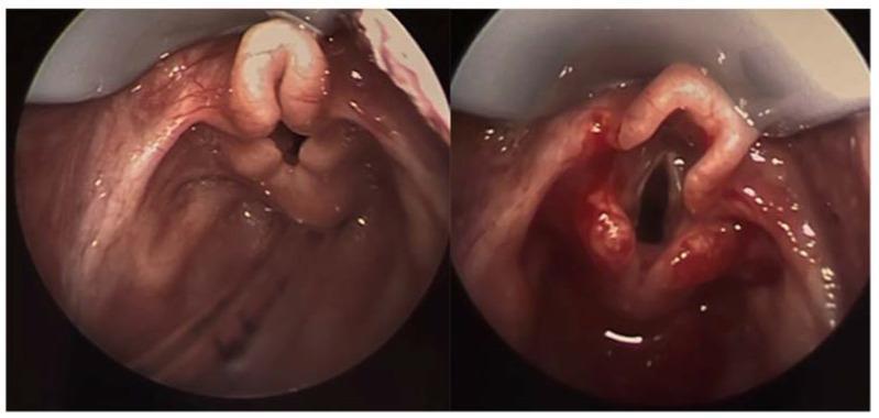
This side-by-side endoscopy comparison displays the laryngeal structures of a patient with laryngomalacia before and after surgical intervention. The left image shows the preoperative state, characterized by an elongated, 'omega-shaped' epiglottis that is collapsed inward and posteriorly. The aryepiglottic folds appear shortened and redundant, contributing to significant supraglottic crowding and obstruction of the glottic opening. The right image illustrates the results immediately following a supraglottoplasty. In this postoperative view, the tension of the aryepiglottic folds has been released, and redundant tissue has been reduced. This surgical modification has resulted in a much wider, more patent airway, with the true vocal cords and glottis now clearly visible and unobstructed. The comparison highlights the typical anatomic defects of congenital laryngomalacia and the effectiveness of surgical supraglottoplasty in restoring upper airway patency. Clinical relevance includes the management of pediatric stridor and respiratory distress caused by supraglottic collapse.
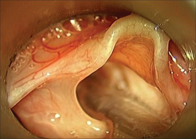
This diagnostic image is an intraoperative endoscopic view of the pediatric larynx during suspension microlaryngoscopy. The clinical photograph demonstrates the hallmark features of laryngomalacia in a 3-month-old infant. The primary finding is the significant shortening and thickening of the aryepiglottic folds, which appear pale, redundant, and edematous. These foreshortened folds contribute to the inward collapse of the supraglottic structures, partially obscuring the laryngeal inlet. The mucosal surface shows a moderate degree of vascularity with fine, branching subepithelial vessels and small, clear fluid-filled vesicles consistent with localized edema or chronic inflammation. Specular reflections from the moist mucosal surface are visible under the endoscopic light source. The educational focus of the image is the anatomical malformation of the supraglottic tissues that leads to inspiratory stridor in pediatric patients, illustrating the surgical landmarks required for supraglottoplasty.
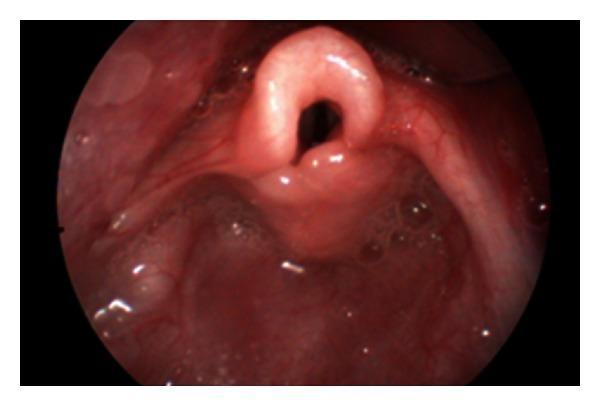
This clinical photograph displays a flexible laryngoscopic view of the infant larynx, demonstrating classic diagnostic features of laryngomalacia. The primary pathological finding is an omega-shaped or tubular epiglottis, which appears tightly curled and rolled upon itself. Surrounding the laryngeal inlet, the aryepiglottic folds are notably shortened, contributing to the constriction of the airway space. Additionally, there is prominent, redundant mucosa over the arytenoid cartilages, appearing as thickened, pinkish-red tissue that can collapse into the glottis during inspiration. The surrounding pharyngeal mucosa is moist and shows no signs of acute inflammation or ulceration. This image serves as a definitive educational example of the structural abnormalities causing congenital inspiratory stridor in pediatric patients. The clinical significance lies in the dynamic obstruction created by these floppy supraglottic structures, which typically manifests as stridor during the first weeks of life.
subglottic stenosis croup steeple sign xray child
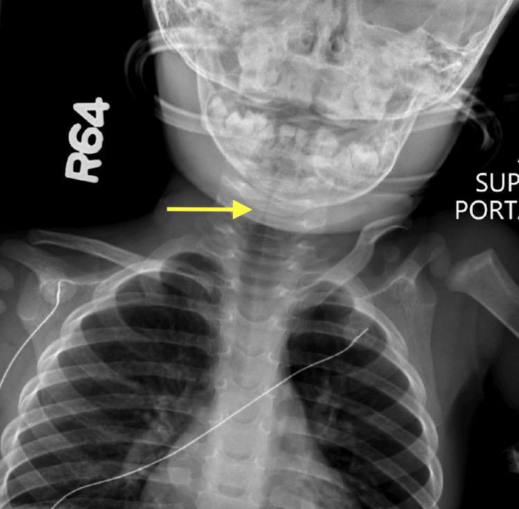
This diagnostic image is a posterior-anterior (PA) neck and upper chest radiograph of a pediatric patient. The central clinical finding is the classic 'steeple sign,' characterized by subglottic narrowing of the airway as it approaches the larynx, indicated by a yellow arrow. This tapering morphology represents edema in the subglottic region, commonly associated with viral laryngotracheobronchitis (croup). Beyond the focal subglottic stenosis, the radiograph demonstrates diffuse tracheal narrowing extending into the upper thoracic portion of the airway. Anatomical structures visible include the base of the skull, the cervical spine, bilateral clavicles, and the superior ribs. The soft tissues of the neck appear unremarkable except for the compromised airway lumen. This image serves as a key educational example of pediatric airway obstruction and the radiological manifestations of inflammatory subglottic stenosis in a clinical setting involving respiratory distress.
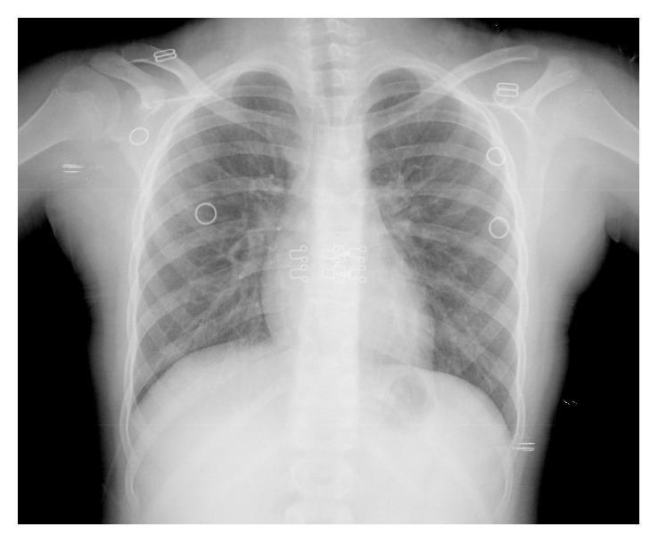
Anteroposterior (AP) chest radiograph of a pediatric patient demonstrating the 'steeple sign,' a diagnostic radiological finding characterized by symmetrical subglottic narrowing of the trachea. The narrowing produces an inverted 'V' or church steeple-like appearance of the airway column, indicative of subglottic edema or stenosis. The image displays the thoracic cage, including the ribs, clavicles, and vertebral column, with clear lung fields and a normal cardiomediastinal silhouette. Several external radiopaque markers and monitoring leads are visible overlying the chest wall and shoulder regions. The visual findings are consistent with conditions causing upper airway obstruction, such as laryngotracheobronchitis (croup) or subglottic stenosis as described in the clinical context of progressive dysphonia and respiratory insufficiency. This diagnostic image serves as a critical educational tool for identifying anatomical manifestations of upper airway narrowing in pediatric respiratory pathology.
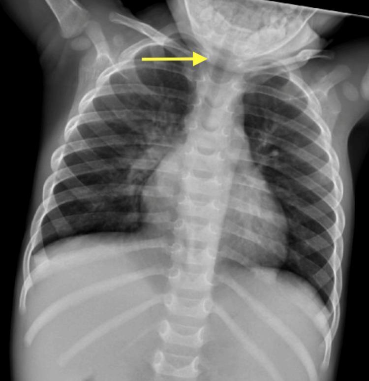
Anteroposterior (AP) pediatric chest radiograph demonstrating findings of subglottic stenosis. The image displays the thoracic cage, including the ribs, clavicles, and mediastinum. A prominent yellow arrow indicates a localized narrowing of the air column in the subglottic region and upper trachea, characteristic of the 'steeple sign' frequently associated with croup (laryngotracheobronchitis) or structural subglottic narrowing. Inferior to the level of narrowing, the mid-to-lower distal trachea appears to maintain a more normalized caliber. The visualized lung fields are relatively clear, without evidence of consolidation, significant opacities, or pleural effusions. This diagnostic image illustrates the radiological assessment of airway diameter in a pediatric patient following critical care for respiratory distress, serving as a teaching tool for identifying upper airway obstruction and evaluating clinical improvement or persistence of tracheal stenosis.
The Paediatric Airway - A Comprehensive Long Answer
1. INTRODUCTION
2. ANATOMY AND PHYSIOLOGY: HOW THE PAEDIATRIC AIRWAY DIFFERS FROM THE ADULT
2a. Structural Differences (TABLE 33.1, Scott-Brown's)
| Feature | Paediatric Airway | Clinical Significance |
|---|---|---|
| Size | Relatively smaller at all levels | Minimal swelling causes maximum obstruction |
| Position of larynx | Higher (C3-C4 in infant vs C5-C6 in adult) | More anterior, making intubation and visualization harder |
| Shape | Curled/omega-shaped epiglottis; shorter vocal folds | Predisposes to supraglottic obstruction |
| Mucosa | More reactive, friable, prone to oedema | Croup common in children, rare in adults |
| Vocal fold laminar structure | Immature in young children (5-layered structure not yet differentiated at birth) | Limits phonosurgical procedures in infants |
| Subglottis | Narrowest point of the paediatric airway (vs glottis in adults) | Critical site for stenosis, croup |
| Cartilage | Softer, more pliable | Predisposes to malacia conditions |
2b. Vocal Fold Development
- Up to age 10, vocal fold length is similar in both sexes (6-8 mm)
- At puberty, the male membranous vocal fold nearly doubles to 14.8-18 mm
- In females the increase is to 8.5-12 mm (a one-third increase)
- The ratio of membranous to cartilaginous vocal fold increases from 1.5 (newborn) to 5.5 (adult male)
- The 5-layered vocal fold structure (epithelium, superficial/intermediate/deep lamina propria, muscle) is not differentiated at birth and matures progressively to puberty
2c. Pitch Changes
3. AIRWAY DYNAMICS: THE PHYSICS (Why Small Matters)
Poiseuille's Law (Hagen-Poiseuille Equation)
Airway resistance is inversely proportional to the FOURTH POWER of the airway radius.
- A 50% reduction in airway radius = 16-fold increase in resistance
- 1 mm of narrowing in a 4 mm diameter infantile airway = 75% reduction in airflow
Bernoulli Principle
- Increased airflow velocity (due to narrowing) creates negative pressure on airway walls → inward collapse
- Smooth (laminar) airflow becomes turbulent (described by Reynolds number)
- Turbulence greatly increases resistance further
- Vibration of closely apposed airway walls produces the sound of stridor
4. STRIDOR: DEFINITION, TYPES, AND LOCALISATION
What is stridor?
- Stertor - low-pitched snoring caused by naso/oropharyngeal obstruction
Types by phase of breathing and what they indicate:
| Type | Site of Obstruction |
|---|---|
| Inspiratory stridor | Supraglottic / glottic (e.g. laryngomalacia, epiglottitis) |
| Biphasic stridor | Subglottic / tracheal (e.g. subglottic stenosis, croup) |
| Expiratory stridor / wheeze | Lower tracheal / bronchial (e.g. tracheomalacia, foreign body) |
Critical Warning Signs
- Recession (subcostal, intercostal, suprasternal, or combined) is a better indicator of obstruction severity than the loudness of stridor
- Nasal flaring and head bobbing are serious signs
- Paradoxically, stridor may become quieter as obstruction worsens (less airflow = less noise)
- Cyanosis is very late - do not wait for it to act
- Children who are working harder to breathe may extend their neck or rotate their head to find a position of comfort
5. CAUSES OF PAEDIATRIC AIRWAY OBSTRUCTION
5a. By Level (Box 28.1/28.2/28.3, Scott-Brown's)
Naso/Oropharyngeal Causes
- Choanal atresia
- Adenotonsillar hypertrophy (most common cause of UAO in children overall)
- Retropharyngeal abscess
- Tonsillitis / glandular fever
- Lingual thyroid
- Macroglossia (Down syndrome, Beckwith-Wiedemann)
- Pierre Robin sequence (micrognathia + glossoptosis)
Supraglottic Causes
- Laryngomalacia (most common cause of neonatal stridor)
- Epiglottitis (Haemophilus influenzae type B - now rare with vaccination)
- Supraglottic cysts
Glottic Causes
- Vocal cord palsy (bilateral)
- Laryngeal web
- Papillomatosis (recurrent respiratory papillomatosis)
- Posterior glottic stenosis
- Laryngeal cleft
Subglottic Causes
- Subglottic stenosis (congenital or acquired - commonest cause being prolonged intubation)
- Subglottic haemangioma
- Croup (laryngotracheobronchitis) - most common acute cause in children 6 months - 3 years
- Bacterial tracheitis
Tracheal / Bronchial Causes
5b. By Age of Onset (Mnemonic Aid)
| Age | Most Likely Cause |
|---|---|
| Birth / first days | Choanal atresia, laryngeal web, vocal cord palsy, vascular ring |
| 1st week - 6 weeks | Laryngomalacia (peaks at 4-6 weeks) |
| 6 months - 3 years | Croup (parainfluenza virus), foreign body |
| 2-6 years | Epiglottitis (pre-vaccination era), foreign body |
| Any age | Tonsillitis, retropharyngeal abscess, foreign body |
6. INDIVIDUAL CONDITIONS IN DETAIL
6a. Laryngomalacia
- Commonest cause of neonatal stridor
- Presents within first few days of life with high-pitched inspiratory stridor
- Caused by collapse of supraglottic structures (omega-shaped epiglottis, foreshortened aryepiglottic folds, redundant arytenoid mucosa) on inspiration
- Stridor worsens with feeding, crying, supine position; improves in prone position and with sleep
- Most cases are self-limiting, resolving by 12-18 months
- Reflux is present in the majority and may be causative or consequential (increased negative intrathoracic pressure during stridor promotes GOR)
- Children with reflux tend to have more severe laryngomalacia requiring surgery
- 10-15% require surgical intervention

- Conservative (majority): reassurance, prone positioning, anti-reflux treatment
- Surgical (10-15%): Supraglottoplasty (laser or cold steel division of aryepiglottic folds, trimming redundant arytenoid mucosa) - indicated for failure to thrive, severe apnoea, severe oxygen desaturation, cor pulmonale
6b. Vocal Cord (Fold) Palsy
- Second most common cause of neonatal stridor
- May be unilateral (weak/breathy cry, aspiration) or bilateral (more severe stridor, risk of respiratory failure)
- Causes: Iatrogenic (cardiac surgery, PDA ligation), neurological disease (Arnold-Chiari malformation, hydrocephalus), idiopathic
- Investigation: Awake flexible nasopharyngoscopy (best to assess dynamic cord movement), MRI brain/spine
- Bilateral palsy often requires tracheostomy; many recover spontaneously
6c. Subglottic Stenosis (SGS)
- Congenital or acquired (most common: prolonged endotracheal intubation in premature neonates)
- Graded by Cotton-Myer classification (Grades I-IV based on % obstruction)
- Presents with biphasic stridor, recurrent croup, voice changes
- Myer-Cotton Grading:
- Grade I: <50% obstruction
- Grade II: 51-70%
- Grade III: 71-99%
- Grade IV: No detectable lumen (100% obstruction)
- Investigation: LTB, CT airway
- Treatment: Depends on grade - endoscopic dilation/laser (Grades I-II), open reconstructive surgery (laryngotracheal reconstruction - LTR or cricotracheal resection - CTR) for Grades III-IV
6d. Croup (Laryngotracheobronchitis)
- Commonest: Parainfluenza virus type 1 (also types 2, 3; RSV, adenovirus)
- Age: 6 months to 3 years (peak 2 years)
- Pathology: Inflammatory oedema of subglottis and trachea (most narrow part of child's airway)
- Classic triad: Barking/seal-like cough + hoarse voice + inspiratory stridor
- Often preceded by 1-2 day URTI
- Steeple sign on AP neck/chest X-ray (subglottic narrowing)

- Mild: Single dose oral dexamethasone (0.15-0.6 mg/kg)
- Moderate-severe: Systemic corticosteroids (dexamethasone/prednisolone) + nebulised adrenaline (1:1000, 0.5 mL/kg, max 5 mL)
- Humidified oxygen if hypoxic
- These are the only Grade A evidence-based treatments in childhood stridor (Scott-Brown's)
- Avoid distressing the child (increases oxygen demand)
6e. Epiglottitis
- Historically caused by Haemophilus influenzae type B (HiB) - now rare after vaccination
- Other organisms: Streptococcus, Staphylococcus
- Age: 2-6 years (pre-vaccination)
- Very rapid onset (hours): high fever, severe sore throat, dysphagia, drooling, "tripod" positioning (leaning forward), toxic appearance, muffled "hot potato" voice
- Inspiratory stridor (less harsh than croup)
- Thumb sign on lateral neck X-ray (swollen epiglottis)
- CRITICAL: Do NOT examine throat with a spatula, do NOT lie child down, do NOT upset the child - can precipitate complete airway obstruction
- Management: Immediate senior involvement; controlled intubation under GA in theatre; IV antibiotics (cefotaxime/ceftriaxone); extubate when clinically improved (usually 24-48 hrs)
6f. Bacterial Tracheitis (Pseudomembranous Croup)
- Caused by: Staphylococcus aureus (most common), also H. influenzae, Streptococcus
- Aggressive illness; often complicates a preceding viral croup
- Toxic appearance, high fever, thick tenacious tracheal secretions/pseudomembrane
- Needs urgent LTB for diagnosis and airway toilet
- Treatment: IV antibiotics, intubation often required
6g. Retropharyngeal Abscess
- 6 months - 3 years typically
- Fever, drooling, neck stiffness, muffled voice, dysphagia, stridor
- Pre-vertebral soft tissue widening on lateral neck X-ray (>6-7 mm at C2, >22 mm at C6)
- CT neck is the investigation of choice
- Treatment: IV antibiotics ± surgical drainage
6h. Foreign Body Inhalation
- Peak age: 1-3 years (hand-to-mouth phase)
- Commonest foreign bodies: peanuts, grapes, small toys, coins
- Classic history: sudden choking/coughing episode while eating/playing, followed by a "silent interval" where symptoms temporarily settle
- Laryngeal/tracheal: immediate severe symptoms (cough, stridor, cyanosis)
- Bronchial (most common - right lower lobe): monophonic wheeze, decreased air entry unilaterally
- Chest X-ray: May be normal; look for unilateral hyperinflation (air trapping), obstructive emphysema, mediastinal shift - inspiratory/expiratory films or lateral decubitus views help
- Treatment: Rigid bronchoscopy under GA for retrieval
6i. Recurrent Respiratory Papillomatosis (RRP)
- Caused by Human Papillomavirus (HPV types 6 and 11)
- Type 11 is more aggressive
- Transmitted vertically (mother to child during vaginal delivery)
- Presents with progressive hoarseness, then stridor, respiratory distress
- Diagnosis: Microlaryngoscopy showing papillomas (usually at glottis, anterior commissure)
- Treatment: Repeated surgical debulking (microdebrider/CO2 laser/KTP laser - never curable)
- Adjuvant therapies: Cidofovir, bevacizumab, HPV vaccination (preventive)
- Tracheostomy avoided if possible (risk of distal spread)
6j. Subglottic Haemangioma
- Presents at 2-3 months of age (neonatal period is symptom-free)
- Biphasic stridor, worsening with crying
- 50% have associated cutaneous haemangioma (especially beard distribution - chin, lip, neck)
- Treatment: Propranolol (oral, first-line - remarkable response), laser ablation, steroids
6k. Tracheomalacia
- Excessive tracheal collapse during expiration due to flaccid walls
- Primary (intrinsic): floppy tracheal cartilages
- Secondary (extrinsic): vascular compression (innominate artery, vascular ring)
- Symptoms: expiratory stridor/wheeze, "barking seal" cough, recurrent chest infections, apnoeas ("dying spells")
- Investigation: Flexible bronchoscopy during spontaneous breathing, echocardiography
- Mild cases resolve spontaneously as cartilage matures
7. EVALUATION OF THE STRIDULOUS CHILD
7a. History
| Parameter | Key Questions |
|---|---|
| Onset | Congenital (birth) vs acquired; age at onset |
| Duration & progression | Improving/stable/worsening |
| Character of stridor | Inspiratory/expiratory/biphasic; pitch; musical vs rough |
| Aggravating/relieving factors | Worse with feeding, crying, supine? Better prone? |
| Voice/cry | Normal cry (not glottic) vs absent/weak cry (glottic) vs muffled (supraglottic) |
| Feeding | Breastfed baby "coming up for air" = significant obstruction; failure to thrive = severe |
| Cough character | Barking = croup/tracheomalacia; bovine = RLN palsy |
| Apnoeas/cyanosis | Severe tracheomalacia |
| Perinatal history | Intubation history (→ subglottic stenosis), maternal HPV (→ RRP), birth trauma (→ VCP) |
| Past medical | Cardiac disease (VCP post-surgery), neurological (VCP, laryngomalacia) |
| Skin lesions | Cutaneous haemangioma → subglottic haemangioma |
7b. Examination
- Observe stridor character and phase
- Assess recession (subcostal, intercostal, suprasternal)
- Look for nasal flaring, head bobbing, abnormal posturing (neck extension)
- Assess colour (cyanosis = very late, severe sign)
- Paradox: Volume of stridor DECREASES as obstruction becomes critical (less airflow)
- Assess for micrognathia (Pierre Robin)
- Examine skin for haemangiomas
- Auscultate for cardiac murmurs (vascular ring) and wheeze
- Check for signs of URTI / tonsil hypertrophy
| Symptom | Probable Pathology |
|---|---|
| Prolonged expiratory phase | Tracheal/bronchial obstruction (tracheomalacia, stenosis) |
| Cough | TOF, VCP, cleft larynx, foreign body, tracheomalacia, reflux |
| Aspiration | TOF, VCP, laryngeal cleft |
| Hoarseness | Laryngeal lesion (VCP, papilloma) |
| Acute obstruction | Retropharyngeal abscess, tonsillitis, glandular fever, foreign body, epiglottitis, croup, bacterial tracheitis |
| Dysphagia / feeding difficulty | Epiglottitis, tonsillitis, retropharyngeal abscess |
| Apnoeas | Tracheobronchomalacia |
8. INVESTIGATIONS
8a. Imaging
| Investigation | Best For |
|---|---|
| Plain X-ray (AP/lateral neck and chest) | Steeple sign (croup), thumb sign (epiglottitis), retropharyngeal widening, foreign body, hyperinflation |
| Fluoroscopy (barium swallow) | Vascular ring, extrinsic tracheal compression, swallowing disorders |
| Contrast tracheobronchogram | Lower airway stenosis, tracheomalacia (measures opening pressures) - high risk in infants |
| Echocardiography | Vascular compression, congenital heart disease |
| Ultrasound larynx | VCP (dynamic), structural lesions (cysts, papilloma) - non-diagnostic |
| CT / MRI | Thoracic vascular anatomy, extrinsic tracheal compression; 3D reconstruction for stenosis ("virtual bronchoscopy") |
| IMPORTANT | CT/MRI do NOT well evaluate dynamic conditions (malacia, cord palsy) - endoscopy is superior |
8b. Respiratory Function Tests
- Flow-volume loops: Distinguish intra- vs extrathoracic obstruction, fixed vs variable obstruction
- Polysomnography (sleep study): Used when obstructive sleep apnoea is suspected
8c. Endoscopy - THE GOLD STANDARD
Awake Flexible Nasopharyngoscopy (Office / Ward)
- First-line dynamic assessment
- Ultra-thin endoscopes (<2 mm) allow evaluation even in neonates
- Best for: Dynamic abnormalities (VCP, laryngomalacia)
- Limitations: No view below glottis; cannot exclude second pathology; no intervention possible
- Most feasible in infants <9-12 months (can be swaddled) and cooperative older children
Laryngotracheobronchoscopy (LTB) - Gold Standard
- Requires experienced team: surgeon + anaesthetist + nursing staff
- Preferred technique: spontaneous respiration (maintains muscle tone, allows detection of dynamic conditions)
- Equipment (Box 28.4): Age-appropriate laryngoscopes, ventilating bronchoscopes with Hopkins rod telescopes, operating microscope, light source, camera/monitor, full range of instruments and tracheostomy tubes
- Assessment: Pharynx → supraglottis → glottis (cord mobility, cricoarytenoid joint) → subglottis → trachea → carina → bronchi
- Posterior laryngeal cleft excluded by probing posterior commissure
- Position: Small sandbag under shoulders, head supported
- Suspension laryngoscope inserted protecting teeth/lips, tongue central
- Hopkins rod (rigid telescope) provides superior image quality
- Tube removed for best laryngeal view - arytenoids probed for mobility assessment
- Age-appropriate bronchoscope passed (always have one size smaller available)
- Entire airway examined systematically to carina and beyond
9. MANAGEMENT OF ACUTE AIRWAY OBSTRUCTION
9a. General Principles
- Do not upset the child - anxiety increases oxygen demand
- Keep parent/carer with child for reassurance
- Sit child upright (unless unconscious) - natural airway-opening position
- Administer humidified oxygen if desaturating
- Senior help immediately - anaesthetist, ENT surgeon
- Do not attempt oropharyngeal examination if epiglottitis is suspected
9b. Specific Interventions
Croup (Evidence Grade A):
- Oral dexamethasone (0.15-0.6 mg/kg single dose) - even for mild/moderate croup
- Nebulised adrenaline (1:1000, 0.5 mL/kg to max 5 mL) for moderate-severe
- Humidified oxygen
- Observe for rebound after adrenaline (effect lasts 2-4 hours)
Epiglottitis:
- Gas induction in theatre with surgeon present and ready for surgical airway
- Nasotracheal intubation under GA, then ICU management
- IV antibiotics: ceftriaxone/cefotaxime
Foreign Body:
- Choking child (conscious, can cough): Encourage coughing
- Choking child (cannot cough/breathe): Back blows + abdominal thrusts (Heimlich >1 year)
- Infant <1 year: Back blows + chest thrusts only
- Rigid bronchoscopy for definitive removal
Surgical Airway:
- Tracheostomy is the definitive surgical airway in children
- Emergency needle cricothyroidotomy as a temporising measure only (very difficult in neonates due to anatomy)
- Surgical cricothyroidotomy is not recommended in children <12 years (risk of subglottic stenosis)
9c. Anaesthesia for Paediatric Airway Endoscopy
- Preference: Spontaneous ventilation (detects dynamic conditions - malacia, cord palsy)
- Paralysis eliminates muscle tone - masks malacia
- Gas induction (sevoflurane - non-irritant, rapid onset) preferred in infants, young children, difficult IV access, precarious airways
- IV induction for older children
- Atropine/glycopyrronium as premedication to reduce secretions
- Pre-operative steroids if significant stenosis or inflammation suspected
- Topical lignocaine to vocal cords (carefully measured - risk of overdose in children)
- Jet ventilation: Not favoured in children (risk of barotrauma; cannot assess dynamic conditions)
10. TRACHEOSTOMY IN CHILDREN
Indications
- Long-term ventilatory support
- Bilateral VCP
- High-grade subglottic stenosis (Grade III-IV)
- RRP with severe lower airway disease (avoid if possible due to distal spread risk)
- Severe craniofacial anomalies with UAO
Key Points
- Performed with greatest care - the short, mobile trachea makes it technically challenging
- Avoid the first tracheal ring (risk of subglottic stenosis)
- Stay tube technique used to facilitate early decannulation if needed
- Tracheostomy tubes must never be disconnected without an immediate replacement and dilators present
- Decannulation requires progressive downsizing, then capping trials
11. REFLUX AND THE PAEDIATRIC AIRWAY
- Causes or exacerbates: hoarseness, laryngomalacia, subglottic stenosis, croup, chronic cough
- Increased work of breathing (from any cause) → negative intrathoracic pressure → promotes reflux → worsens airway disease (self-perpetuating cycle)
- Aspiration of refluxate → pulmonary disease → multilevel airway pathology (tracheobronchomalacia)
- Term "reflux-induced airway disease" is clinically useful as a unifying concept
- Anti-reflux treatment is important in managing these children
12. SUMMARY: KEY EXAM POINTS
| Point | Detail |
|---|---|
| Narrowest part of child's airway | Subglottis (vs glottis in adults) |
| Commonest neonatal stridor | Laryngomalacia |
| 2nd commonest neonatal stridor | Bilateral vocal cord palsy |
| Commonest acute stridor 6m-3y | Viral croup (parainfluenza 1) |
| Gold standard investigation | LTB (laryngotracheobronchoscopy) |
| Only Grade A evidence treatments | Dexamethasone + nebulised adrenaline (for croup) |
| Poiseuille's law | Resistance ∝ 1/r⁴ (1 mm oedema in 4 mm airway = 75% reduction in flow) |
| Dangerous sign | Stridor getting quieter (impending complete obstruction) |
| HPV types in RRP | Types 6 and 11 (type 11 more aggressive) |
| SGS treatment indicator | Myer-Cotton Grade - Grades III-IV need open surgery |
| Subglottic haemangioma Rx | Oral propranolol (first-line) |
| DO NOT in epiglottitis | Examine with spatula / lay child down / upset child |
| Jet ventilation risk in children | Barotrauma + cannot assess dynamic conditions |
| Surgical cricothyroidotomy | Not recommended <12 years (use needle technique as bridge only) |