
Tendon transfer for foot drop
"tendon transfer" AND "foot drop"
tendon transfer foot drop tibialis anterior posterior surgery
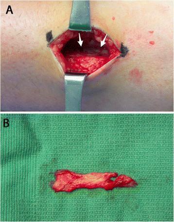
This composite clinical photograph depicts a key step in a posterior tibialis tendon (PTT) transfer surgical procedure for foot drop. Image A shows an intraoperative view of the lower leg where a surgical incision is held open by metal retractors. The extensor digitorum longus muscle is elevated medially to expose the crural interosseous membrane (indicated by white arrows), which appears as a white, fibrous, translucent tissue plane. Image B displays a 4-cm excised segment of the interosseous membrane resting on a green sterile surgical drape. The specimen is irregularly rectangular, exhibiting a dense fibrous texture with focal areas of blood staining and small amounts of adherent muscle tissue. This excision is performed to create a surgical window, allowing the PTT to pass from the posterior to the anterior compartment without entrapment or mechanical scarring. This visual material is intended for orthopedic and plastic surgery education, focusing on surgical anatomy and tendon transfer techniques.
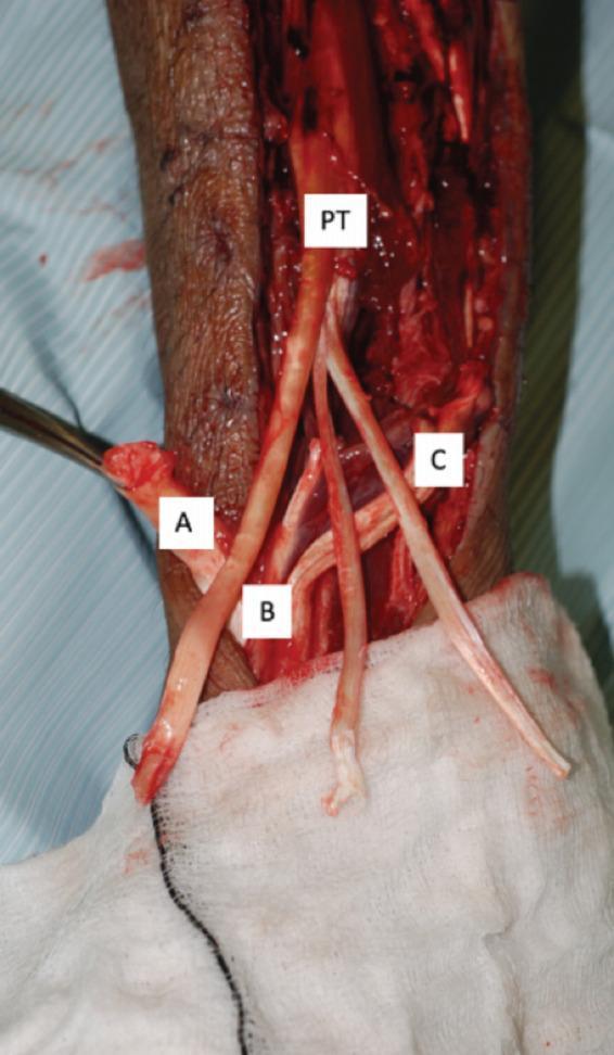
This clinical photograph shows an intra-operative view of a tendon transfer procedure in the lower extremity, likely following compartment syndrome or traumatic muscle loss. The image captures the dissection of key anatomical structures labeled for educational purposes. A thick Tibialis Posterior (PT) tendon is prominently displayed, split distally into three separate strands for transfer. Other identified tendons include the Tibialis Anterior (A), Extensor Hallucis Longus (B), and Extensor Digitorum Longus (C). These tendons appear as glistening, off-white, fibrous cords against a backdrop of deep red muscle tissue and a bloody surgical field. The surgical environment is characterized by the presence of metallic retractors, blue sterile drapes, and white gauze. This visual demonstrates the surgical approach to correcting foot drop or flexible deformities by utilizing the tibialis posterior as a motor unit to restore dorsiflexion. It is a critical resource for orthopedic and reconstructive surgery education regarding tendon harvesting and repositioning techniques.
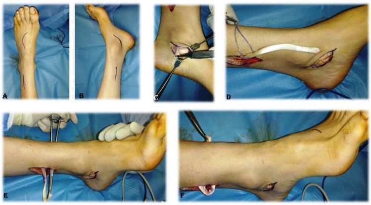
A series of clinical photographs (labeled A-F) documenting the surgical stages of a tibialis posterior tendon transfer for the correction of foot drop. Subfigures A and B show preoperative skin markings on the lower leg and dorsum of the foot, indicating the planned surgical approaches for tendon harvesting and insertion. Subfigure C demonstrates the identification and exposure of the tibialis posterior tendon through a medial incision using surgical retractors and forceps. Subfigure D illustrates the proximal delivery of the harvested tendon, showing its characteristic white, cord-like appearance as it is pulled through a proximal leg incision. Subfigures E and F depict the tunneling of the tendon through the interosseous membrane from the posterior compartment to the anterior compartment of the leg, utilizing surgical clamps and sutures for guidance. This procedure is common in orthopedics and rehabilitation medicine for restoring active dorsiflexion by repurposing the tibialis posterior muscle. The imagery demonstrates essential steps including marking, harvesting, and rerouting of the donor tendon.
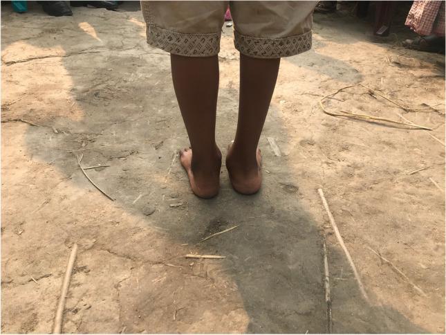
Clinical photograph showing a posterior view of the lower legs and feet of a pediatric patient, specifically a 5-year-old boy, two years following a right posterior medial release (PMR) with tibialis anterior tendon transfer (TATT). The image illustrates post-surgical foot alignment and morphology. Key visual findings include a bilateral valgus deformity of the hindfoot, with a more pronounced abduction and eversion on the right side compared to the left. The right medial malleolus appears prominent, indicating weight-bearing distribution favoring the medial aspect. The skin and soft tissues around the ankles appear well-healed without significant scarring, edema, or discoloration, suggesting successful recovery from complex orthopedic intervention for congenital or developmental foot deformity. This visual serves as an educational example of post-operative outcomes in pediatric orthopedic surgery for conditions like clubfoot (talipes equinovarus).
Tendon Transfer for Foot Drop
What is Foot Drop?
- Common peroneal (fibular) nerve injury - most common cause (compression at fibular head, knee dislocation, trauma)
- Sciatic nerve injury
- L4-L5 radiculopathy / disc prolapse
- Spinal cord injury / stroke
- Peripheral neuropathies (e.g. Charcot-Marie-Tooth disease, leprosy)
- Compartment syndrome sequelae
Principles of Tendon Transfer
- Wait at least 12 months after nerve injury to allow spontaneous recovery
- Assess with EMG/NCS to confirm permanent denervation
- Trial of foot-drop splint/ankle-foot orthosis (AFO)
- Tendon transfer is offered when the splint is inadequate and the nerve shows no recovery
- Donor muscle must have adequate power (MRC grade 4-5)
- Joints must be supple (no fixed contracture)
- Skin and soft tissue must be healthy
- The donor must be expendable
The Standard Procedure: Tibialis Posterior Tendon Transfer (TPTT)
- It is innervated by the tibial nerve (unaffected in common peroneal palsy)
- It has sufficient excursion and power
- Its loss from the medial side is relatively well tolerated
Routes of Transfer
| Route | Description | Notes |
|---|---|---|
| Interosseous membrane (transpositional) | Through a window in the interosseous membrane | Most direct; provides best line of pull for dorsiflexion |
| Circumtibial (subcutaneous) | Routed around the medial border of the tibia under skin | Simpler but less efficient line of pull; may bowstring |
| Circumfibular | Around the lateral border | Rarely used |
Surgical Steps (TPTT via Interosseous Membrane)
- Medial incision - Identify and detach the tibialis posterior tendon from its insertion at the navicular, with as long a length as possible
- Proximal delivery - Withdraw the tendon proximally into the deep posterior compartment
- Interosseous membrane window - Create a ~4 cm window in the interosseous membrane (the extensor digitorum longus is elevated medially to expose it)
- Anterior passage - The tendon is passed through the membrane into the anterior compartment
- Insertion - Fixed to the dorsum of the foot, most commonly:
- Medial cuneiform (restores dorsiflexion + inversion balance) - Gray's Anatomy for Students
- Third cuneiform / dorsolateral foot - for more neutral pull
- Some surgeons split the tendon and attach to both the medial cuneiform and fibularis brevis
- Tensioning - Fixed with the foot in slight dorsiflexion under appropriate tension
- Immobilisation - Short leg cast in dorsiflexion for 6 weeks, followed by physiotherapy
Insertion Sites and Their Rationale
| Insertion | Muscle action restored | Context |
|---|---|---|
| Medial cuneiform | Dorsiflexion + inversion | CMT disease, isolated foot drop |
| Dorsum of foot (2nd-3rd cuneiform) | Neutral dorsiflexion | Peroneal palsy, leprosy |
| Fibularis brevis (split transfer) | Dorsiflexion + eversion | When eversion is also needed |
Other Donor Tendons (Less Common)
- Flexor digitorum longus (FDL) - alternative posterior compartment donor
- Flexor hallucis longus (FHL) - rarely used for this purpose
- Peroneus longus - in partial palsies where eversion is preserved
Adjunct Procedures
- Posterior capsulotomy / Achilles tendon lengthening - if equinus contracture is present
- Triple arthrodesis - in rigid, long-standing deformity with joint destruction
- Calcaneal osteotomy - in cavovarus (CMT)
- Ankle-foot orthosis (AFO) - initial non-surgical management, or post-operative adjunct
Outcomes
- In peroneal nerve palsy: good to excellent results in 60-80% of patients with a compliant, rehabilitated patient
- In CMT: tibialis posterior transfer with osteotomies significantly reduces foot drop and improves gait
- In post-traumatic foot drop: results are best when transfer is done early (1-2 years post injury), with mobile joints and no fixed equinus
Summary
| Feature | Detail |
|---|---|
| Primary indication | Persistent foot drop >12 months, failed conservative Rx |
| Main donor | Tibialis posterior (tibial nerve innervated) |
| Preferred route | Through interosseous membrane |
| Main insertion | Medial cuneiform or dorsum of foot |
| Key adjunct | Equinus release if needed |
| Immobilisation | 6 weeks cast in dorsiflexion |
| Contraindications | Fixed joint contracture, flail limb, poor skin/soft tissue |