
Question Analysis
- Has no cough impulse
- Does not reduce on compression or lying down
- Is absent in the morning, but appears by evening
- (a) To leave it alone (masterly inactivity) ✅ CORRECT
- (b) Herniotomy
- (c) Eversion of sac
- (d) Hernioplasty
Why the Answer is (a) - Masterly Inactivity
Diagnosis: Congenital (Communicating) Hydrocele
| Feature | Explanation |
|---|---|
| Absent in the morning | Fluid drains back into the peritoneal cavity when child lies flat overnight |
| Appears by evening | Peritoneal fluid accumulates in the scrotum during the day (upright position) |
| No cough impulse | The communication is too small to allow significant pressure transmission |
| Does not reduce on pressure | The processus vaginalis opening is narrow - fluid doesn't rush back easily |
Why wait (masterly inactivity)?
- The majority resolve on their own
- Surgery carries anaesthetic and operative risks
- Bailey and Love's confirms: "Congenital hydroceles are treated by ligation of the patent processus vaginalis (herniotomy) if they do not resolve spontaneously."
- The child is older (typically >2 years) and it has not resolved, OR
- There is concern about hernia
Why the Other Options Are Wrong
| Option | Procedure | Why Wrong Here |
|---|---|---|
| (b) Herniotomy | Ligation of patent processus vaginalis | Done when hydrocele persists beyond ~2 years; this is the eventual surgery if it doesn't resolve, but NOT the first-line in this context |
| (c) Eversion of sac | Jaboulay's operation - everting the tunica vaginalis | Used for adult acquired hydroceles with a thin sac, not for congenital communicating hydroceles in children |
| (d) Hernioplasty | Mesh repair of the posterior inguinal wall | Used for inguinal hernias in adults; completely inappropriate here as there is no hernia |
Key Teaching Points
- Communicating hydrocele = patent processus vaginalis + intermittent scrotal swelling (positional)
- No cough impulse = not a hernia (small communication only)
- Management in children: Watchful waiting first; herniotomy only if it persists beyond 2 years of age or enlarges significantly
- Eversion of sac (Jaboulay's) and herniotomy are operations for different indications - never confuse them in exams
- Hernioplasty (mesh) has NO role in pediatric hydrocele
Question 5 (UPSC 2007 I Q-89)
- (a) Percutaneous embolization of gonadal veins ✅ CORRECT
- (b) Radio frequency ablation of testicular veins
- (c) Laparoscopic excision of affected testes
- (d) Excision of pampiniform plexus
Diagnosis: Varicocele
| Feature | Significance |
|---|---|
| Dragging pain in scrotum | Classic symptom - worse on standing, end of day |
| "Bag of worms" on examination | Dilated, tortuous pampiniform plexus veins |
| Disappears on lying down | Veins drain by gravity in supine position |
| Young adult male (25 years) | Typical age of presentation |
Why Option (a) is Correct
"When the discomfort is significant, then percutaneous embolisation of the gonadal veins is the usual first-line..." - Bailey and Love's Short Practice of Surgery, 28th Ed.
- It is minimally invasive (interventional radiology procedure)
- Done under local anaesthesia
- Coils or sclerosant are introduced via the femoral/jugular vein into the testicular vein to block retrograde blood flow
- Faster recovery, lower morbidity than surgery
- Comparable success rates to surgical ligation
Why the Other Options Are Wrong
| Option | Problem |
|---|---|
| (b) Radiofrequency ablation of testicular veins | Not a standard treatment for varicocele; may damage testicular vasculature |
| (c) Laparoscopic excision of affected testes | Completely wrong - you NEVER excise the testes for varicocele |
| (d) Excision of pampiniform plexus | The plexus contains the testicular artery and other vital structures; excision would cause testicular ischaemia and atrophy |
Summary of Varicocele Management
| Indication | Treatment |
|---|---|
| Symptomatic (dragging pain) | Percutaneous embolization (first-line) |
| Infertility + abnormal semen | Varicocelectomy (surgical - Palomo/Ivanissevich) or embolization |
| Subclinical / asymptomatic | Observe; no treatment needed |
| Adolescent with small varicocele | Watch and wait (risk of overtreatment) |
What is salmon patch
salmon patch nevus simplex newborn birthmark
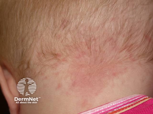
Imaging modality: Clinical photography of neonatal skin. The primary subject is a nevus simplex, commonly called a salmon patch, located on the posterior nape of the neck (occipital region). This flat, pink patch is blanchable with gentle pressure, well-demarcated, and non-palpable. The lesion lies within the dermis as a superficial capillary malformation with slow vascular filling, typical of nevus simplex. There is no surface atrophy, ulceration, or scaling. The surrounding skin shows normal neonatal hair distribution over the scalp. The appearance is a benign vascular lesion; in most infants it fades spontaneously within the first 1-2 years, though patches on the eyelids or glabella may persist longer. Clinically this lesion must be differentiated from more extensive capillary malformations such as port-wine stains, which tend to be darker, thicker, and persist or widen with time. The diagnostic significance is that this is a common, benign skin finding requiring no treatment. If the patch persists beyond early childhood, or is associated with other malformations (Sturge-Weber, etc.), further evaluation may be considered. This image is useful for dermatology education, newborn examination references, and image-based clinical decision support for capillary malformations. This supports standardized documentation and future comparative dermatologic research and training for clinicians.
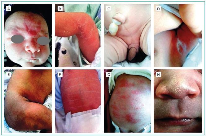
A composite of eight clinical photographs (A-H) demonstrating various common neonatal dermatoses. (A) Shows a salmon patch (nevus simplex), appearing as a midline, flat, reddish-purple vascular macule on the forehead and glabella. (B, E) Exhibit physiological desquamation and erythema of the extremities, characterized by dry, peeling skin and flaky textures. (C) Displays a clamped umbilical cord stump and neonatal genitalia. (D) Shows intertriginous erythema with possible excoriation in a skin fold. (F) Depicts generalized erythema with prominent horizontal skin creases, often seen in vernix caseosa or physiological skin shedding. (G) Shows scattered erythematous macules and papules consistent with erythema toxicum neonatorum. (H) Provides a close-up of milia, presenting as multiple, 1-2 mm pearly white or yellowish subepidermal keratin cysts on the nose and upper lip. This educational resource illustrates normal physiological skin changes and benign transient dermatoses in the newborn period, useful for pediatric clinical training.
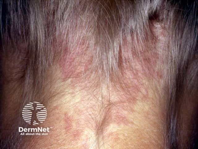
Imaging modality: Clinical photography of a cutaneous lesion. Subject is an adult with a patch on the posterior neck consistent with naevus simplex (salmon patch). The view is a close-up, posterior neck photograph intended to document color, borders, and surface characteristics. In this image the patch is flat and blanchable, range pink to light red, with an oval-to-irregular contour and well-defined margins. The surface appears smooth with no scale, crust, or induration; there is no nodularity or edema. The lesion resides within the integumentary system, involving the superficial dermis and dilated capillaries. Laterality is midline on the posterior neck. The patch is non-palpable and non-tender. This presentation aligns with classic sun-exposed naevi simplex findings in adults, a benign capillary malformation that commonly persists from infancy in some individuals. Diagnostic significance: benign, with no malignant potential; distinguishes from pigmented nevi only by vascular coloration and blanchability. Differential considerations include port-wine stain (nevus flammeus), small vascular malformations, and chronic inflammatory patches; clinical correlation and, if indicated, dermoscopy or vascular imaging can aid confirmation. Management is observational; treatment is typically unnecessary unless cosmetic concern arises or if lesion changes. Clinical education and documentation recommended.
Salmon Patch (Nevus Simplex)
Definition
Other Names
| Name | Location |
|---|---|
| "Stork bite" | Nape of neck / occiput |
| "Angel's kiss" | Glabella / forehead (V-shaped) |
| Nevus simplex | General medical term |
| Nevus flammeus neonatorum | Older term |
Characteristic Locations
- Nape of neck (most common - "stork bite")
- Glabella (between the eyebrows - "angel's kiss")
- Upper eyelids
- Nasal philtrum and alae
- Lumbosacral region
- Temples and vertex scalp
Pathophysiology
Natural History
| Location | Outcome |
|---|---|
| Facial (glabella, eyelids) | Fade spontaneously by 1-3 years of age |
| Nape of neck / extrafacial | Often persist into adulthood (covered by hair) |
Key Clinical Features
- Flat (macular) - not raised or palpable
- Pink-red color
- Blanchable on pressure (glass slide test)
- Indistinct borders, somewhat irregular ("splattered" appearance)
- No surface change (no nodularity, scaling, or ulceration)
- Become more prominent with crying or Valsalva
Differentiating from Port-Wine Stain (PWS)
| Feature | Salmon Patch | Port-Wine Stain |
|---|---|---|
| Color | Light pink-red | Deeper red-purple |
| Borders | Indistinct | Well-demarcated |
| Location | Midline / neural axis | Unilateral, any site |
| Fades? | Yes (facial > nape) | Never fades; darkens with age |
| Pathology | Persistent fetal circulation | Somatic GNAQ mutation |
| Association | Rarely syndromic | Sturge-Weber syndrome (V1 PWS) |
Associated Syndromes (rare)
- Beckwith-Wiedemann syndrome
- Megalencephaly-capillary malformation (MCAP) syndrome
- Roberts SC syndrome
- Nova syndrome
Treatment
Question 18 - Colorectal Carcinoma
- Left-sided Colorectal Carcinoma presents with bleeding per rectum.
- Right-sided Colorectal Carcinoma presents with iron deficiency anaemia.
- Right-sided CRC is more common than left-sided CRC.
- Colonoscopy is the investigation of choice for suspected CRC.
Evaluating Each Statement
✅ Statement 1 - TRUE
✅ Statement 2 - TRUE
❌ Statement 3 - FALSE
- Rectum: ~35-40% (most common site)
- Sigmoid colon: ~25%
- Descending/left colon: ~15%
- Right colon/caecum: ~15-20%
✅ Statement 4 - TRUE
- Biopsy/histological confirmation possible
- Can detect and remove synchronous polyps
- Can detect synchronous cancers (3-5%)
- Gold standard for diagnosis
Summary
| Statement | Verdict |
|---|---|
| 1. Left-sided CRC → bleeding per rectum | ✅ TRUE |
| 2. Right-sided CRC → iron deficiency anaemia | ✅ TRUE |
| 3. Right-sided CRC more common | ❌ FALSE - left-sided is more common |
| 4. Colonoscopy = investigation of choice | ✅ TRUE |
Exam Memory Aid
| Side | Lumen | Stool consistency | Tumour type | Presentation |
|---|---|---|---|---|
| Right (caecum, ascending) | Wide | Liquid | Polypoid/exophytic | Iron deficiency anaemia, palpable mass |
| Left (descending, sigmoid, rectum) | Narrow | Solid | Annular/constricting | Rectal bleeding, obstruction, altered bowel habit |
Question 19 - Oesophageal Perforation
- Perforation by a flexible endoscope
- Perforation into mediastinum
- Perforation with a small septic load
- Perforation of the abdominal oesophagus
Evaluating Each Statement
✅ Statement 1 - FAVOURS non-operative management
❌ Statement 2 - FAVOURS operative management (NOT non-operative)
✅ Statement 3 - FAVOURS non-operative management
✅ Statement 4 - FAVOURS non-operative management
| Location | Outcome tendency |
|---|---|
| Cervical / pharyngeal | Best for conservative management |
| Abdominal oesophagus | Also amenable to conservative management |
| Intrathoracic / mediastinal | Requires surgical intervention |
Classic Criteria Favouring Non-Operative Management of Oesophageal Perforation
| Criterion | Why it helps |
|---|---|
| Iatrogenic / flexible endoscope | Small, clean perforation |
| Contained perforation (no free leak) | Limited contamination |
| Small septic load | No systemic sepsis |
| Cervical or abdominal location | Not in mediastinum |
| Minimal pleural/mediastinal contamination | Avoids mediastinitis |
| Clinically stable patient | Can tolerate conservative course |
| Early diagnosis | Less soiling over time |
Summary
| Statement | Favours non-operative? |
|---|---|
| 1. Flexible endoscope perforation | ✅ YES |
| 2. Perforation into mediastinum | ❌ NO - favours surgery |
| 3. Small septic load | ✅ YES |
| 4. Abdominal oesophagus perforation | ✅ YES |
Question 29
- (a) Anaerobic bacterial infection
- (b) Colovesical fistula ✅ CORRECT
- (c) Tubercular cystitis
- (d) Urethrocutaneous fistula
Diagnosis: Colovesical Fistula
Key Clinical Clue - Pneumaturia
What is a Colovesical Fistula?
Classic Triad of Colovesical Fistula
| Feature | Explanation |
|---|---|
| Pneumaturia (bubbles/frothy urine) | Gas from colon enters bladder |
| Fecaluria | Faecal particles pass into urine |
| Recurrent polymicrobial UTIs | Mixed flora from gut colonizes bladder |
Common Causes
| Cause | % |
|---|---|
| Diverticular disease (most common) | ~65% |
| Colorectal carcinoma | ~20% |
| Crohn's disease | ~7% |
| Radiation injury | ~5% |
| Other (trauma, foreign body) | rare |
Diagnosis
- CT scan - investigation of choice: shows air in the bladder (in absence of prior instrumentation = diagnostic of fistula), bladder wall thickening, adherent sigmoid colon
- Cystoscopy - shows localized inflammatory reaction; bowel contents may be visible
- Colonoscopy - to exclude colorectal cancer
Why the Other Options Are Wrong
| Option | Why wrong |
|---|---|
| (a) Anaerobic bacterial infection | Gas-forming bacteria CAN cause pneumaturia, but this would NOT cause recurrent UTIs with the specific pattern described. A one-time infection is not "recurrent episodes." This is the classic distractor. |
| (c) Tubercular cystitis | Causes dysuria, frequency, haematuria - NOT pneumaturia/bubbles in urine |
| (d) Urethrocutaneous fistula | Communication between urethra and skin - causes urine leaking THROUGH SKIN, not bubbles in urine |
Treatment
Exam pearl: Any middle-aged/elderly patient with recurrent UTIs + pneumaturia (bubbles, gas, frothy urine) = Colovesical fistula until proven otherwise. The most common cause is diverticular disease.
Does larynx akd fixation kf chest kr swallowing of food