
Human papilloma virus
"human papillomavirus"[MeSH Terms] AND (vaccine OR cervical cancer OR carcinogenesis)
HPV vaccine cervical cancer prevention
human papillomavirus HPV pathogenesis types oncogenesis vaccine overview 2024
https://pmc.ncbi.nlm.nih.gov/articles/PMC11277246
Human Papillomavirus (HPV)
Overview
Classification
By Oncogenic Risk
| Risk Category | HPV Types | Associated Diseases |
|---|---|---|
| High-risk (HR) | 16, 18, 26, 31, 33, 35, 39, 45, 51, 52, 53, 56, 58, 59, 66, 68, 73, 82 | Cervical, anal, oropharyngeal, vulvar, penile carcinomas |
| Low-risk (LR) | 6, 11, 40, 42, 44, 54, 55, 61, 62, 71, 74, 81, 84, 89, 90 | Condylomata acuminata (genital warts), recurrent respiratory papillomatosis |
| Cutaneous | 1, 2, 3, 7, 10, 27, 57, 73 | Common warts, plantar warts, flat warts |
| EV-associated | 5, 8, 9, 10, 14, 17, 20-25, 37, 38 | Epidermodysplasia verruciformis (risk of skin SCC) |
Genome Structure and Key Proteins
- Long Control Region (LCR) - non-coding regulatory region controlling transcription and replication
- Early genes (E1, E2, E4, E5, E6, E7) - expressed early in infection; regulate replication and oncogenesis
- Late genes (L1, L2) - encode the major (L1) and minor (L2) capsid proteins
Oncoproteins - the Key Cancer Drivers
- E6 - binds and promotes ubiquitin-mediated degradation of p53 (the "guardian of the genome"), blocking apoptosis and allowing DNA damage to accumulate
- E7 - binds and inactivates pRb (retinoblastoma protein), releasing transcription factor E2F and pushing cells into uncontrolled S-phase entry
- E5 - enhances EGFR signaling, promotes immune evasion
- E1 / E2 - regulate viral DNA replication; loss of E2 (via chromosomal integration) removes the brake on E6/E7 transcription, a critical step toward malignancy
Pathogenesis and Oncogenesis
Steps to Infection
- HPV enters through microabrasions in stratified squamous epithelium (skin/mucosa)
- Infects basal keratinocytes (the only dividing cells in the epithelium)
- Viral genome maintained as episome (extrachromosomal); E1/E2 regulate replication
- As cells differentiate and migrate upward, late genes (L1, L2) are expressed and virions assembled in superficial layers
- Viral shedding occurs from superficial cells
From Infection to Cancer
- Viral integration into host chromosomes disrupts E2, upregulating E6 and E7
- E6 degrades p53 → loss of apoptosis and cell cycle arrest
- E7 inactivates Rb → uncontrolled cell proliferation
- Genomic instability accumulates → CIN (Cervical Intraepithelial Neoplasia) progression: CIN I → CIN II → CIN III → invasive carcinoma
Transformation Zone
Clinical Manifestations
Anogenital Disease
- Condylomata acuminata (genital warts) - soft, flesh-colored, cauliflower-like papules; caused by HPV 6 and 11; highly contagious; rarely malignant
- Cervical cancer - usually squamous cell carcinoma; HPV 16, 18 predominate; preceded by CIN
- Vulvar/vaginal intraepithelial neoplasia (VIN/VaIN) - HPV 16 major type
- Anal/rectal carcinoma - high incidence in MSM with HIV; HPV 16, 18
- Penile carcinoma - HPV 16, 18 involved in ~40-50% of cases
- Bowenoid papulosis - pigmented papules with carcinoma-in-situ histology; HPV 16, 18
Non-Genital Cutaneous Disease
| Disease | Types |
|---|---|
| Common warts (verruca vulgaris) | 1, 2, 4, 27 |
| Plantar warts (verruca plantaris) | 1, 2, 4, 57 |
| Flat warts (verruca plana) | 3, 10, 27, 28 |
| Butcher's warts | 1-4, 7, 10, 28 |
| Epidermodysplasia verruciformis | 5, 8 (malignant potential) |
Oropharyngeal / Head & Neck Disease
- Recurrent respiratory papillomatosis (RRP) - laryngeal/tracheal papillomas from HPV 6, 11; can be life-threatening; juvenile form acquired perinatally
- Oropharyngeal SCC (tonsil, base of tongue) - HPV 16; incidence rising sharply in developed countries; better prognosis than HPV-negative HNSCC
- Oral leukoplakia, oral carcinoma - HPV 16, 18
Diagnosis
- Cytology (Pap smear) - screening for cervical precancer; koilocytes (perinuclear halo, nuclear atypia) are the hallmark HPV-infected cells
- HPV DNA testing (co-test) - detects high-risk HPV types; used alone (primary screening) or with Pap smear
- Colposcopy + biopsy - for abnormal cytology/HPV results; biopsy for histological grading
- p16/Ki-67 immunostaining - surrogate marker for high-risk HPV transcriptional activity; used in histopathology and cytology triage
- Genotyping - identifies specific types (e.g., HPV 16/18 separately)
Vaccines (Prophylactic)
| Vaccine | Types Covered | Trade Name |
|---|---|---|
| Bivalent | 16, 18 | Cervarix (no longer sold in the US) |
| Quadrivalent | 6, 11, 16, 18 | Gardasil |
| 9-valent | 6, 11, 16, 18, 31, 33, 45, 52, 58 | Gardasil-9 (current standard) |
Efficacy
- Efficacy against CIN, VIN, and PIN caused by covered types: >90% (approaching 100% in HPV-naive recipients)
- Efficacy against genital warts (Gardasil/Gardasil-9): 89-98%
- HPV infections for vaccine-covered types decreased >80% among US women aged 14-24 between 2003-2006 and 2015-2018
- Scottish national data (2024) showed zero invasive cervical cancers among 448,000 women vaccinated at ages 12-13
- Anal cancers/precancers decreased ~70% in young women vaccinated before age 17 (Denmark 2024)
- Cervical cancer rates fell 69% among US women aged 20-24 between 2013-2021
Vaccination Schedule (US - CDC 2024)
- Ages 9-14: 2-dose series (0, 6-12 months)
- Ages 15-26: 3-dose series (0, 1-2, 6 months)
- Ages 27-45: Shared clinical decision-making (less benefit due to likely prior exposure)
- Recommended for all genders
- Best given before sexual debut (no therapeutic effect on existing infection)
Treatment
Genital Warts
- Patient-applied: Imiquimod (5%), podophyllotox (0.5%), sinecatechins (15% ointment)
- Provider-applied: Trichloroacetic acid (TCA), cryotherapy, surgical excision, laser
- High recurrence rates (~30-70%) due to latent virus in surrounding tissue
Cervical Precancer (CIN)
- CIN I: Observation (most regress spontaneously)
- CIN II/III: LEEP (loop electrosurgical excision procedure), cold knife conization, cryotherapy
- Post-treatment HPV vaccination may reduce recurrence (PMID: 40919695 - 2025 Cochrane review on HPV vaccination after conisation)
Invasive Cervical Cancer
- Surgery (radical hysterectomy), radiotherapy, cisplatin-based chemotherapy depending on stage
- Pembrolizumab (anti-PD-1) now approved for recurrent/metastatic cervical cancer with PD-L1 expression
Recurrent Respiratory Papillomatosis
- Serial surgical debulking (CO2 laser, microdebrider); adjuvant intralesional cidofovir or bevacizumab in refractory cases; no cure
Therapeutic Vaccines (Investigational)
Epidemiology
- ~340 million prevalent HPV infections worldwide at any time
- Cervical cancer: ~660,000 new cases and ~350,000 deaths annually (global; predominantly in low- and middle-income countries with limited screening access)
- HPV is responsible for virtually 100% of cervical cancers, ~90% of anal cancers, ~70% of oropharyngeal cancers, ~40-50% of penile/vulvar cancers
- Oropharyngeal HPV-related cancers are increasing in high-income countries despite declining cervical cancer rates
Key Points Summary
- HPV is a DNA virus with >200 types; high-risk types (especially 16, 18) drive most HPV-related cancers via E6 (degrades p53) and E7 (inactivates Rb)
- Most infections are cleared; persistent infection plus co-factors leads to neoplasia
- 9-valent vaccine (Gardasil-9) is the current standard, with near-100% efficacy in HPV-naive individuals
- Screening (Pap + HPV co-test) and vaccination together are the pillars of cervical cancer prevention
- No antiviral exists; treatment targets lesions (excision, ablation, topical agents)
Morphology of acute rheumatic disease and major complications
acute rheumatic fever Aschoff body histology pathology
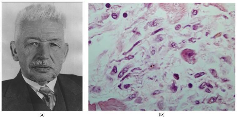
This composite image includes a portrait of Karl Albert Ludwig Aschoff and a high-magnification histological micrograph of a myocardial tissue section stained with Haematoxylin and Eosin (H&E). The micrograph (b) demonstrates the characteristic pathognomonic features of rheumatic myocarditis. It displays a granulomatous 'Aschoff body' within the cardiac interstitium. Key cellular components visible include 'Aschoff cells'—large, multinucleated or mononucleated cells with abundant eosinophilic cytoplasm and prominent 'owl-eye' nucleoli—and 'Anitschkow cells'. The Anitschkow cells, also known as 'caterpillar cells', are identified by their elongated nuclei with a central band of chromatin and fine lateral projections. The surrounding myocardial matrix shows interstitial edema and a mixed inflammatory infiltrate. This image is an essential educational resource for pathology and cardiology, illustrating the classic histopathological hallmarks used to diagnose acute rheumatic fever and its manifestation in the heart muscle.
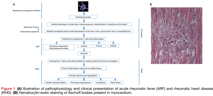
Summary : This figure illustrates the pathophysiology and clinical presentation of acute rheumatic fever (ARF) and rheumatic heart disease (RHD), and shows a histological image of Aschoff bodies in myocardium. flowchart and photo: # Panel A: Pathophysiology and Clinical Presentation Flowchart ## Nodes : • Streptococcus pyogenes infection (text node) • Tonsillopharyngitis (rectangle) • Antibody develops to molecules in bacterial group A carbohydrate or streptococcal M protein (rectangle) • Antibody recognizes same molecules present on the heart, brain, joints and/or skin in susceptible host (rectangle) • Infiltration of autoreactive T cells and inflammation (rectangle) • Erythema marginatum, Subcutaneous nodules (rectangle, branch) • Carditis (rectangle, branch) • Chorea (rectangle, branch) • Arthritis (rectangle, branch) • Valvulitis (rectangle) • Autoreactive T cells continually activated by cardiac proteins that serve as antigens (rectangle) • Chronic inflammatory response to cardiac tissue (rectangle) • Valvular stenosis and regurgitation (rectangle) • Cardiac chamber dilation (rectangle, branch) • Pulmonary hypertension (rectangle, branch) • Arrhythmia (rectangle, branch) • Heart failure (rectangle, branch) ## Connectors : • Linear downward arrows connect each main step. • Branching arrows from "Infiltration of autoreactive T cells and inflammation" to four clinical features: erythema marginatum/subcutaneous nodules, carditis, chorea, arthritis. • Branching arrows from "Valvular stenosis and regurgitation" to four complications: cardiac chamber dilation, pulmonary hypertension, arrhythmia, heart failure. ## Layout : • Vertical, left-to-right flow with two main phases: ARF (2–4 weeks) and RHD (years). • Branches at clinical presentation and complications. • Timeline markers on the right: "2–4 weeks" for ARF, "Years" for RHD. # Panel B: Histological Image ## Scene Overview : • Main subject: Myocardial tissue section stained with hematoxylin-eosin. • Central focus: Cluster of cells (Aschoff body) within pink-stained cardiac muscle fibers. • Colour palette: Pink (muscle), purple/blue (nuclei and inflammatory infiltrate). ## Technical Details : • Staining: Hematoxylin-eosin. • Magnification and scale bar: Not specified. • Tissue: Myocardium. ## Spatial Relationships : • Aschoff body is centrally located, surrounded by elongated muscle fibers. • Dense cellular infiltrate contrasts with the striated background. # Analysis : • The flowchart in Panel A details the progression from streptococcal infection to autoimmune-mediated heart damage, highlighting both acute and chronic clinical manifestations and complications. • The histological image in Panel B visually confirms the presence of Aschoff bodies, a hallmark of rheumatic carditis, within the myocardium.
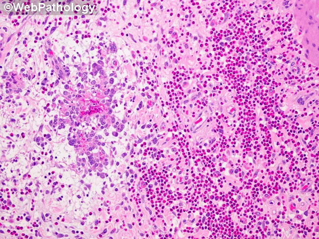
Imaging modality and technique: Light microscopy of a paraffin-embedded gallbladder mucosa section stained with Hematoxylin and Eosin (H&E). Primary subject: eosinophilic cholecystitis with destruction of Rokitansky-Aschoff sinus glands by an intense eosinophilic infiltrate. The visible lamina propria demonstrates dense clusters of eosinophils (bilobed nuclei with bright pink granules) intermixed with other inflammatory cells, accompanied by glandular distortion and gland rupture within Rokitansky-Aschoff sinuses. Mucosal damage is evident as focal erosion and edema, with disruption of normal mucosal architecture and crypt-like glands in the gallbladder mucosa. The eosinophilic inflammation drives tissue destruction, contributing to gallbladder dyskinesia and potential biliary colic. Notable features include perivascular eosinophils, tissue edema, and preservation of downstream serosa in this field, though transmural involvement may occur in advanced cases. This histology is diagnostic for eosinophilic cholecystitis, a rare inflammatory variant of cholecystitis linked to systemic eosinophilia, atopy, parasitic infection, or drug hypersensitivity. Diagnostic significance rests on recognizing eosinophil predominance and mucosal injury rather than neutrophilic invasion. Differential considerations include acute bacterial cholecystitis, chronic cholecystitis with eosinophils, parasitic inflammation, and drug-induced hypersensitivity. Clinically, correlate with peripheral eosinophil count, allergic history, and imaging findings; guide management and immunologic workup; educate pathology trainees and researchers about eosinophil-mediated biliary disease.
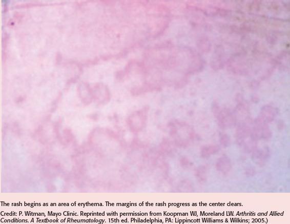
This clinical photograph displays a dermatological manifestation of erythema marginatum, a hallmark cutaneous sign of acute rheumatic fever. The image reveals multiple pinkish-red, erythematous lesions characterized by a distinct annular and serpiginous configuration. Key visual features include a vibrant, well-defined advancing periphery with significant central clearing, where the skin returns to a paler or near-normal hue. The rash appears non-pruritic and non-elevated, typically found on the trunk and proximal extremities. This visual serves as a primary diagnostic indicator for clinicians identifying systemic inflammatory conditions following a Group A streptococcal infection. The morphology demonstrates the classic 'smoke ring' appearance associated with this evanescent rash, emphasizing its migratory nature and peripheral progression.
rheumatic heart disease mitral stenosis valve pathology gross
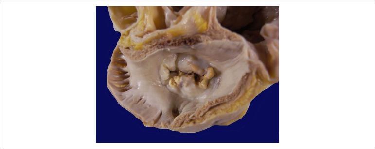
This clinical photograph shows a gross pathological specimen of a human heart, specifically an atrial view of the mitral valve. The image demonstrates hallmark signs of chronic rheumatic heart disease. Key features include significant thickening and fibrosis of the valve leaflets, resulting in a 'fish-mouth' or 'buttonhole' appearance of the valvular orifice. There is evidence of extensive commissural fusion and multiple focal areas of yellowish-white calcification along the leaflet margins and within the valve body. These structural changes characterize severe mitral stenosis, where the restricted opening of the valve impairs blood flow from the left atrium to the left ventricle. The surrounding atrial endocardium appears thickened, consistent with chronic pressure overload. This visual is highly representative for educational purposes in cardiology and pathology to illustrate the long-term sequelae of rheumatic fever on cardiac valves.
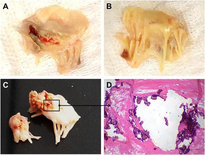
A multi-panel figure illustrating the gross and microscopic pathology of rheumatic heart valve disease (RHVD) in human mitral valves. Panels A and B present clinical photographs of excised mitral valve tissue from a 49-year-old female, showing marked leaflet thickening, retraction, and an opaque, yellowish appearance consistent with chronic fibrosis. Panel A specifically shows focal areas of reddish-dark discoloration, suggesting hemorrhage or tissue damage. Panel C displays gross pathology from a 61-year-old male, highlighting advanced calcification with rigid, cauliflower-like deposits on the valve structure. Panel D is a high-magnification light microscopy image (Hematoxylin and Eosin stain) of the anterior mitral valve leaflet. It demonstrates nodular calcification, appearing as irregular, dark purple hematoxylin-positive deposits embedded within the eosinophilic (pink) fibrotic collagenous matrix. A scale bar of 500 μm is provided. This figure serves as an educational resource for identifying the progression from chronic inflammation and fibrosis to end-stage valvular calcification in rheumatic heart disease.
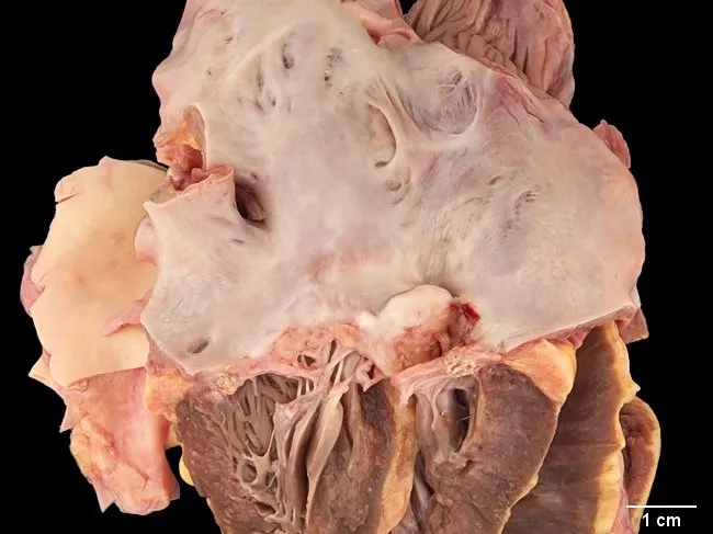
Gross pathology photograph of an excised mitral valve from a 55-year-old woman with long-standing systemic lupus erythematosus, illustrating Libman-Sacks endocarditis. The valve is stenotic with restricted leaflet mobility and marked left atrial dilation. Vascularized scar tissue is evident on the mitral leaflets near the line of closure, and the chordae tendineae are scarred and thickened. The surfaces exhibit irregular, pale to tan areas consistent with prior vegetations and repair, without visible bacterial organisms. The specimen demonstrates sterile vegetations and fibrinous thrombi embedded with immune complexes and mononuclear cells—hallmarks of nonbacterial thrombotic endocarditis in autoimmune disease. These changes produce impaired valvular function, typically mitral stenosis with or without regurgitation, and contribute to chronic left atrial enlargement. Clinically, Libman-Sacks endocarditis indicates active systemic lupus erythematosus with cardiac involvement and may increase risk of embolization and heart failure. The image supports the differential diagnosis against infectious endocarditis, rheumatic valvulitis, and marantic endocarditis, emphasizing autoimmune valvulopathy. This gross finding correlates with histopathology showing sterile vegetations; echocardiography often complements diagnosis in vivo. The case underscores the importance of multidisciplinary management combining immunosuppression and, when needed, valvular intervention. This image is suitable for educational, surgical planning, and pathophysiology research contexts and supports standardized reporting and citation.
Gross and microscopy features of acute rheumatic disease
Morphology of Acute Rheumatic Disease (ARF / Acute Rheumatic Fever)
Pathogenesis (Brief - Underpins Morphology)
I. CARDIAC MORPHOLOGY (Pancarditis)
A. ENDOCARDITIS (Valvulitis) - Most Clinically Significant
Gross Features
- Edema and swelling of valve leaflets (earliest change)
- Small (1-2 mm), warty vegetations called verrucae - appear along the lines of closure of the valve leaflets
- Verrucae are most common on the mitral valve (left-sided, high-pressure), followed by the aortic valve
- These vegetations are small, firm, and bead-like, NOT large or destructive (unlike infective endocarditis)
- MacCallum plaques - irregular, roughened thickenings on the posterior wall of the left atrium, caused by regurgitant jet injury to the subendocardium
"Small vegetations (verrucae) are visible along the line of closure of the mitral valve leaflet. Previous episodes of rheumatic valvulitis have caused fibrous thickening and fusion of the chordae tendineae." - Robbins & Kumar Basic Pathology
Microscopic Features
- Fibrinoid necrosis within the valve cusps and tendinous cords
- Overlying necrotic foci are covered by platelet-rich sterile microthrombi (the verrucae)
- Verrucae do NOT embolize (firmly adherent, unlike infective endocarditis vegetations)
- Lymphocytic and macrophage infiltration of the valve stroma
- In healing: neovascularization and fibrosis of the leaflets
B. MYOCARDITIS - Pathognomonic Lesion: The Aschoff Body
Gross Features
- Heart may appear pale, flabby, and edematous with dilated chambers
- Aschoff nodules may be visible grossly as tiny foci, especially beneath the endocardium near valves, but are often only appreciated microscopically
Microscopic Features - The ASCHOFF BODY
| Component | Description |
|---|---|
| Central zone | Fibrinoid necrosis - eosinophilic, homogeneous debris |
| Anitschkow cells ("caterpillar cells") | Plump activated macrophages; central ovoid/round nucleus with chromatin condensed into a central slender wavy ribbon - resembling a caterpillar. Most characteristic cell |
| Aschoff giant cells | Multinucleated macrophages (fusion of Anitschkow cells) with "owl-eye" nucleoli |
| Peripheral infiltrate | Predominantly CD4+ T lymphocytes, occasional CD8+ T cells, plasma cells |
| Stage | Features |
|---|---|
| Stage 1 - Exudative/Degenerative | Central fibrinoid necrosis, edema, nonspecific lymphocytic and plasma cell infiltrate |
| Stage 2 - Granulomatous | Accumulation of characteristic Aschoff giant cells and Anitschkow cells - the specific diagnostic stage |
| Stage 3 - Healing/Sclerosing | Diminution of cellular infiltrate, replacement by scar tissue (fibrosis) |
"The myocardial inflammatory lesions - called Aschoff bodies - are pathognomonic for rheumatic fever; these are collections of lymphocytes (primarily T cells), scattered plasma cells, and plump activated macrophages called Anitschkow cells associated with zones of fibrinoid necrosis." - Robbins & Kumar Basic Pathology
C. PERICARDITIS
Gross Features
- Fibrinous pericarditis - "bread and butter" pericarditis
- Shaggy, rough, fibrinous exudate coating both pericardial surfaces
- May cause a friction rub clinically
- Usually resolves completely without significant scarring (unlike tuberculous pericarditis)
Microscopic Features
- Fibrinous exudate with acute inflammatory cells
- Reactive mesothelial cells
- Rarely progresses to constrictive pericarditis
II. JOINT MORPHOLOGY (Arthritis)
Gross Features
- Warm, swollen, erythematous joints with joint effusion
- Large joints predominantly affected (knees 76%, ankles 50%, elbows/wrists 12-15%)
- Classic migratory polyarthritis - inflammation moves from joint to joint
- No gross joint destruction
Microscopic Features
- Synovial fluid: sterile inflammatory exudate
- Reduced complement components C1q, C3, C4 (immune complex consumption)
- No erosions on radiography (non-destructive arthritis)
- Aschoff bodies are NOT found in joint synovium
The arthritis is completely reversible and leaves no permanent joint deformity (unlike rheumatoid arthritis).
III. SKIN MORPHOLOGY
Erythema Marginatum
- Gross/Clinical: Pink-red, annular/serpiginous ("smoke ring") lesions with central clearing; evanescent, migratory; found on trunk and proximal extremities; non-pruritic, non-elevated
- Microscopic: Perivascular lymphocytic/plasma cell infiltrate around superficial dermal vessels; no vasculitis
Subcutaneous Nodules (Aschoff Nodules of Skin)
- Gross: Small (0.5-2 cm), firm, painless nodules over bony prominences (elbows, wrists, occiput, vertebral spinous processes)
- Microscopic: Central fibrinoid necrosis surrounded by palisading histiocytes - identical to Aschoff bodies in the heart; also resembles rheumatoid nodules histologically
- Usually appear weeks after arthritis; resolve completely
IV. BRAIN - Sydenham's Chorea
- No specific gross pathology is reliably identified
- Microscopic: Arteritis of cerebral vessels; degenerative changes in the caudate nucleus, subthalamic nucleus, and cerebellum
- Clinically: involuntary, purposeless choreiform movements ("St. Vitus' dance")
Summary Table: Key Morphologic Features
| Organ | Gross | Microscopy |
|---|---|---|
| Heart - Endocardium | Small warty verrucae on valve lines of closure; MacCallum plaques in LA | Fibrinoid necrosis + sterile platelet thrombi; no destruction |
| Heart - Myocardium | Pale, flabby heart; dilated chambers | Aschoff bodies (fibrinoid necrosis + Anitschkow caterpillar cells + Aschoff giant cells) - PATHOGNOMONIC |
| Heart - Pericardium | "Bread and butter" fibrinous pericarditis | Fibrinous exudate, acute inflammation |
| Joints | Warm, swollen, effusion; no erosions | Sterile exudate, complement consumption |
| Skin | Erythema marginatum (ring rash); subcutaneous nodules | Perivascular infiltrate; nodules = Aschoff-like lesions |
| Brain | No reliable gross change | Cerebral arteritis, caudate/subthalamic degeneration |
Major Complications (Chronic Rheumatic Heart Disease)
1. Mitral Stenosis (Most Common - "Pure" Mitral Stenosis = RHD Until Proven Otherwise)
- Gross: Leaflet thickening and fibrosis; commissural fusion; thickening and shortening of chordae tendineae; calcification in advanced disease; classic "fish-mouth" or "buttonhole" valve orifice; marked left atrial dilatation
- Microscopic: Fibrosis, neovascularization, and calcification of leaflets; loss of normal valve architecture
- Mitral valve involved in virtually 100% of chronic RHD cases (isolated in ~2/3; combined with aortic in ~1/4)
2. Mitral Regurgitation
- Leaflet retraction and scarring prevents proper coaptation
- Often coexists with mitral stenosis
3. Aortic Valve Disease
- Second most commonly involved valve
- Aortic stenosis or regurgitation depending on pattern of scarring/commissural fusion
- Combined mitral + aortic disease in ~25% of cases
4. Left Atrial Enlargement and Atrial Fibrillation
- Consequence of mitral valve obstruction/regurgitation
- AF predisposes to left atrial thrombus (especially in the left atrial appendage) and systemic embolism (stroke)
5. Infective Endocarditis
- Scarred, deformed valves are highly susceptible to secondary bacterial seeding
6. Heart Failure
- End-stage consequence of untreated valvular disease: cardiac chamber dilation, pulmonary hypertension, right heart failure
7. Tricuspid and Pulmonary Valve Involvement
- Tricuspid is infrequently affected; pulmonary valve only rarely
"The cardinal anatomic changes of the mitral valve in chronic RHD are leaflet thickening, commissural fusion and shortening, and thickening and fusion of the tendinous cords." - Robbins, Cotran & Kumar Pathologic Basis of Disease
Prozone phenomenon in vdrl/rpr test
prozone phenomenon antibody excess antigen antibody flocculation diagram
Prozone Phenomenon in VDRL/RPR Tests
What is the Prozone Phenomenon?
"Patients with advanced HIV infection may have negative test results, and some patients have such high titers of antibody that they are in antibody excess; dilution of their serum paradoxically results in conversion of a negative test result to a positive one - the so-called prozone reaction." - Goldman-Cecil Medicine
Immunological Basis
Three Zones of Precipitation
| Zone | Condition | Result |
|---|---|---|
| Prozone | Antibody excess | No lattice - false negative |
| Zone of equivalence | Optimal Ab:Ag ratio | Large lattice - positive/reactive |
| Postzone | Antigen excess | No lattice - false negative (less common in VDRL/RPR) |
Why VDRL/RPR Are Specifically Vulnerable
- Lipid antigen particles remain dispersed in normal serum
- When reagin (antibody) is present at the right concentration, particles flocculate (VDRL requires microscopy; RPR uses colored particles visible to the naked eye)
- At very high antibody titers, the flocculation is inhibited (prozone)
"There may be a prozone phenomenon, particularly in secondary syphilis - antibody excess produces a negative result at low serum dilutions but positive results at higher dilutions." - Jawetz Medical Microbiology 28e
When Does Prozone Occur? - Clinical Settings
1. Secondary Syphilis (Most Classic Setting)
- Secondary syphilis generates the highest reagin antibody titers in the entire course of disease (often exceeding 1:32, sometimes 1:256 or higher)
- The prozone phenomenon is classically associated with this stage
- Reported in fewer than 1% of all secondary syphilis cases when using standard undiluted serum
- "All tests are reactive during secondary syphilis. Fewer than 1% of patients with high titers have a lipoidal test that is nonreactive or weakly reactive with undiluted serum but is reactive with diluted serum - the prozone phenomenon." - Harrison's Principles of Internal Medicine 22e
2. HIV Co-infection
- The prozone phenomenon is observed more often in HIV-coinfected individuals
- HIV alters immune regulation, potentially driving extreme polyclonal B-cell activation and very high antibody titers
- This is an important reason not to dismiss a negative VDRL/RPR in an HIV-positive patient with clinical suspicion for syphilis
3. Other High-Titer States
- Early latent syphilis with high titers
- Re-infection superimposed on already-high titers
Frequency
| Source | Reported Frequency |
|---|---|
| Harrison's 22e | <1% of secondary syphilis cases |
| Goldman-Cecil | Recognized but rare; more common in HIV+ |
| Red Book 2021 | Not quantified; specifically flagged for HIV co-infection |
How to Detect and Correct It
Step 1 - Suspect Prozone
- Patient with signs/symptoms of secondary syphilis (rash, condylomata lata, mucosal patches) but negative VDRL/RPR
- HIV-positive patient with symptoms of syphilis
- Strongly positive treponemal test (FTA-ABS, TPPA) with negative nontreponemal test
Step 2 - Dilute the Serum
Step 3 - Confirm with Treponemal Test
VDRL vs RPR - Key Comparison
| Feature | VDRL | RPR |
|---|---|---|
| Antigen | Cardiolipin-lecithin-cholesterol | Same, with charcoal/colored particles |
| Reading | Microscopy required | Naked eye (macroscopic) |
| Specimen | Serum (heated); CSF standard | Unheated serum or plasma |
| Use | CSF diagnosis of neurosyphilis; monitoring | Rapid clinical screening |
| Prozone | Equally susceptible | Equally susceptible |
| Titers | Not interchangeable with RPR titers | Not interchangeable with VDRL titers |
"VDRL titers do not correspond directly to RPR titers, and sequential quantitative testing must employ a single test." - Harrison's 22e
Prozone vs. Other Causes of False-Negative VDRL/RPR
| Cause | Mechanism | Stage |
|---|---|---|
| Prozone | Antibody excess blocks flocculation | Secondary syphilis, HIV+ |
| Very early primary syphilis | Antibody not yet produced | Primary (<1 week of chancre) |
| Late latent / tertiary syphilis | Declining antibody titers over time | Latent, cardiovascular, late neurosyphilis |
| Biological false-negative (rare) | Non-syphilitic causes | Any |
Key Points to Remember
- Prozone = antibody excess → false-negative VDRL/RPR
- Classically in secondary syphilis (highest antibody titers); also HIV co-infection
- Occurs in <1% of secondary syphilis cases
- Fix: dilute the serum - the test turns positive at appropriate dilution
- A negative VDRL/RPR does NOT exclude secondary syphilis - always correlate with clinical findings and confirm with treponemal tests (FTA-ABS, TPPA)
- VDRL remains the gold standard for CSF (neurosyphilis); RPR is preferred for serum screening
- Monitor treatment response with the same test, same laboratory (VDRL titers ≠ RPR titers)
For 3 marks what we have to write
Prozone Phenomenon in VDRL/RPR (3 Marks)
- VDRL/RPR depend on flocculation - visible clumping of cardiolipin-lecithin-cholesterol antigen particles when they cross-link with reagin antibodies (IgG + IgM)
- When antibody is in gross excess, all antigen sites are saturated - no cross-linking, no flocculation → test appears negative
- On serial dilution of serum, the antibody:antigen ratio reaches the zone of equivalence → test becomes positive
- Classically seen in secondary syphilis (highest reagin titers, >1:32)
- More frequent in HIV co-infected individuals
- Occurs in <1% of secondary syphilis cases
Writing Tip for 3 Marks:
- What - false-negative due to antibody excess
- Why - inhibition of flocculation/lattice formation
- When + Fix - secondary syphilis / HIV co-infection; corrected by dilution
Here test should be negative right
The Confusion Explained
| What actually is the patient? | What the VDRL/RPR shows | What we call it |
|---|---|---|
| Has syphilis (secondary) | Negative (no flocculation) | False-negative |
- The test should be positive (patient truly has syphilis)
- But it appears negative (antibody excess prevents the reaction)
- This is why it is called a false-negative, not a true negative
Simple Analogy
- Antigen = parking spaces
- Antibodies = cars
- At normal levels, cars fill spaces and form an organized cluster (visible flocculation ✓)
- When there are too many cars, they crowd each other out, no organized pattern forms (no flocculation ✗)
Key Point to Remember
In prozone, the test is falsely negative despite the patient having active syphilis with very high antibody levels.
Its for university exam what's the prozone phenomenon in vdrl rpr test and describe
Prozone Phenomenon in VDRL/RPR Test
Definition
Background — How VDRL/RPR Works
- VDRL (Venereal Disease Research Laboratory) and RPR (Rapid Plasma Reagin) are nontreponemal flocculation tests
- They detect reagin — a mixture of IgG and IgM antibodies against the cardiolipin-lecithin-cholesterol antigen complex
- A positive test = antibodies cross-link antigen particles → visible flocculation/clumping
- A negative test = no flocculation
Mechanism of Prozone Phenomenon
- Every antigen site is individually saturated by antibody molecules
- No cross-linking between antigen particles occurs
- No lattice/flocculate is formed
- The test reads negative — even though the patient truly has syphilis
The Three Zones (Important for Exams)
| Zone | Condition | Result |
|---|---|---|
| Prozone | Antibody excess | False-negative ✗ |
| Zone of Equivalence | Optimal Ab:Ag ratio | True positive ✓ |
| Postzone | Antigen excess | False-negative ✗ |
When Does It Occur?
- Secondary syphilis — highest reagin titers in the entire course of syphilis (often >1:32); most classic setting
- HIV co-infection — polyclonal B-cell activation drives extremely high antibody titers; prozone more frequent here
- Occurs in <1% of secondary syphilis cases overall
How to Detect and Correct It
Clinical Importance
- A negative VDRL/RPR does NOT rule out secondary syphilis
- Always suspect prozone in a patient with clinical signs of secondary syphilis (generalized rash, condylomata lata, mucous patches) but negative VDRL/RPR
- Always confirm with a treponemal test (FTA-ABS or TPPA) when clinical suspicion is high
One-Line Summary for Exams
Prozone phenomenon = false-negative VDRL/RPR due to antibody excess, classically in secondary syphilis and HIV co-infection, corrected by serial dilution of serum.