
Treatment for vasogenic oedema
vasogenic edema treatment corticosteroids
vasogenic edema brain MRI blood brain barrier
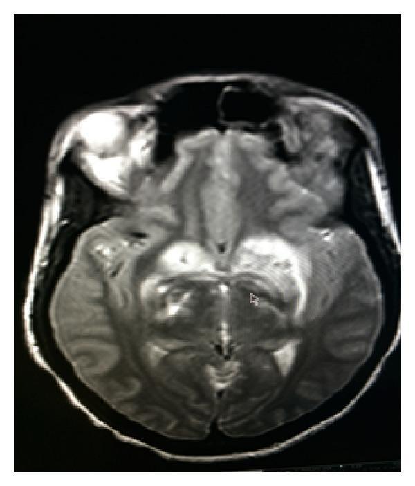
This diagnostic image is an axial T2-weighted Magnetic Resonance Image (MRI) of the brain at the level of the basal ganglia. The image reveals diffuse areas of high signal intensity (hyperintensity), particularly prominent within the basal ganglia and extending into the surrounding white matter. These findings are highly characteristic of vasogenic edema, indicating extracellular fluid accumulation often associated with blood-brain barrier disruption. There is evidence of mild to moderate mass effect in the left hemisphere, manifesting as subtle expansion of the involved tissue and slight compression of the adjacent cerebral structures, although the midline appears relatively stable in this specific slice. The hyperintensities are asymmetric, appearing more extensive in the left hemisphere. This imaging is clinically significant for identifying secondary complications of intracranial pathology, such as neuroinfection or inflammatory processes, and is used to monitor response to anti-edema treatments like dexamethasone or mannitol.
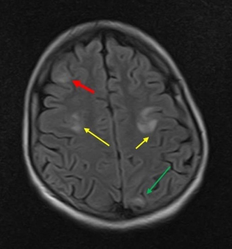
This diagnostic image is an axial T2-Fluid Attenuated Inversion Recovery (FLAIR) MRI scan of the brain. The image displays multiple, multifocal hyperintense lesions within the cortical and subcortical white matter, representing vasogenic edema. Colored arrows highlight the specific anatomical distribution of these findings: a red arrow points to a hyperintense lesion in the right frontal lobe; yellow arrows indicate bilateral hyperintensities in the parietal lobes; and a green arrow identifies a smaller area of increased signal in the left occipital lobe. These asymmetrical, high-signal regions are characteristic of conditions causing disruption of the blood-brain barrier, such as Posterior Reversible Encephalopathy Syndrome (PRES) or other metabolic encephalopathies. The image serves as a clinical teaching tool for identifying typical signal intensities and lobar involvement in neurological disorders presented via MRI sequencing.
Treatment of Vasogenic Oedema
1. Corticosteroids (Primary Treatment)
Dexamethasone
- Mechanism: Rapidly closes the BBB; effect on permeability can be seen as early as 1 hour after administration.
- Indication: Oedema surrounding brain tumours (primary or metastatic), cerebral abscess, multiple sclerosis exacerbations.
- Dose (brain tumour): Start at 8-12 mg/day, adjusted to the lowest effective dose. Clinical improvement often begins within minutes to hours; dramatic improvement occurs within 24-48 hours (Goldman-Cecil Medicine).
- Harrison's protocol: Dexamethasone 4 mg q6h for vasogenic oedema from tumour or abscess.
- Contraindications: Corticosteroids are not effective in cytotoxic oedema and are contraindicated in:
- Head trauma (worsens outcome)
- Ischaemic stroke
- Intracerebral/subarachnoid haemorrhage (systemic complications can worsen the patient's condition)
- Vasogenic oedema from trauma also typically does not respond.
Methylprednisolone (MS Exacerbations)
- Dose: 1 g/day for 3-5 days for acute MS exacerbations with vasogenic oedema component.
- Dramatic reduction in BBB enhancement on MRI may be seen. Effect is, however, lost after several months.
Side Effects to Monitor
- Insomnia, steroid myopathy, GI complications, hyperglycaemia (monitor blood glucose closely).
- If corticosteroids are required for >6 weeks, give prophylaxis against Pneumocystis jirovecii.
2. Osmotic Therapy (Acute ICP Reduction)
Mannitol
- Dose: 0.25-1 g/kg IV (low doses as effective as older high doses of 3 g/kg, with fewer electrolyte disturbances).
- Mechanisms: Increases serum osmolality, reduces brain tissue volume (particularly non-infarcted tissue), reduces CSF and interstitial fluid secretion by ~50%, rheological blood changes, antioxidant effect.
- Target: Serum osmolality 300-315 mOsm/L; do not exceed 320 mOsm/L.
- Cautions: Avoid in ICH, aneurysms, or AVMs before craniotomy (can expand haematoma as normal brain shrinks); may precipitate pulmonary oedema in patients with cardiac or renal impairment; rebound oedema may occur on discontinuation.
Hypertonic Saline
- Dose: 30 mL of 23.4% NaCl bolus; or 3% NaCl infusion.
- Increasingly advocated especially in patients where mannitol is contraindicated.
- Caution: Monitor serum sodium and osmolality carefully; risk of osmotic demyelination syndrome if used in hyponatraemic patients.
3. General Measures for Raised ICP
| Step | Intervention |
|---|---|
| 1 | Elevate head of bed to 30°; avoid hypotonic IV fluids |
| 2 | Intubate and hyperventilate to PaCO₂ 30-35 mmHg (acute measure only - effects are short-lived, may worsen ischaemia) |
| 3 | Drain CSF via ventriculostomy if in place |
| 4 | Osmotherapy (mannitol or hypertonic saline) |
| 5 | Dexamethasone for vasogenic oedema (tumour/abscess) |
| 6 | Sedation (morphine, propofol, midazolam) ± neuromuscular paralysis |
| 7 | Vasopressors to maintain MAP and CPP ≥60 mmHg |
4. Second-Line / Refractory ICP
- Decompressive craniectomy - shown to reduce mortality in TBI with refractory ICP.
- High-dose barbiturate ("pentobarb coma") - metabolic suppression.
- Hypothermia to 33°C.
5. Anti-VEGF Therapy (Tumour-Associated Oedema)
- Bevacizumab (anti-VEGF antibody) is effective for symptomatic cerebral oedema in brain tumours and can significantly reduce corticosteroid requirements.
- Particularly useful in recurrent glioblastoma or radiation-induced oedema.
6. Treat the Underlying Cause (Definitive)
| Cause | Definitive Treatment |
|---|---|
| Brain tumour | Surgery, radiotherapy, chemotherapy |
| Abscess | Antibiotics ± surgical drainage |
| MS exacerbation | High-dose methylprednisolone |
| Spinal cord compression | Dexamethasone 0.1 mg/kg (max 10 mg) IV + urgent decompression |
| Bacterial meningitis | Antibiotics + dexamethasone (also targets BBB disruption via MMP inhibition) |
Key Distinctions
| Feature | Vasogenic Oedema | Cytotoxic Oedema |
|---|---|---|
| BBB | Disrupted | Intact |
| Fluid type | Protein-rich plasma | Intracellular water |
| Location | White matter | Grey + white matter |
| Corticosteroids | Effective | Not effective |
| Osmotic agents | Useful temporarily | More appropriate first-line |
| Causes | Tumours, abscess, MS, trauma | Stroke, hypoxia, metabolic |
Treatment of PRES
"posterior reversible encephalopathy syndrome" AND treatment
PRES posterior reversible encephalopathy syndrome MRI FLAIR white matter
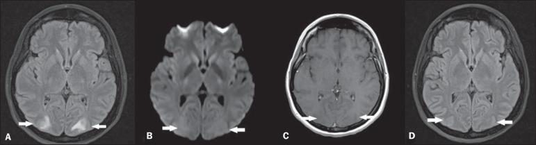
Diagnostic axial brain MRI series illustrating Posterior Reversible Encephalopathy Syndrome (PRES). Panel A (FLAIR) shows hallmark bilateral, symmetric hyperintensities in the white matter of the occipital lobes, indicative of vasogenic edema. Panel B (Diffusion-Weighted Imaging) demonstrates no restricted diffusion in these areas, confirming the absence of cytotoxic edema or acute infarction. Panel C (T1-weighted post-contrast) shows no pathological gadolinium enhancement, highlighting the lack of blood-brain barrier breakdown. Panel D (Follow-up FLAIR at 4 weeks) shows complete radiological resolution of the previous hyperintense lesions. This comparison demonstrates the classic reversible neuroimaging features of vasogenic edema associated with PRES, typically seen in clinical contexts such as hypertensive crisis, preeclampsia, or immunosuppressive therapy.
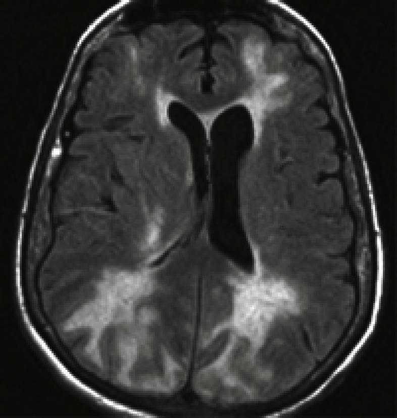
This diagnostic image is an axial T2-weighted or Fluid-Attenuated Inversion Recovery (FLAIR) MRI scan of the brain, demonstrating characteristic findings of Posterior Reversible Encephalopathy Syndrome (PRES). The image reveals prominent, bilateral, patchy areas of hyperintense signal within the cortical and subcortical white matter. These hyperintensities are most pronounced in the posterior regions, specifically the occipital and parietal lobes, though they also extend into the frontal lobes. The lesions represent vasogenic edema and appear as irregular, ill-defined clusters. The ventricular system, including the lateral ventricles, maintains a relatively normal morphology without significant mass effect or midline shift despite the adjacent parenchymal changes. The sulcal and gyral patterns are largely preserved. This imaging modality is essential for identifying the symmetric vasogenic edema typical of PRES, which often presents clinically with encephalopathy, seizures, and visual disturbances associated with acute hypertension or drug toxicity.
PMID: 40818477
Treatment of PRES (Posterior Reversible Encephalopathy Syndrome)
Core Principles
- Blood pressure control
- Remove/treat the precipitating cause
- Seizure management
1. Blood Pressure Control
- Agents of choice: IV labetalol or IV nicardipine (both recommended by Harrison's 22E as first-line IV agents)
- Target: Lower the MAP by approximately 20% initially - do NOT aim for rapid normalisation, as overshoot below the patient's autoregulatory lower limit can cause secondary cerebral ischaemia and infarction.
- Monitoring: Continuous BP monitoring; repeat neuroimaging if clinical worsening occurs despite treatment.
- Hypertensive crisis is the most common trigger, but blood pressure may not always be markedly elevated at presentation (especially in drug-induced or eclampsia-related PRES).
2. Remove the Precipitating Cause
| Cause | Action |
|---|---|
| Calcineurin inhibitors (cyclosporine, tacrolimus) | Reduce dose or discontinue; switch to sirolimus (very few cases of neurotoxicity) |
| Chemotherapy / immunotherapy (rituximab, bevacizumab, tyrosine kinase inhibitors, CAR-T therapy) | Withhold or discontinue offending agent |
| Eclampsia / pre-eclampsia | Magnesium sulfate + delivery (definitive treatment) |
| Erythropoietin (especially in ESKD) | Dose reduction; manage fluid overload and BP |
| ESKD / fluid overload | Urgent dialysis to achieve euvolaemia |
| Autoimmune disease / vasculitis | Treat the underlying condition (but note steroids are generally not used for PRES itself) |
Note from Harrison's: Calcineurin inhibitor neurotoxicity can occur even at normal serum drug levels, so normal levels do not exclude them as the cause.
3. Seizure Management
- Seizures occur in up to 90% of patients with PRES (Brenner & Rector).
- Non-convulsive seizures are common - maintain a low threshold for continuous EEG monitoring.
- Standard anticonvulsants are effective (levetiracetam is commonly used as it avoids hepatic metabolism and has fewer drug interactions).
- Special case - Eclampsia: Magnesium sulfate is preferred over conventional anticonvulsants for seizure control, with evidence supporting its superiority in this context.
- Duration of anticonvulsant therapy: Can typically be weaned once the PRES resolves radiologically, as seizures do not usually persist after the underlying cause is treated.
4. General Supportive Care
- Elevate head of bed to 30°
- Avoid hypotonic IV fluids (worsen cerebral oedema)
- Correct any metabolic disturbances (hyponatraemia, hypomagnesaemia, hypocalcaemia) that lower the seizure threshold
- Maintain airway if consciousness is impaired; ICU-level monitoring for severe cases
5. Monitoring and Prognosis
- Repeat MRI typically shows complete radiological resolution after BP control and removal of the offending agent.
- Prognosis is generally favourable, but a 2025 Lancet Neurology review by Fugate, Hawkes & Rabinstein highlights that complications including epilepsy and stroke can follow severe or recurrent PRES, and that novel agents (CAR-T, tyrosine kinase inhibitors) are expanding the spectrum of presentations.
- Emerging biomarkers (IL-6, IL-10, VEGF) may help predict severity and outcome.
Quick Reference Summary
| Step | Intervention |
|---|---|
| 1 | IV labetalol or nicardipine - lower MAP by ~20% |
| 2 | Remove offending drug (calcineurin inhibitor, chemotherapy) |
| 3 | Treat eclampsia - MgSO₄ + delivery |
| 4 | Dialysis if ESKD with fluid overload |
| 5 | Anticonvulsants for seizures; continuous EEG if encephalopathic |
| 6 | ICU monitoring; repeat MRI to confirm resolution |
| Avoid | Rapid BP normalisation (risk of ischaemia); corticosteroids are not standard treatment for PRES |