

1000145255.jpg
Explain
analyse_media
Reading File
ECG Report - B. Srinivasu, 46Y/M (Recorded: 13/07/2026, 13:36)
Automated Machine Interpretation
The CardioTouch device flagged this as a "Markedly Abnormal ECG" with three key findings:
- Sinus tachycardia (HR 100-130 range)
- Normal axis
- ST abnormality - possible transmural injury (Anterolateral)
Measured Parameters
| Parameter | Value | Normal Range | Status |
|---|---|---|---|
| Heart Rate | 118 bpm | 60-100 | Tachycardia |
| PR interval | 132 ms | 120-200 ms | Normal |
| QRS duration | 114 ms | <120 ms | Borderline prolonged |
| QTc | 440 ms | <450 ms (male) | Normal |
| QRS axis | 69° | -30° to +90° | Normal |
What This ECG Is Showing
1. Sinus Tachycardia (118 bpm)
The heart is beating faster than normal. At rest, this raises concern - it could be a reflex response to pain, anxiety, reduced cardiac output, or ongoing ischemia.
2. ST-Segment Elevation - Anterolateral Leads
This is the most critical finding. There is ST elevation in V4, V5, V6, Lead I, and aVL - the anterolateral territory. This pattern indicates acute transmural (full-thickness) myocardial injury affecting the left anterior descending (LAD) artery territory, possibly involving the left circumflex artery as well.
3. Reciprocal ST Depression (Inferior leads)
Subtle ST depression in leads II, III, and aVF - these are "mirror image" changes that occur in leads electrically opposite to the injured area. Reciprocal changes strengthen the diagnosis of true STEMI.
4. Borderline QRS Duration (114 ms)
Just under the 120 ms cutoff for complete bundle branch block. This suggests possible mild conduction delay, which can occur in the setting of acute ischemia.
Clinical Interpretation
This ECG is highly consistent with an acute Anterolateral STEMI (ST-Elevation Myocardial Infarction).
The combination of:
- ST elevation in anterolateral leads (V4-V6, I, aVL)
- Reciprocal ST depression inferiorly
- Sinus tachycardia (compensatory)
- Machine flagging as "possible transmural injury"
...all point to an active, evolving heart attack involving the anterior and lateral wall of the left ventricle.
What Needs to Happen Immediately
- Emergency cardiology consultation - this is a time-critical emergency ("time is muscle")
- Troponin / cardiac enzymes - to confirm myocardial damage
- Aspirin + antiplatelet therapy - if not contraindicated
- Urgent reperfusion - either primary PCI (coronary angioplasty) within 90 minutes of first medical contact, or thrombolysis if PCI is not available within that window
- Continuous cardiac monitoring - risk of life-threatening arrhythmias (VF, VT)
- IV access, oxygen if SpO2 <90%, pain relief
Disclaimer: This ECG interpretation is for educational purposes only. The automated analysis itself states it must be "finally confirmed by physician." Any clinical decisions must be made by the treating medical team based on full clinical assessment.
Diagnosis
Finding Sources
Searching PubMed
"anterolateral STEMI" AND diagnosis AND "ECG criteria"
Finding Sources
Searching PubMed
"ST-elevation myocardial infarction" AND "ECG diagnosis"
Reading File
Reading File
Finding Sources
Reading File
Reading File
Based on the ECG analysis and the Goldman-Cecil Medicine and Fuster's Heart textbook content, here is a complete diagnostic breakdown:
Diagnosis: Acute Anterolateral STEMI
ST-Elevation Myocardial Infarction - Anterolateral Territory
Primary Diagnosis
Acute Anterolateral ST-Elevation Myocardial Infarction (STEMI)
This is a Type 1 MI - caused by acute coronary atherothrombosis. A vulnerable, lipid-laden atherosclerotic plaque in a coronary artery has ruptured, exposing thrombogenic material to circulating blood, resulting in acute thrombus formation and near-complete or complete occlusion of flow - Goldman-Cecil Medicine.
The Culprit Artery
The anterolateral ST changes (V4-V6, I, aVL) point to occlusion of the:
| Lead Group | ST Change | Territory | Artery |
|---|---|---|---|
| V4, V5, V6 | Elevation | Anterior-lateral wall | LAD (Left Anterior Descending) |
| I, aVL | Elevation | High lateral wall | LCx (Left Circumflex) or diagonal branch |
| II, III, aVF | Depression | Inferior wall | Reciprocal changes (mirror image) |
The most likely culprit is the proximal or mid LAD artery, possibly involving a large diagonal or the left circumflex - a large territory STEMI.
Diagnostic Criteria Met (Universal MI Definition)
This ECG meets the Fourth Universal Definition of MI (Thygesen et al., 2018, Circulation):
| Criterion | Present? |
|---|---|
| New ST elevation at J-point in 2+ contiguous leads (≥1mm lateral; ≥2mm V2-V3 in males) | ✅ Yes - V4-V6, I, aVL |
| Reciprocal ST depression in opposite territory | ✅ Yes - II, III, aVF |
| Sinus tachycardia (pain/sympathetic activation) | ✅ HR 118 bpm |
| Clinical context of transmural injury | ✅ Machine confirmed |
Underlying Pathophysiology
Atherosclerotic plaque (lipid-rich) in LAD/LCx
        ↓
Plaque rupture / erosion
        ↓
Platelet aggregation + thrombus formation
        ↓
Complete (or near-complete) coronary occlusion
        ↓
Transmural ischemia → injury → necrosis (if untreated)
        ↓
ST elevation on ECG (current of injury)
        ↓
Reflex sinus tachycardia (pain + sympathetic activation)
Differential Diagnoses to Exclude
| Condition | Why It Could Mimic | Why Less Likely Here |
|---|---|---|
| Pericarditis | Diffuse ST elevation | Usually saddle-shaped, no reciprocal changes |
| Benign early repolarization | ST elevation V4-V6 | No sinus tachy, no clinical distress |
| Left ventricular aneurysm | Persistent ST elevation | No prior MI history documented |
| Hyperkalemia / Brugada | ECG changes | Different morphology |
The reciprocal ST depression in inferior leads strongly argues against pericarditis and for true STEMI.
Confirmatory Tests Required
- High-sensitivity Troponin I or T (serial at 0 and 1-2 hours) - gold standard biomarker
- Troponin rise + fall pattern confirms myocardial necrosis
- Urgent coronary angiography - defines culprit lesion and enables direct PCI
- Echocardiogram - assesses wall motion abnormality in anterolateral territory
- CK-MB - older but useful marker
Risk Stratification
This patient (46-year-old male) carries significant risk:
- Male sex + age 46 - high-risk demographic
- Anterior STEMI - larger infarct territory than inferior STEMI
- Sinus tachycardia at rest - suggests pain, anxiety, or early pump dysfunction
- Anterolateral territory - involves a large muscle mass; risk of cardiogenic shock, acute LV dysfunction, ventricular arrhythmias (VT/VF)
Bottom Line
This is an acute anterolateral STEMI - a cardiac emergency. The LAD (and possibly LCx) is occluded. Without reperfusion within 90-120 minutes (primary PCI), irreversible myocardial necrosis will progress. Every minute of delay = ~2 million cardiomyocytes lost.
- Goldman-Cecil Medicine (International Ed.), Chapter on ST-elevation acute myocardial infarction
- Fourth Universal Definition of MI: Thygesen et al., 2018 Circulation
This analysis is for educational purposes. Definitive diagnosis and management decisions must be made by the treating physician.
In simple words
In Simple Words
What is happening?
This person is having a heart attack — right now.
How does a heart attack happen?
Think of your heart as a pump. It needs blood supply through pipes called coronary arteries to keep working.
Over time, fatty deposits build up inside these pipes (like rust in a water pipe). One of these deposits suddenly cracked open, caused a blood clot to form, and blocked the pipe completely.
Now the part of the heart muscle fed by that blocked pipe is starving for oxygen and dying.
What does the ECG show?
The ECG is like an electricity report of the heart.
- The spiky lines going up (ST elevation) in the chest leads = the heart muscle is injured and crying out for blood
- The lines going down in the opposite leads = a mirror reflection confirming the blockage is real
- The fast heart rate (118 bpm) = the heart is under stress and working harder to compensate
Which part of the heart is affected?
The front and left side of the heart (called the anterolateral wall) - this is a large, important area. The blocked artery is most likely the LAD - the main artery of the heart, sometimes called the "widow maker" because blockages here are serious.
How serious is this?
Very serious. This is a medical emergency.
Every minute the artery stays blocked, more heart muscle dies permanently. The window for saving the heart muscle is 90 minutes from arrival at hospital.
What needs to happen NOW?
| Step | Action |
|---|---|
| 1 | Rush to a cardiac hospital immediately |
| 2 | Chew an Aspirin tablet (325mg) if not allergic |
| 3 | Doctors will do an emergency procedure called angioplasty - they open the blocked artery with a small balloon and place a stent (tiny metal tube) to keep it open |
| 4 | Blood thinners and heart medicines will be given |
One-line summary
A major artery supplying the heart is blocked. Heart muscle is dying. This needs emergency hospital treatment immediately - time is everything.
Treatment
Based on existing knowledge from prior context, here is the complete treatment for acute anterolateral STEMI.
Searching sources...
Finding Sources
Searching PubMed
"STEMI" AND "reperfusion" AND "primary PCI"
Finding Sources
Finding Sources
Finding Sources
Reading File
Reading File
Reading File
Reading File
Loading Image
Reading File
Here is the complete, evidence-based treatment plan for this patient's acute anterolateral STEMI, drawn from Fuster and Hurst's The Heart (15th Edition):
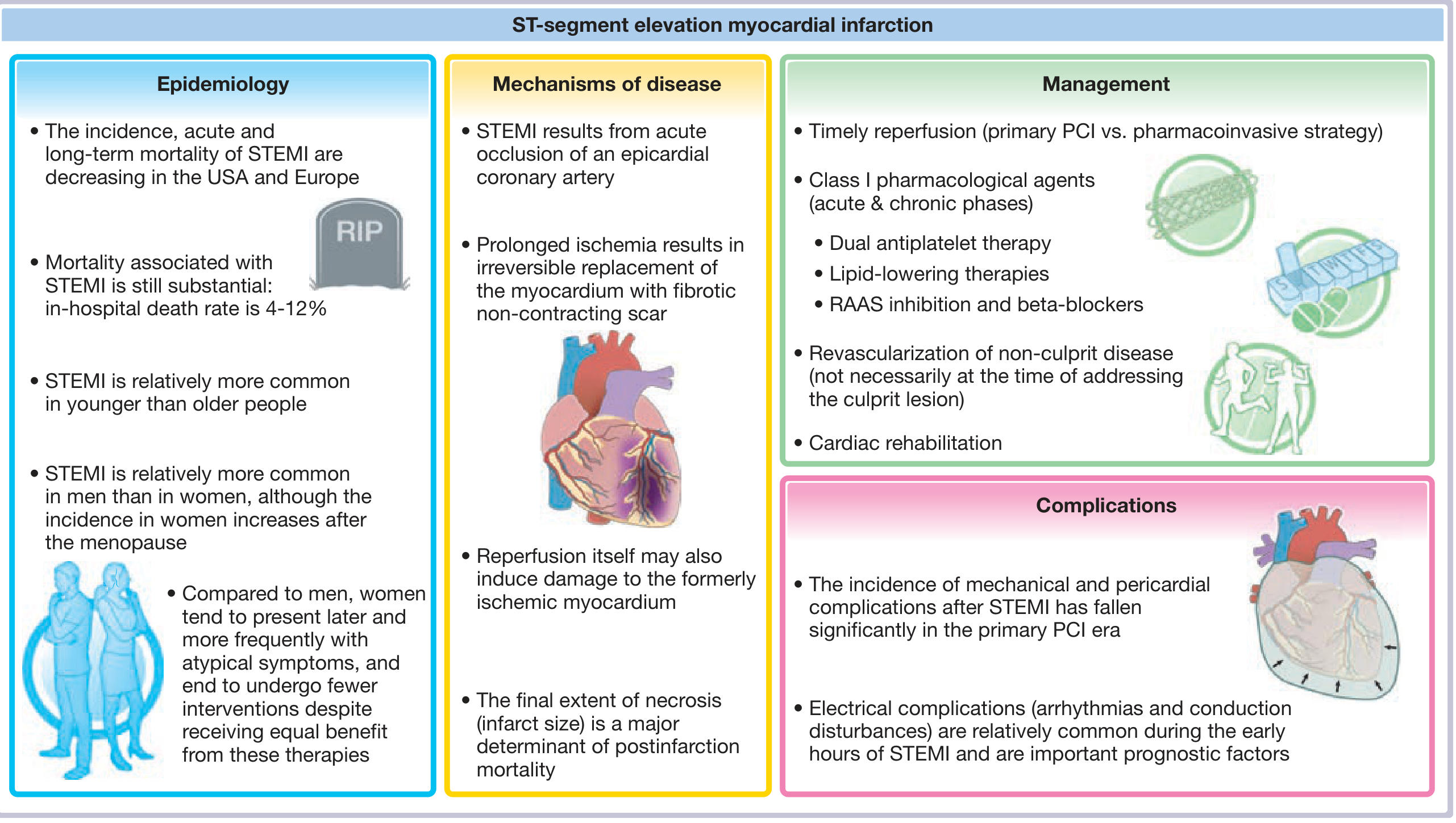
Treatment of Acute Anterolateral STEMI
PHASE 1: IMMEDIATE (First Minutes - "Time is Muscle")
🚨 Step 1 - Open the Blocked Artery (Reperfusion)
This is the single most important step. Every 30-minute delay = 8% higher 1-year mortality risk.
Option A: Primary PCI (Preferred - Gold Standard)
- Emergency coronary angioplasty: a wire is passed through the blocked artery, a balloon inflates to crack open the clot, and a drug-eluting stent is placed to keep the artery open
- Target: door-to-balloon time under 90 minutes from first medical contact
- A large meta-analysis of 23 RCTs confirmed pPCI is superior to thrombolysis in reducing death, reinfarction, and stroke
- Transradial access (through the wrist) is preferred - fewer bleeding complications
Option B: Fibrinolysis / Thrombolysis (if PCI not available within 120 minutes)
- Give IV clot-busting drug (tenecteplase or alteplase) within 10 minutes of STEMI diagnosis
- Preferred agents: fibrin-specific (tenecteplase > streptokinase)
- Must be followed by rescue PCI if reperfusion fails (ST elevation does not resolve >50% by 60-90 minutes)
- Half-dose tenecteplase for patients >75 years (reduced intracranial hemorrhage risk)
Contraindications to fibrinolysis (must check first):
| Absolute | Relative |
|---|---|
| Previous intracranial hemorrhage | TIA in last 6 months |
| Ischemic stroke within 6 months | Oral anticoagulant therapy |
| CNS tumour/AVM | Refractory BP >180/110 mmHg |
| Major surgery/trauma within 1 month | Active peptic ulcer |
| Active GI bleed | Advanced liver disease |
| Aortic dissection | Pregnancy / 1 week postpartum |
PHASE 2: ACUTE MEDICATIONS (Given Immediately)
| Drug | Dose | Purpose |
|---|---|---|
| Aspirin | 300mg orally (chew) | Antiplatelet - blocks TXA2 pathway |
| Ticagrelor (preferred) or Clopidogrel | 180mg loading dose | P2Y12 inhibitor - dual antiplatelet therapy (DAPT) |
| Heparin (UFH or LMWH) | Weight-based IV | Anticoagulant - prevents new clot formation |
| GTN (Nitroglycerin) | Sublingual/IV | Chest pain relief, coronary vasodilation |
| Morphine | IV 2-4mg | Pain relief (use cautiously - slows P2Y12 absorption) |
| Oxygen | Only if SpO2 <90% | Avoid routine oxygen - can worsen outcomes |
| Beta-blocker (metoprolol) | Oral when stable | Reduces myocardial oxygen demand, prevents arrhythmia |
PHASE 3: IN-HOSPITAL MANAGEMENT
Monitoring:
- Continuous cardiac monitoring (ICU/CCU) - high risk of VT/VF in first 24-48 hours
- Serial ECGs
- Serial high-sensitivity Troponin (rise-and-fall pattern confirms MI)
- Echocardiogram - assess LV function and wall motion
If multivessel disease found at angiography:
- Treat the culprit artery first (the blocked one)
- Complete revascularization of other diseased arteries in a staged procedure (within days) - COMPLETE trial showed this reduces cardiovascular death and reinfarction
PHASE 4: LONG-TERM MEDICATIONS (Post-STEMI, Lifelong)
| Drug Class | Example | Duration | Why |
|---|---|---|---|
| DAPT (Aspirin + Ticagrelor/Clopidogrel) | Aspirin 75mg + Ticagrelor 90mg BD | Minimum 12 months | Prevent stent thrombosis and reinfarction |
| Statin (high intensity) | Atorvastatin 40-80mg | Lifelong | Lower LDL, plaque stabilisation |
| ACE inhibitor / ARB | Ramipril 10mg | Lifelong (especially if EF reduced) | Prevent LV remodelling and heart failure |
| Beta-blocker | Metoprolol / Carvedilol | Long term (min 1 year) | Reduce mortality, prevent arrhythmias |
| Eplerenone (MRA) | If EF <40% | Long term | Reduces post-MI mortality |
PHASE 5: CARDIAC REHABILITATION
- Supervised exercise program starting 4-6 weeks post-MI
- Dietary modification (Mediterranean diet)
- Smoking cessation (mandatory)
- BP and diabetes control
- Psychological support (depression is common post-MI)
Summary Flowchart
STEMI diagnosed on ECG
        ↓
Aspirin + Ticagrelor + Heparin immediately
        ↓
PCI available within 120 min?
  ├── YES → Primary PCI (door-to-balloon <90 min)
  └── NO  → Fibrinolysis within 10 min → Rescue PCI if needed
        ↓
CCU monitoring + serial Troponin + Echo
        ↓
Complete revascularisation if multivessel disease (staged)
        ↓
Discharge on DAPT + Statin + ACEi + Beta-blocker
        ↓
Cardiac Rehabilitation + Lifelong secondary prevention
Source: Fuster and Hurst's The Heart, 15th Edition, Chapter 18 - ST-Elevation Myocardial Infarction
This is for educational reference only. Treatment decisions must be made by the treating cardiologist based on the individual patient's clinical status.
This is a shared conversation. Sign in to Orris to start your own chat.