
osgood-schlatter disease
Osgood-Schlatter disease tibial tubercle apophysitis knee
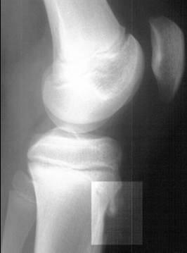
This lateral knee radiograph of a pediatric patient demonstrates the classic radiographic features of Osgood-Schlatter disease. The imaging reveals a prominent tibial tubercle with clear evidence of fragmentation and heterotopic ossification within the patellar tendon insertion site. In addition to the bony changes, there is distinct soft tissue swelling and thickening of the patellar tendon overlying the apophysis. The overall joint alignment is maintained, with normal positioning of the patella relative to the distal femur and a well-preserved tibiofemoral joint space. The growth plates are visible, confirming the adolescent stage of skeletal development. This diagnostic image is used to illustrate traction apophysitis of the tibial tubercle, a common cause of anterior knee pain in active adolescents. The clinical significance of these findings relates to the repetitive strain on the patellar tendon at its insertion point, leading to microtrauma and subsequent fragmentation of the developing bone.
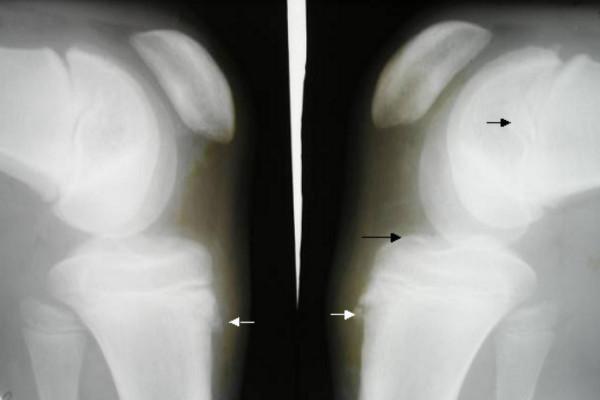
This lateral knee radiograph comparison displays bilateral skeletal structures of the knee joints, including the distal femur condyles, patellae, and proximal tibiae. The images demonstrate classic features of Osgood-Schlatter disease, characterized by bilateral irregularity and fragmentation of the tibial tuberosity apophyses. Distinct white arrows highlight the areas of bony avulsion or separation where the patellar tendon inserts into the tibial tubercle. Black arrows point to associated osteochondral changes and the articulation space. The radiographs were captured with 10-20 degrees of internal rotation. Visible hallmarks include the prominence and fragmentation of the tibial tubercle and soft tissue thickening in the infrapatellar region. These findings are clinically significant for diagnosing traction apophysitis in adolescent patients presenting with anterior knee pain and point tenderness over the tibial tuberosity.
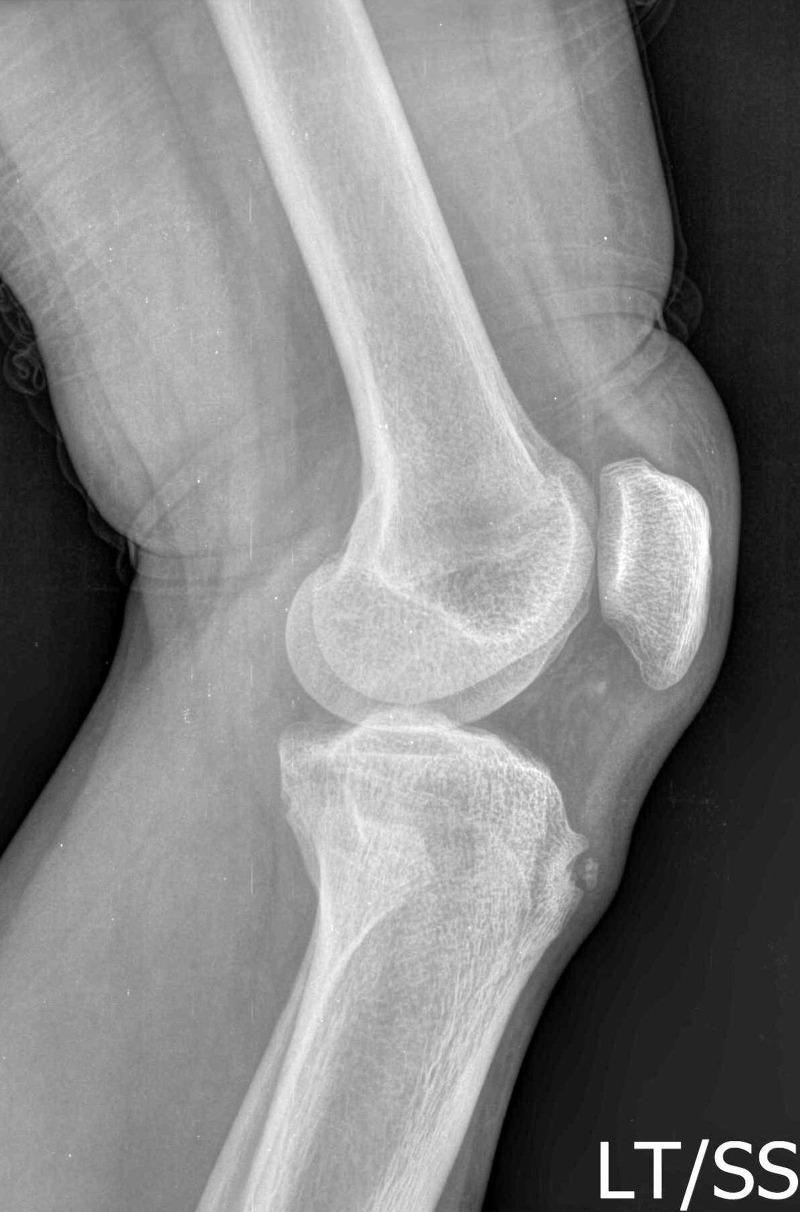
This diagnostic image is a lateral radiograph (X-ray) of the human knee joint, illustrating hallmark features of Osgood-Schlatter disease (OSD). The radiograph clearly depicts the distal femur, the patella, and the proximal tibia. The primary pathology is localized at the tibial tuberosity, where there is a visible bony prominence and irregularity. Notably, a small, distinct, separated ossicle is located anterior to the tibial tuberosity within the distal portion of the patellar tendon. These findings represent traction apophysitis or fragmentation of the tibial tubercle common in the sub-acute or chronic stages of OSD. The joint space appears preserved, and there is no evidence of acute fracture of the femoral condyles or the patella. This visual is significant for medical education in pediatrics and orthopedics for diagnosing adolescent knee pain and distinguishing traction apophysitis from acute avulsion fractures.
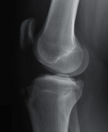
This lateral view X-ray radiography of the knee joint demonstrates the characteristic features of Osgood-Schlatter disease. The imaging focuses on the proximal tibia, distal femur, and patella. A prominent, irregular bony enlargement is visible at the tibial tuberosity, which serves as the insertion site for the patellar ligament. This area shows significant anterior protrusion and evidence of bony fragmentation, with small ossicles or fragments separated from the main tibial cortex. The surrounding bone tissue exhibits mild sclerosis. The joint space and patellar positioning appear otherwise normal. This diagnostic image is representative of chronic traction apophysitis in an adolescent patient, illustrating the sequelae of repetitive stress on the tibial tubercle apophysis. It serves as an educational example for orthopedic radiology, particularly in the context of pediatric sports medicine and growth-related musculoskeletal conditions.
"Osgood-Schlatter disease"[MeSH Terms]