
Nitric oxide - synthesis and secondary causes
I'll start with a quick answer, then consult the medical library for detailed information.
Nitric oxide (NO) is synthesized from L-arginine by nitric oxide synthase (NOS) enzymes, producing NO and L-citrulline. There are three NOS isoforms: endothelial (eNOS), neuronal (nNOS), and inducible (iNOS). NO acts as a vasodilator, neurotransmitter, and immune mediator. In pathology, excess NO production (especially via iNOS) contributes to conditions like septic shock, while deficient NO underlies hypertension and endothelial dysfunction.
Searching sources...
Reading File
Now let me search the medical library in parallel for both synthesis and secondary causes of nitric oxide.
Finding Sources
Finding Sources
Excellent results. Now let me read the key sections in parallel - Medical Physiology for synthesis, Lippincott Biochemistry for the detailed synthesis pathway, and Braunwald's for cardiovascular context.
Reading File
Reading File
Reading File
Let me now read the Lippincott Biochemistry section more carefully (the synthesis pathway) and also check the Kaplan Psychiatry and Rheumatology sections, plus search for NOS isoforms and secondary causes.
Reading File
Finding Sources
Finding Sources
Excellent. Now let me read the key dedicated NOS sections from Basic Medical Biochemistry and Goldman-Cecil for secondary causes.
Reading File
Reading File
Now let me fetch the synthesis diagram from Lippincott and also read the NO toxicity section plus search for more secondary causes (hypertension, erectile dysfunction, etc.).
Loading Image
Reading File
Finding Sources
Now let me also read the Kaplan Psychiatry section on nitric oxide and get information on secondary hypertension/erectile dysfunction connections.
Reading File
Finding Sources
I now have comprehensive information to write a detailed response. Let me also fetch the NOS synthesis diagram from Basic Medical Biochemistry.
Loading Image
Loading Image
I now have everything needed for a thorough, well-illustrated answer. Here is the complete response:
Nitric Oxide (NO) - Synthesis and Secondary Causes
1. Chemistry and Basic Properties
Nitric oxide (NO•) is a gaseous free radical - the dot denotes its single unpaired electron, which makes it highly reactive. It should not be confused with nitrous oxide (N₂O, the anesthetic) or nitrogen dioxide (NO₂, a pollutant). Because NO is a small, uncharged gas, it freely diffuses across water and lipid membranes, acting in a paracrine rather than endocrine fashion - its effects are restricted to the immediate site of release. Haemoglobin avidly binds NO, which limits its diffusion distance and confines its action locally. The physiological target of NO is soluble guanylyl cyclase, which contains an iron-heme group (just as Hb does) that binds NO and converts GTP to cyclic GMP (cGMP). - Medical Physiology, Boron & Boulpaep
2. Synthesis Pathway
Biochemical Reaction
L-Arginine + O₂ + NADPH → Nitric Oxide (NO•) + L-Citrulline + NADP⁺
This reaction is catalysed by nitric oxide synthase (NOS), which is a complex enzyme similar in mechanism to cytochrome P450.
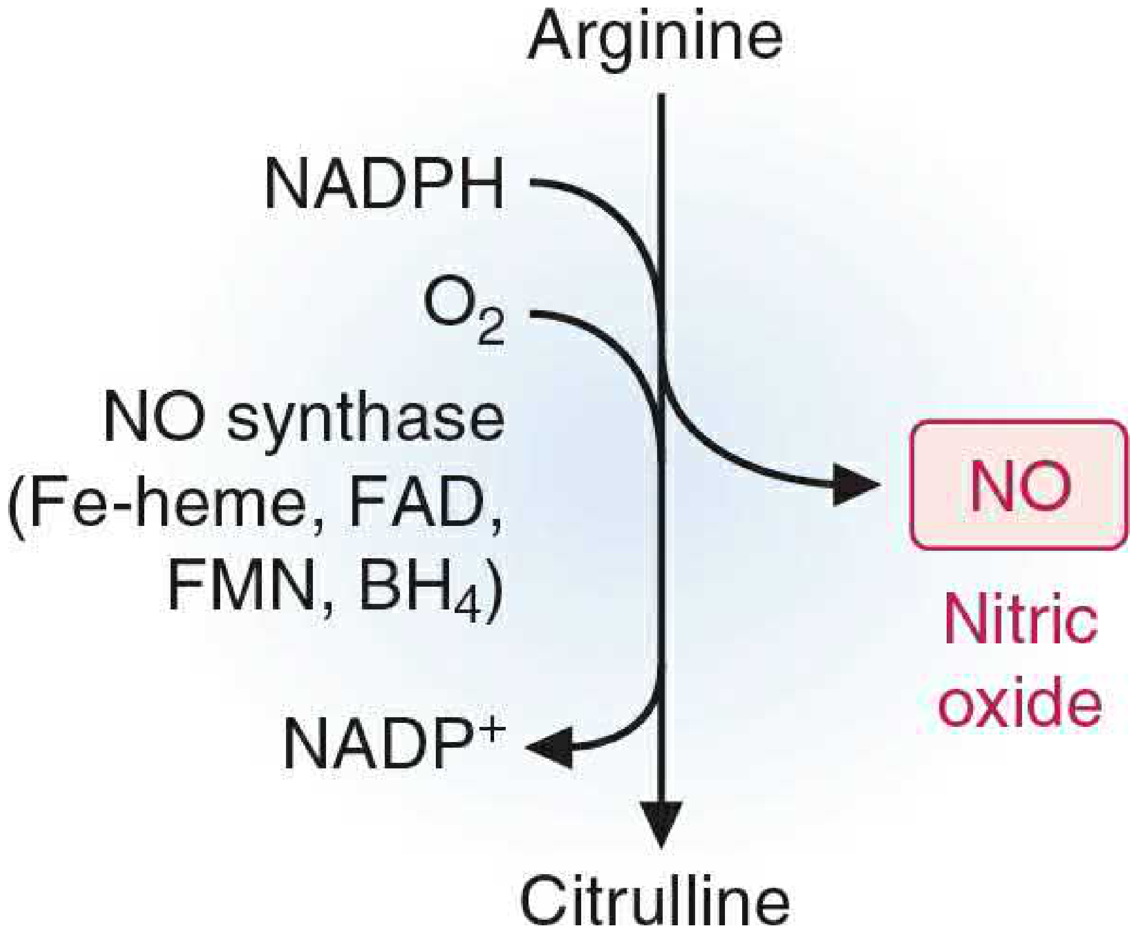
Fig. NOS reaction - Arginine → NO + Citrulline. Cofactors: Fe-heme, FAD, FMN, BH₄ (Basic Medical Biochemistry, 6e)
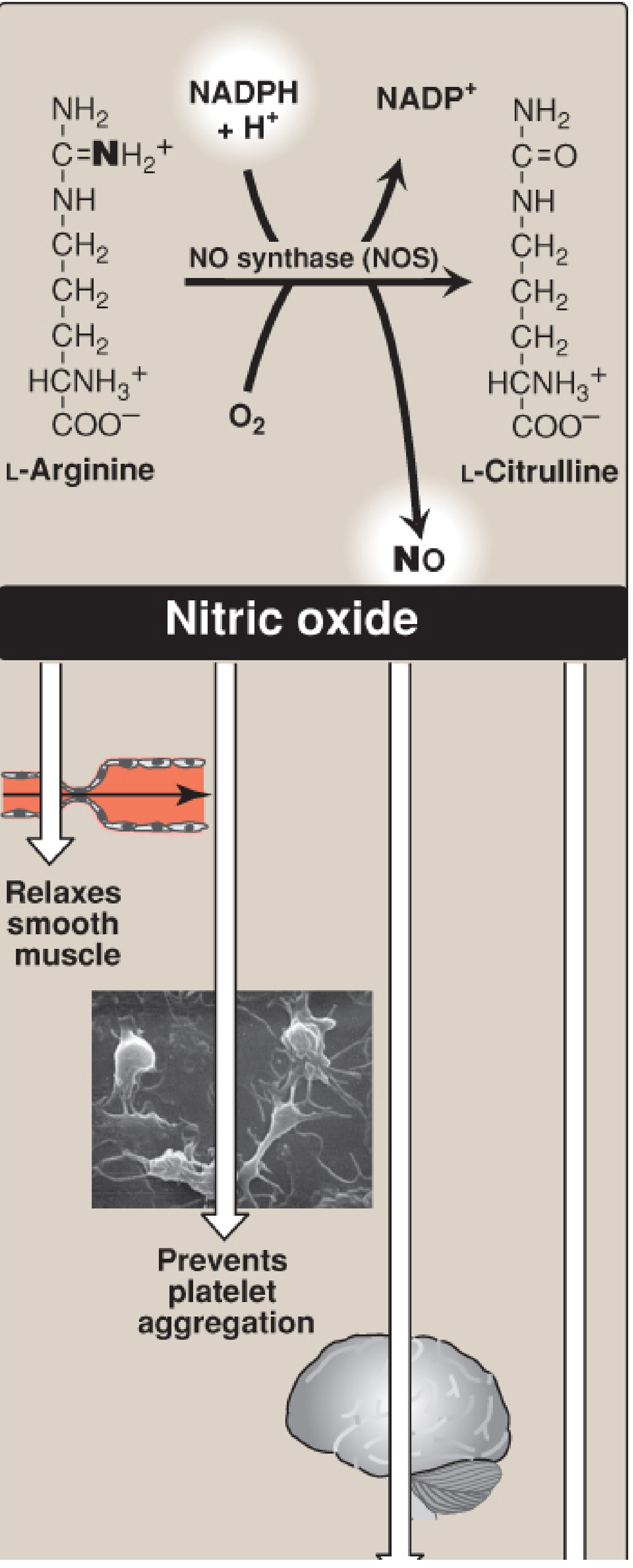
Fig. NO synthesis and downstream actions: smooth muscle relaxation and inhibition of platelet aggregation (Lippincott Biochemistry, 8e)
Cofactors Required by NOS
| Cofactor | Role |
|---|---|
| Fe-heme | Electron transfer (like CYP450) |
| FAD (Flavin adenine dinucleotide) | Electron carrier |
| FMN (Flavin mononucleotide) | Electron carrier |
| BH₄ (Tetrahydrobiopterin) | Essential for coupling; without it, NOS produces O₂⁻ instead of NO ("uncoupled NOS") |
| NADPH | Reducing equivalents |
| O₂ | Incorporated into NO and citrulline |
3. NOS Isoforms
There are three genetically distinct isoforms of nitric oxide synthase, each encoded by a separate gene. - Basic Medical Biochemistry, 6e; Kaplan & Sadock's Psychiatry
| Isoform | Name | Also Called | Location | Regulation | Output Level |
|---|---|---|---|---|---|
| NOS I | nNOS (neuronal) | bNOS | Neurons (cortex, hippocampus, cerebellum, striatum), skeletal muscle | Ca²⁺/calmodulin-dependent; activated by neuronal depolarization | Low, physiological |
| NOS II | iNOS (inducible) | - | Macrophages, astroglia, neutrophils, many immune cells | Gene transcription - induced by LPS, IFN-γ, TNF-α; NOT Ca²⁺-dependent | Very high, sustained |
| NOS III | eNOS (endothelial) | - | Vascular endothelium | Ca²⁺/calmodulin + phosphorylation (e.g., by Akt/shear stress) | Low, physiological |
Key distinction: nNOS and eNOS are tightly regulated by intracellular Ca²⁺ to produce small, physiological quantities of NO. iNOS is regulated by cytokine induction of gene transcription and produces far higher, potentially toxic quantities. - Basic Medical Biochemistry, 6e
4. Downstream Signalling
- NO diffuses into target cells (smooth muscle, platelets, neurons)
- Binds Fe-heme group of soluble guanylyl cyclase (sGC)
- sGC activated → converts GTP to cGMP
- cGMP activates protein kinase G (PKG)
- PKG phosphorylates MLCK (myosin light chain kinase) and ion channels → smooth muscle relaxation → vasodilation
- cGMP is degraded by PDE-5 (phosphodiesterase type 5) - the target of sildenafil
5. Physiological Functions of NO
| System | Function |
|---|---|
| Vascular | Vasodilation (tonic regulation of blood pressure), inhibits platelet adhesion and aggregation, inhibits smooth muscle proliferation |
| Neural (nNOS) | Neurotransmitter; involved in long-term potentiation (LTP), learning and memory, neurogenesis |
| Immune (iNOS) | Bactericidal: activated macrophages produce NO that combines with superoxide → peroxynitrite → hydroxyl radical (OH•) → kills pathogens |
| Penile erection | nNOS/eNOS in penile endothelium and nerves causes cavernous smooth muscle relaxation enabling erection |
| GI | Non-adrenergic, non-cholinergic (NANC) neurotransmission; gut motility |
| Hypoxia | Under hypoxic conditions, nitrite (NO₂⁻) is reduced to NO → binds deoxyhemoglobin → released to cause vasodilation and increase O₂ delivery |
6. Reactive Nitrogen-Oxygen Species (RNOS) and NO Toxicity
When iNOS is massively upregulated, very high NO concentrations lead to formation of toxic species:
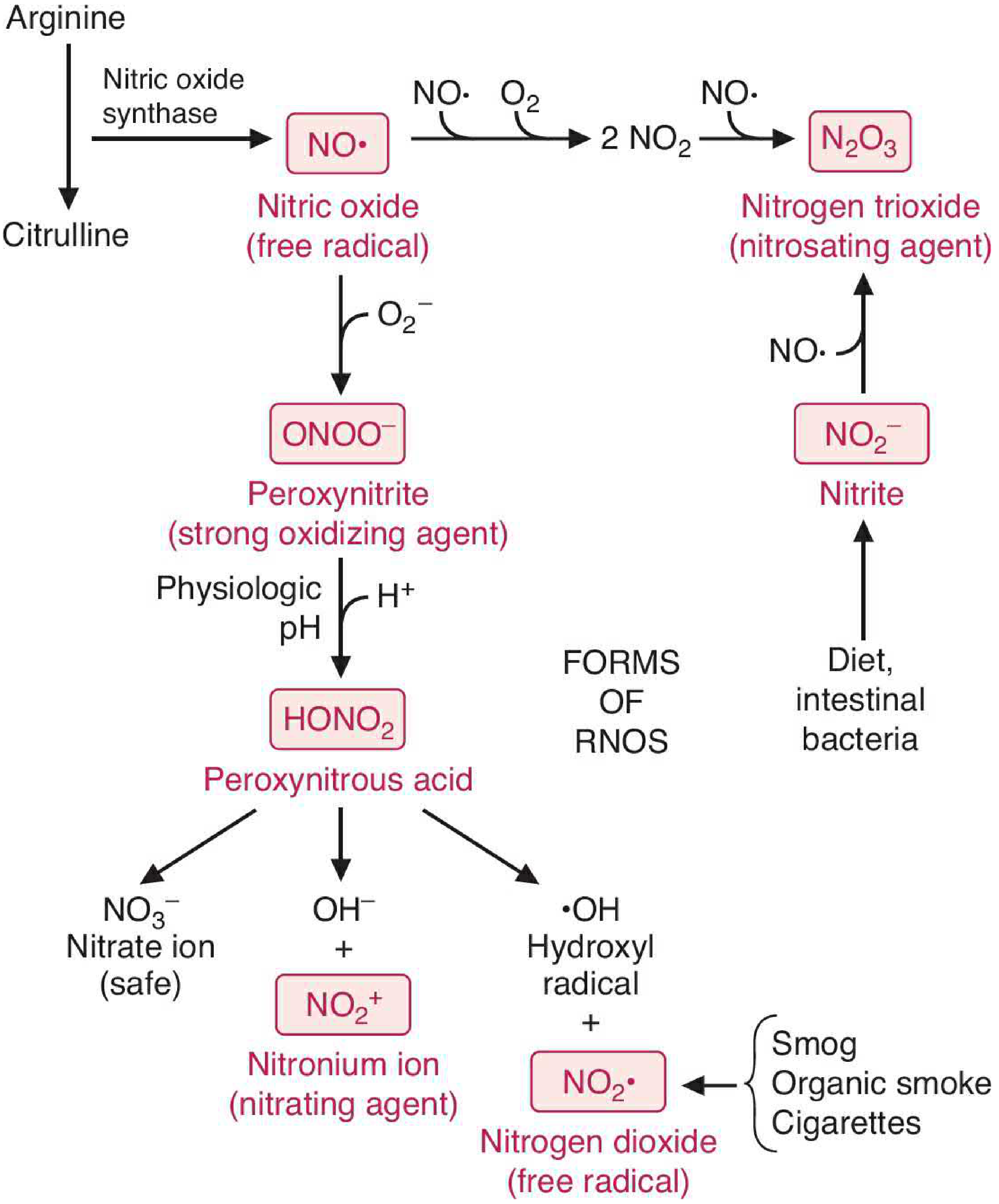
Fig. RNOS formation from excess NO. Peroxynitrite (ONOO⁻) and derived species cause oxidative, nitrating, and nitrosylating damage (Basic Medical Biochemistry, 6e)
- NO + O₂⁻ → ONOO⁻ (peroxynitrite): strong oxidising agent; nitrates tyrosine residues (nitrotyrosine is a biomarker of oxidative/nitrosative stress)
- NO + O₂ → N₂O₃: nitrosating agent; attacks -SH groups and sulfhydryl groups
- Downstream effects: inhibition of mitochondrial ETC complexes, DNA strand breaks, lipid peroxidation, enzyme dysfunction
7. Secondary Causes (Disease Conditions) Linked to NO Dysregulation
These can be divided into conditions of excess NO (iNOS-driven pathology) and deficient NO (eNOS dysfunction).
A. Conditions of EXCESS NO (iNOS-mediated)
| Condition | Mechanism | Consequence |
|---|---|---|
| Septic shock | Bacterial LPS + cytokines (IFN-γ, TNF-α) massively induce iNOS in vascular wall and macrophages → unregulated, massive NO production | Profound refractory vasodilation, hypotension; main mediator of distributive/vasodilatory shock; contributes to multiple organ dysfunction |
| Tissue hypoxia in sepsis | Hypoxia activates NF-κB → upregulates iNOS; VEGF released in hypoxia also increases iNOS | Amplifies vasodilation; inhibits HIF-1α beneficial effects (erythropoietin upregulation) |
| Neurodegenerative diseases | Excess NO + O₂⁻ → peroxynitrite → neuronal damage | Implicated in Parkinson disease, ALS |
| Chronic inflammation | iNOS induction in inflammatory states | Rheumatoid arthritis, IBD - RNOS cause tissue injury |
| HFpEF (Heart Failure with preserved EF) | iNOS upregulation in myocardium → increased nitrosylation of proteins | Myocardial dysfunction |
Clinically: In septic shock, NO is the primary mediator of peripheral vasodilation. Vasodilation is mediated by increased synthesis of NO and prostacyclin; catecholamines (norepinephrine, epinephrine) are elevated but peripheral responsiveness to vasoconstrictors is decreased. - Goldman-Cecil Medicine
B. Conditions of DEFICIENT NO (eNOS dysfunction / reduced bioavailability)
| Condition | Mechanism | Consequence |
|---|---|---|
| Essential and secondary hypertension | Reduced eNOS activity, decreased NO bioavailability | Loss of vasodilatory tone → increased peripheral resistance |
| Pulmonary arterial hypertension (PAH) | Impaired eNOS activity OR reactive oxygen species scavenge NO → form peroxynitrite (ONOO⁻) | Loss of pulmonary vasodilation, vascular remodeling, increased PVR |
| Atherosclerosis / endothelial dysfunction | Oxidative stress (ROS) scavenges NO; eNOS uncoupling (BH₄ deficiency) produces O₂⁻ instead of NO | Endothelial dysfunction, platelet activation, smooth muscle proliferation - proatherogenic |
| Erectile dysfunction (ED) | Dysregulation of nNOS/eNOS in penile endothelium; atherosclerosis reduces NO; general endothelial dysfunction | Failure of cavernous smooth muscle relaxation - basis for PDE-5 inhibitor therapy (sildenafil, tadalafil) |
| Diabetes mellitus | Glycation and oxidative stress impair eNOS; BH₄ oxidised → eNOS uncoupling | Accelerated atherosclerosis, nephropathy, erectile dysfunction |
| Uremia / Renal failure | Impaired NO synthesis in endothelium contributes to hypertension and bleeding tendency | Platelet dysfunction (bleeding), vascular disease |
8. Pharmacological Exploitation of the NO Pathway
| Drug / Intervention | Mechanism |
|---|---|
| Nitroglycerin / organic nitrates | Metabolised to NO in blood → coronary and venous vasodilation; relieves angina |
| Sodium nitroprusside | Releases NO directly → potent vasodilation; used in hypertensive emergencies |
| Sildenafil / PDE-5 inhibitors | Inhibit cGMP breakdown → potentiate NO-mediated smooth muscle relaxation; used in ED and PAH |
| Riociguat | sGC stimulator - directly activates guanylyl cyclase and sensitises it to endogenous NO; used in PAH |
| Inhaled NO | Selective pulmonary vasodilator; used in persistent pulmonary hypertension of newborn (PPHN) and ARDS |
| NOS inhibitors (e.g., L-NMMA) | Block iNOS in septic shock (experimental; non-selective inhibitors trialled but not in routine use) |
Summary Table: NO at a Glance
| Feature | Detail |
|---|---|
| Source amino acid | L-Arginine |
| Product co-formed | L-Citrulline |
| Enzyme | Nitric oxide synthase (NOS) |
| Key cofactors | NADPH, O₂, FAD, FMN, Fe-heme, BH₄ |
| Isoforms | nNOS (Ca²⁺-dep), eNOS (Ca²⁺/phospho-dep), iNOS (transcription-induced) |
| Receptor | Soluble guanylyl cyclase (sGC) → cGMP |
| Terminated by | PDE-5; binding to Hb; reaction with O₂ and O₂⁻ |
| Too little NO | Hypertension, PAH, atherosclerosis, ED |
| Too much NO | Septic shock, neurodegeneration, chronic inflammation |
Sources:
- Basic Medical Biochemistry: A Clinical Approach, 6e - NOS isoforms, RNOS toxicity
- Lippincott's Illustrated Reviews: Biochemistry, 8e - Synthesis pathway, iNOS and macrophage killing
- Kaplan & Sadock's Comprehensive Textbook of Psychiatry - nNOS, neural roles, cGMP pathway
- Goldman-Cecil Medicine - Septic shock vasodilation
- Braunwald's Heart Disease - PAH, eNOS, cGMP-PDE5 pathway
- Goodman & Gilman's Pharmacological Basis of Therapeutics - eNOS signalling, HFpEF, PDE-5
- Medical Physiology (Boron & Boulpaep) - NO and Hb, paracrine action
Protein biosynthesis
Protein biosynthesis (translation) is the process of decoding mRNA to assemble a polypeptide chain. It occurs on ribosomes and proceeds through four stages: aminoacyl-tRNA synthetase activation, initiation, elongation, and termination. In eukaryotes, the ribosome is 80S (40S + 60S subunits); in prokaryotes, 70S (30S + 50S). Energy is consumed at multiple steps (ATP for tRNA charging, GTP for elongation factors).
Searching sources...
Reading File
Finding Sources
Finding Sources
Good. The Histology textbook has a comprehensive dedicated section. Let me read that and simultaneously search Lippincott Biochemistry for the detailed translation steps.
Reading File
Finding Sources
Excellent content. Now let me search for the detailed Lippincott/Basic Biochem sections on translation steps (initiation, elongation, termination) and also look for antibiotic inhibition.
Finding Sources
Finding Sources
Finding Sources
Finding Sources
Good hits. Let me now read the key sections in parallel - Basic Medical Biochemistry for aminoacyl-tRNA and translation steps, Lippincott chapter summary for translation overview, and the Histology section continuation for signal peptide and post-translational details.
Reading File
Reading File
Reading File
Excellent content. Now let me read the main translation steps (initiation, elongation, termination) from Basic Medical Biochemistry and fetch some key diagrams.
Reading File
Loading Image
Excellent content from Basic Medical Biochemistry. I got the elongation and termination text as well. Let me also fetch the initiation diagram and check what text I got on elongation/termination.
Loading Image
Reading File
I have all the elongation and termination text in the truncated output. Let me read the elongation/termination section fully and also get the antibiotic inhibition table.
Reading File
Finding Sources
I now have comprehensive, detailed information from multiple authoritative textbooks. Let me also fetch the elongation diagram for illustration.
Loading Image
I now have everything needed. Let me compile the comprehensive, well-illustrated response.
Protein Biosynthesis (Translation)
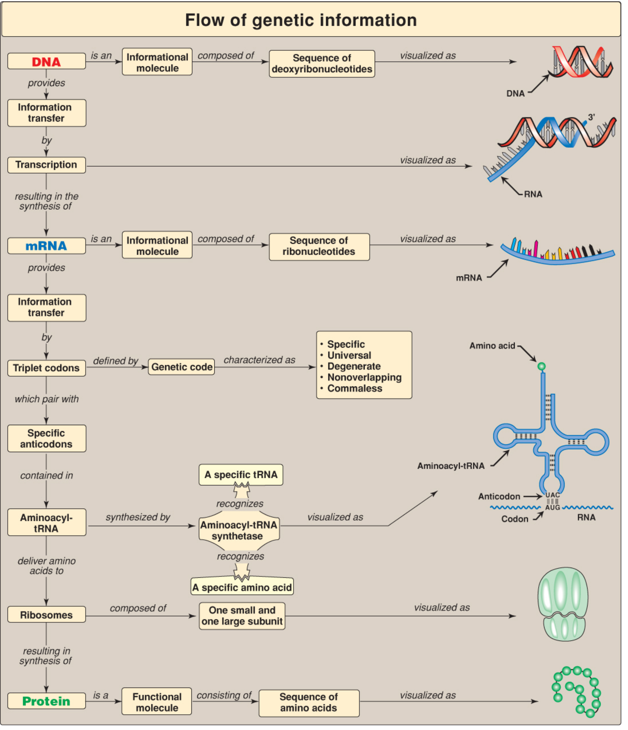
Protein biosynthesis is the process by which the genetic information encoded in mRNA is decoded to synthesize a specific polypeptide chain. It requires ribosomes, tRNAs, mRNA, and numerous protein factors, and proceeds through four major phases: activation (aminoacyl-tRNA formation), initiation, elongation, and termination, followed by post-translational processing.
Overview: From DNA to Protein
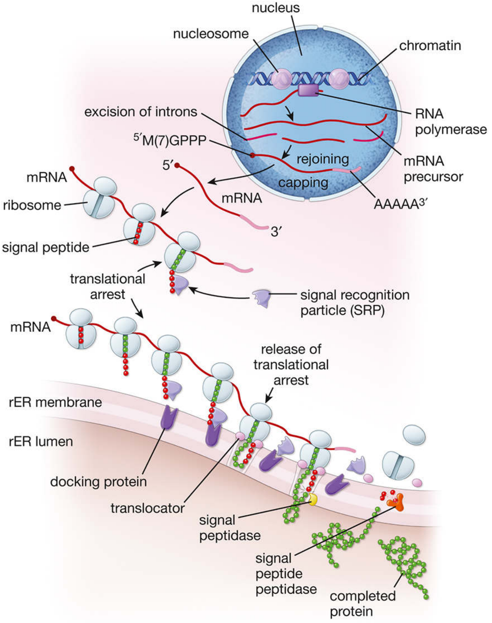
Fig. Complete overview of protein synthesis - from transcription in the nucleus to translation at the rER (Histology: A Text and Atlas, 9e)
The genetic code is first transcribed in the nucleus: DNA → pre-mRNA → mature mRNA (after splicing of introns, 5' methylguanosine capping [M(7)GPPP], and 3' poly-A tail addition). The mRNA travels to the cytoplasm where translation occurs. A single mRNA molecule is read simultaneously by many ribosomes (forming a polysome or polyribosome), spaced as close as 80 nucleotides apart, enormously amplifying output. - Histology: A Text and Atlas, 9e
Phase 1: Activation - Aminoacyl-tRNA Formation
Before translation can begin, each amino acid must be covalently attached to its specific tRNA - a process called aminoacylation or charging.
Enzyme: Aminoacyl-tRNA synthetase (one specific enzyme per amino acid; 20 synthetases total)
Reaction (two steps):
- Step 1 - Activation: Amino acid + ATP → Aminoacyl-AMP-enzyme complex + PPi (pyrophosphate)
- Cleavage of a high-energy bond of ATP provides energy
- Pyrophosphatase cleaves PPi, driving the reaction forward
- Step 2 - Transfer: Aminoacyl-AMP + tRNA → Aminoacyl-tRNA + AMP
- The amino acid is linked via an ester bond to the 2'- or 3'-OH of the ribose at the 3'-terminal A of tRNA (all tRNAs end in -CCA)
Net energy cost: 2 high-energy bonds (equivalent to 2 ATP)
Proofreading: Each synthetase has an editing site. If the wrong amino acid is attached to the tRNA, it is hydrolysed and the process is retried - this is the first fidelity checkpoint in translation. - Basic Medical Biochemistry, 6e
Recognition: Some synthetases recognise tRNA via the anticodon; others use nucleotide sequences elsewhere in the tRNA (so-called "identity elements").
Phase 2: Initiation
The ribosome assembles around the mRNA at the start codon.
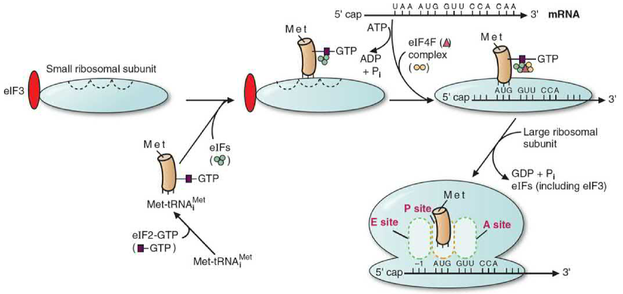
Fig. Eukaryotic translation initiation complex assembly (Basic Medical Biochemistry, 6e)
In Eukaryotes (80S ribosomes):
| Step | Event |
|---|---|
| 1 | Met-tRNA(i)^Met forms complex with eIF2-GTP |
| 2 | This complex binds the small 40S subunit (aided by eIF3, which also blocks premature 60S joining) |
| 3 | The 5' cap of mRNA is recognised by the eIF4F complex (comprising eIF4E, eIF4A, eIF4G) |
| 4 | mRNA + eIF4F binds to the 40S-Met-tRNAi complex; ATP hydrolysis (helicase activity of eIF4A) unwinds secondary structures |
| 5 | The complex scans 5'→3' until it finds the AUG start codon within the Kozak consensus sequence (A/G-CCAUGG) |
| 6 | GTP is hydrolysed → eIFs released → 60S subunit joins → complete 80S ribosome |
| 7 | Met-tRNA(i)^Met sits in the P site; the A site is empty and positioned at the second codon |
In Prokaryotes (70S ribosomes):
- 70S ribosome = 30S + 50S subunits
- mRNA is not capped; instead a purine-rich Shine-Dalgarno (SD) sequence upstream of AUG base-pairs with complementary sequence at the 3'-end of 16S rRNA in the 30S subunit
- Only 3 initiation factors (IF1, IF2, IF3) required (vs. 12+ in eukaryotes)
- Initiating amino acid is N-formylmethionine (fMet), not simple methionine
Ribosome Sites:
- A site (Aminoacyl) - incoming aminoacyl-tRNA
- P site (Peptidyl) - tRNA carrying the growing polypeptide chain
- E site (Exit/Ejection) - discharged tRNA leaves here
Regulation of Initiation:
- Insulin activates eIF4E by phosphorylating 4E-binding protein (4E-BP), releasing eIF4E to stimulate general protein synthesis
- Starvation, heat shock, viral infection activate kinases that phosphorylate eIF2 → inactivates it → blocks translation initiation
- Heme in reticulocytes: heme deficiency leads to phosphorylation of eIF2 → reduced globin synthesis (elegant feedback ensuring globin and heme are balanced) - Basic Medical Biochemistry, 6e
Phase 3: Elongation
Each elongation cycle adds one amino acid to the growing polypeptide. Three sub-steps repeat cyclically:
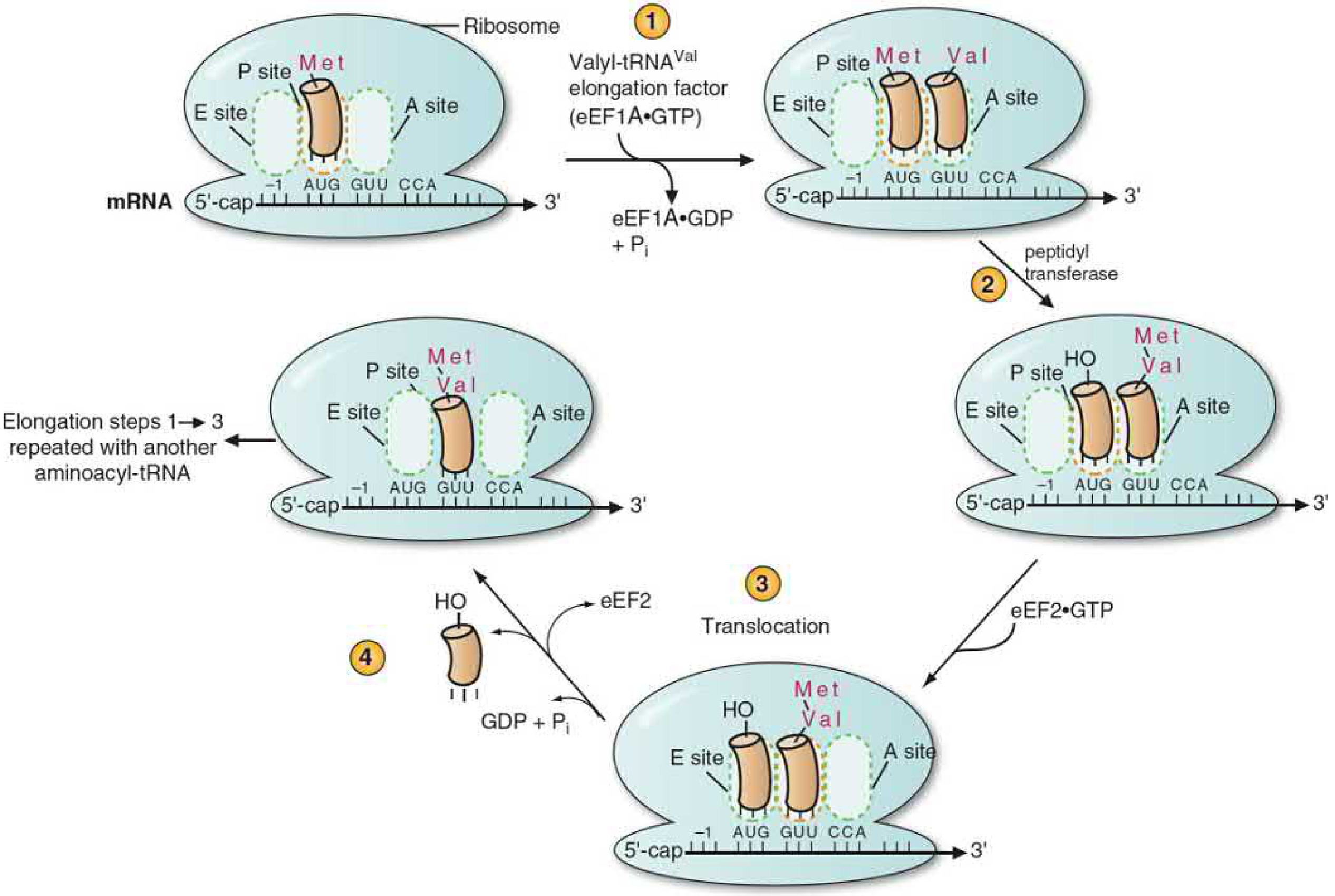
Fig. Elongation cycle showing A, P, E sites and peptidyl transferase activity (Basic Medical Biochemistry, 6e)
Step 1: Aminoacyl-tRNA binding to A site
- The aminoacyl-tRNA complementary to the mRNA codon in the A site arrives as a complex with eEF1A-GTP (prokaryotic equivalent: EF-Tu-GTP)
- The ribosome activates the GTPase activity of eEF1A → GTP hydrolysed to GDP + Pi → eEF1A-GDP dissociates → aminoacyl-tRNA released into A site
- Second fidelity checkpoint: If the wrong aminoacyl-tRNA arrives, GTPase activation does not occur and the complex leaves, preventing misincorporation
Step 2: Peptide bond formation
- Peptidyl transferase catalyses formation of a peptide bond between:
- The amino group of the aminoacyl-tRNA in the A site
- The carbonyl of the peptidyl-tRNA in the P site
- The polypeptide chain is transferred from the P site tRNA to the A site aminoacyl-tRNA
- Peptidyl transferase is a ribozyme - the catalytic activity resides in the 23S rRNA (prokaryotes) / 28S rRNA (eukaryotes) of the large ribosomal subunit - this is the enzymatic activity of RNA itself
- Energy: the energy in the aminoacyl-tRNA ester bond is used; no additional ATP/GTP needed for this step
Step 3: Translocation
- eEF2-GTP (prokaryotic: EF-G-GTP) binds the ribosome
- GTP hydrolysis drives the ribosome to advance 3 nucleotides (one codon) in the 5'→3' direction along the mRNA
- The peptidyl-tRNA (now bearing the growing chain) moves from A site → P site
- The empty/deacylated tRNA moves from P site → E site → exits
- The A site is now empty and positioned on the next codon, ready for the next cycle
Energy cost per elongation cycle: 2 GTP (one for aminoacyl-tRNA delivery, one for translocation) - Lippincott Illustrated Reviews: Biochemistry, 8e; Basic Medical Biochemistry, 6e
Phase 4: Termination
- When a stop (nonsense) codon (UAA, UAG, UGA) enters the A site, no tRNA has an anticodon that matches it
- Instead, release factors (RFs) recognise the stop codon and occupy the A site
- Prokaryotes: RF1 (reads UAA, UAG), RF2 (reads UAA, UGA), RF3 (GTPase)
- Eukaryotes: eRF1 (recognises all three stop codons), eRF3 (GTPase)
- Release factors stimulate peptidyl transferase to act as a hydrolase: the ester bond between the polypeptide and the P-site tRNA is hydrolysed → polypeptide is released
- The ribosome dissociates from the mRNA; ribosomal subunits are recycled - Basic Medical Biochemistry, 6e; Lippincott Biochemistry, 8e
Summary: Energy Consumed in Translation
| Step | Energy Source | Cost |
|---|---|---|
| Aminoacyl-tRNA formation | ATP | 2 high-energy bonds (ATP → AMP + PPi) |
| Initiation (mRNA unwinding) | ATP | 1 ATP (eIF4A helicase) |
| Aminoacyl-tRNA → A site | GTP | 1 GTP |
| Translocation | GTP | 1 GTP |
| Total per amino acid added | ~4 high-energy bonds |
Prokaryotes vs. Eukaryotes: Key Comparison
| Feature | Prokaryotes | Eukaryotes |
|---|---|---|
| Ribosome size | 70S | 80S |
| Small subunit | 30S (16S rRNA) | 40S (18S rRNA) |
| Large subunit | 50S (23S + 5S rRNA) | 60S (28S + 5.8S + 5S rRNA) |
| mRNA 5' structure | No cap; Shine-Dalgarno sequence | 5' methylguanosine cap; Kozak sequence |
| Initiating amino acid | N-formylmethionine (fMet) | Methionine (Met) |
| Initiation factors | 3 (IF1, 2, 3) | 12+ (eIFs) |
| Transcription/translation | Coupled (simultaneous) | Uncoupled (nuclear/cytoplasmic separation) |
| Elongation factors | EF-Tu, EF-G | eEF1A, eEF2 |
| Release factors | RF1, RF2, RF3 | eRF1, eRF3 |
Post-Translational Modifications and Protein Targeting
After the polypeptide is released, it undergoes numerous modifications:
| Modification | Description | Example |
|---|---|---|
| N-terminal methionine removal | Met often cleaved by aminopeptidase | Most cytoplasmic proteins |
| Phosphorylation | Addition of phosphate to Ser, Thr, Tyr - activates or inactivates proteins | Glycogen phosphorylase, kinase cascades |
| Glycosylation | Addition of oligosaccharides in rER/Golgi - roles in protein targeting, cell recognition | IgG, glycoproteins |
| Hydroxylation | Proline → hydroxyproline; Lysine → hydroxylysine | Collagen (requires Vit C) |
| Disulfide bond formation | -SH groups oxidised in rER lumen | Antibodies, insulin |
| Proteolytic cleavage | Signal peptide removed by signal peptidase | Secreted proteins; proinsulin → insulin |
| Ubiquitination | Misfolded/destined-for-degradation proteins tagged | Targets protein to proteasome for degradation |
Signal Peptide and Protein Targeting
Proteins destined for secretion or membrane insertion carry a signal sequence (15-60 hydrophobic amino acids) at the N-terminus - analogous to an airline luggage tag. The sequence of events:
- Signal sequence emerges from ribosome → binds Signal Recognition Particle (SRP)
- SRP arrests elongation (translational arrest)
- SRP-ribosome complex docks to SRP receptor (docking protein) on rER membrane
- Ribosome aligns with translocator channel in rER membrane; SRP dissociates; translation resumes
- Growing polypeptide is threaded into the rER lumen
- Signal peptidase (on cisternal face) cleaves the signal sequence
- Protein is modified in rER (core glycosylation, folding aided by chaperones) → packaged into vesicles → Golgi apparatus → final destination - Histology: A Text and Atlas, 9e
Chaperones and Proteasome
- Protein folding can be spontaneous or facilitated by molecular chaperones (e.g., HSP70, HSP90)
- Misfolded or short-lived proteins are tagged with ubiquitin chains and degraded by the 26S proteasome - Lippincott Biochemistry, 8e
Inhibitors of Translation (Antibiotics and Toxins)
The structural and functional differences between prokaryotic (70S) and eukaryotic (80S) ribosomes are exploited by many clinically important antibiotics.
| Agent | Target | Mechanism | Spectrum |
|---|---|---|---|
| Aminoglycosides (streptomycin, gentamicin) | 30S subunit (prokaryote) | Binds 16S rRNA; causes misreading of mRNA; disrupts initiation | Gram-negative bacteria; TB |
| Tetracyclines | 30S subunit (prokaryote) | Blocks binding of aminoacyl-tRNA to A site | Broad-spectrum bacteriostatic |
| Chloramphenicol | 50S subunit (prokaryote) | Inhibits peptidyl transferase (23S rRNA) | Broad-spectrum; bone marrow toxicity |
| Macrolides (erythromycin, azithromycin) | 50S subunit (prokaryote) | Blocks translocation; causes early release of peptide chain | Gram-positive and atypicals |
| Clindamycin | 50S subunit (prokaryote) | Acts near peptidyl transferase site (overlaps with chloramphenicol/erythromycin binding) | Anaerobes, Gram-positives |
| Linezolid | 50S + 23S rRNA | Blocks initiation complex formation | MRSA, VRE |
| Fusidic acid | EF-G (EF-2 analog) | Prevents translocation by locking EF-G on ribosome | Staphylococci |
| Puromycin | Both 70S and 80S | Structural analog of aminoacyl-tRNA; causes premature chain termination | Non-selective (experimental only) |
| Cycloheximide | 80S (eukaryotic) | Inhibits peptidyl transferase and translocation | Eukaryotes only; too toxic for clinical use |
| Diphtheria toxin | eEF2 (eukaryotic) | ADP-ribosylates eEF2 → inactivates translocation | Eukaryotic cells only |
| Ricin | 28S rRNA | Depurinates a specific adenosine in the large ribosomal subunit → blocks EF binding | Extremely potent; eukaryotes |
- Basic Medical Biochemistry, 6e; Medical Microbiology, 9e; Goodman & Gilman's; Histology: A Text and Atlas
Summary Flow
DNA (nucleus)
↓ Transcription
pre-mRNA
↓ Splicing, 5' cap, poly-A tail
Mature mRNA
↓ Nuclear export
Cytoplasm
↓
Aminoacylation (aminoacyl-tRNA synthetases, 2 ATP/amino acid)
↓
INITIATION [80S/70S ribosome assembled at AUG; Met/fMet in P site]
↓
ELONGATION [A site binding (GTP) → Peptidyl transferase → Translocation (GTP)]
↓ (cycle repeats ~300-3000 times for average protein)
TERMINATION [Stop codon → Release factors → Polypeptide released]
↓
Post-translational modifications (phosphorylation, glycosylation, etc.)
↓
Protein folding (chaperones) → Functional protein
Energy total: approximately 4 high-energy phosphate bonds per amino acid incorporated (2 ATP for charging + 2 GTP during elongation), making protein synthesis one of the most energy-intensive cellular processes.
Sources:
- Basic Medical Biochemistry: A Clinical Approach, 6e - Aminoacyl-tRNA, initiation, elongation, termination in full detail
- Lippincott's Illustrated Reviews: Biochemistry, 8e - Chapter summary, post-translational modifications, ubiquitin-proteasome
- Histology: A Text and Atlas with Correlated Cell and Molecular Biology, 9e - Transcription-to-translation overview, signal peptide, rER processing
- Medical Microbiology, 9e; Goodman & Gilman's; Katzung's - Antibiotic mechanisms
Biochemical role of arginine & compounds synthesized from it
Arginine is a conditionally essential amino acid with an extraordinary range of biochemical roles. It is the most nitrogen-rich amino acid and serves as a precursor for nitric oxide, creatine, polyamines, urea, agmatine, and proline. It is also a key intermediate in the urea cycle itself.
Searching sources...
Reading File
Finding Sources
Finding Sources
Good. Let me now read the Harper's dedicated sections on Arginine and also search Lippincott and Basic Biochemistry for the arginine metabolism map.
Reading File
Reading File
Finding Sources
Let me now look for the Harper's metabolic fates of arginine figure and fetch images, then search for creatine synthesis, polyamines, and the urea cycle role of arginine in parallel.
Finding Sources
Finding Sources
Finding Sources
Now let me read the key sections - Basic Biochemistry urea cycle/arginine section and search for creatine and polyamine synthesis from arginine.
Reading File
Finding Sources
Finding Sources
Excellent. Now let me read the Harper's Figure 30-1 context (arginine/ornithine/proline metabolism diagram), the creatine synthesis section in Basic Biochemistry, and fetch key diagrams.
Reading File
Reading File
Loading Image
Excellent. Now let me fetch the Harper's arginine/ornithine/proline metabolism diagram (Figure 30-1) and the NOS reaction diagram, and also search for the polyamine synthesis details.
Loading Image
Loading Image
Finding Sources
Now let me read the polyamine section from Harper's and the creatine synthesis diagram.
Reading File
Finding Sources
I now have comprehensive information from multiple authoritative sources. Let me also fetch the creatine synthesis figure.
Loading Image
I now have all the information needed for a comprehensive, well-illustrated answer. Let me compose it.
Biochemical Role of Arginine and Compounds Synthesized from It
1. Arginine - Structure and Classification
L-Arginine is a conditionally essential amino acid - non-essential under normal physiological conditions but essential during rapid growth, stress, critical illness, and in premature infants. It is the most nitrogen-rich of the 20 standard amino acids, containing 4 nitrogen atoms (1 α-amino, 1 α-carboxyl, and the distinctive guanidinium group with 3 nitrogens at its side chain).
Classification: Basic amino acid (along with lysine and histidine); positively charged at physiological pH due to the guanidinium group (pKa ~12.5).
Conditionally essential in: Premature infants, surgical/critical illness, cancer, burns, sepsis (listed alongside cysteine, glutamine, glycine, proline, tyrosine). - Sabiston Textbook of Surgery; Sleisenger and Fordtran's GI and Liver Disease
2. Overview: Metabolic Fates of Arginine
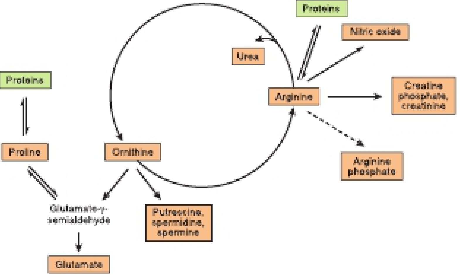
Fig. Metabolic fates of arginine, ornithine, and proline. Arginine sits at the centre of multiple biosynthetic pathways. (Harper's Illustrated Biochemistry, 32e)
Arginine is at the metabolic hub of nitrogen metabolism. Its major fates are:
| Pathway | Product(s) | Key Enzyme |
|---|---|---|
| Urea cycle | Urea + Ornithine | Arginase |
| NO synthesis | Nitric oxide + Citrulline | NOS (eNOS/nNOS/iNOS) |
| Creatine synthesis | Guanidinoacetate → Creatine | Arginine:glycine amidinotransferase (AGAT) |
| Polyamine synthesis | Putrescine, Spermidine, Spermine | Ornithine decarboxylase, synthases |
| Proline synthesis | Proline | via Ornithine → Glutamate-γ-semialdehyde |
| Protein synthesis | Incorporated into proteins | Aminoacyl-tRNA synthetase |
| Catabolism | α-Ketoglutarate | via Ornithine → Glutamate-γ-semialdehyde |
3. Compound 1: UREA (Urea Cycle)
The Urea Cycle
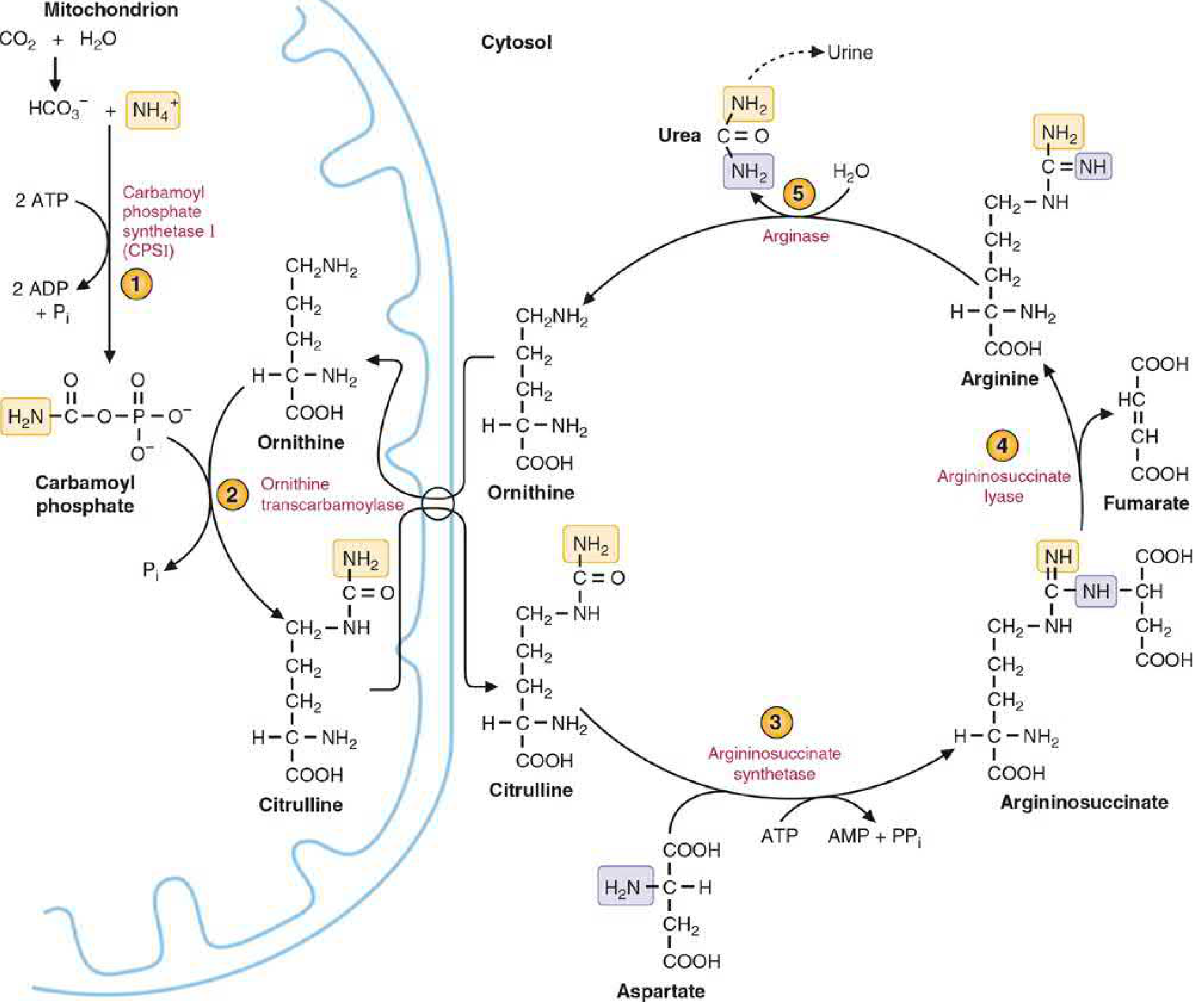
Fig. Urea cycle - arginine is both a product of and substrate for the cycle (Basic Medical Biochemistry, 6e)
Arginine is the immediate precursor of urea and is central to the cycle:
| Step | Reaction | Location | Enzyme |
|---|---|---|---|
| 1 | CO₂ + NH₄⁺ + 2ATP → Carbamoyl phosphate + 2ADP + Pi | Mitochondria | CPSI (Carbamoyl phosphate synthetase I) |
| 2 | Carbamoyl phosphate + Ornithine → Citrulline | Mitochondria | OTC (Ornithine transcarbamoylase) |
| 3 | Citrulline + Aspartate + ATP → Argininosuccinate | Cytosol | Argininosuccinate synthetase |
| 4 | Argininosuccinate → Arginine + Fumarate | Cytosol | Argininosuccinate lyase |
| 5 | Arginine + H₂O → Urea + Ornithine | Cytosol | Arginase |
Key points:
- Ornithine is the regenerated carrier (like OAA in TCA cycle)
- Two nitrogens enter urea: one from NH₄⁺ (via carbamoyl phosphate), one from aspartate (linking urea cycle to TCA)
- Fumarate produced links to TCA cycle (connects N metabolism to energy metabolism)
- Energy cost: 3 ATP equivalents per urea molecule (2 ATP for CPSI, 1 ATP for argininosuccinate synthetase)
- Regulation: N-acetylglutamate (NAG) is an obligate allosteric activator of CPSI; high protein intake → increased NAG → increased urea cycle flux
- Location: Liver (primarily); small intestine also has partial urea cycle
- Deficiency: Arginase deficiency → Hyperargininemia (spastic diplegia, intellectual disability, elevated blood arginine). Red bar in the figure marks this site. - Basic Medical Biochemistry, 6e; Harper's, 32e
4. Compound 2: NITRIC OXIDE (NO)
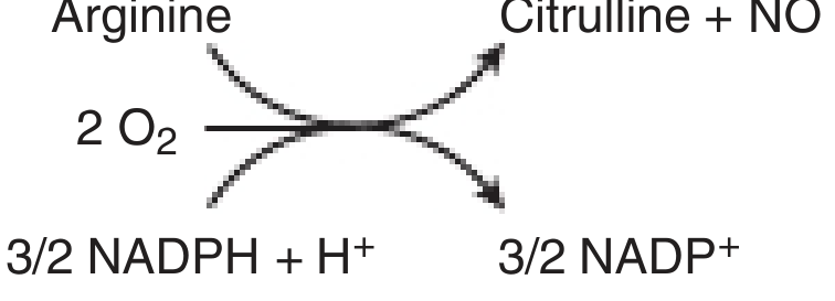
Fig. NOS reaction - a five-electron oxidoreductase (Harper's, 32e)
Reaction:
L-Arginine + 2O₂ + 3/2 NADPH + H⁺ → L-Citrulline + NO + 3/2 NADP⁺
L-Arginine + 2O₂ + 3/2 NADPH + H⁺ → L-Citrulline + NO + 3/2 NADP⁺
Enzyme: Nitric oxide synthase (NOS) - a five-electron oxidoreductase requiring Fe-heme, FAD, FMN, tetrahydrobiopterin (BH₄), and NADPH as cofactors. Converts one nitrogen of the guanidino group to NO. - Harper's, 32e
Three NOS isoforms:
| Isoform | Location | Regulation | Function |
|---|---|---|---|
| nNOS (NOS I) | Neurons, skeletal muscle | Ca²⁺/calmodulin | Neurotransmitter, LTP, memory |
| iNOS (NOS II) | Macrophages, astroglia | Gene transcription (LPS, IFN-γ, TNF-α) | Bactericidal, innate immunity |
| eNOS (NOS III) | Vascular endothelium | Ca²⁺/calmodulin + phosphorylation | Vasodilation, anti-platelet aggregation |
Roles of NO:
- Vasodilation - diffuses to vascular smooth muscle → binds soluble guanylyl cyclase → ↑cGMP → PKG activation → relaxation
- Neurotransmitter - non-conventional gaseous transmitter in CNS/PNS
- Bactericidal - iNOS-derived NO + O₂⁻ → ONOO⁻ (peroxynitrite) → kills pathogens
- Anti-platelet - inhibits platelet adhesion and aggregation
Clinical: Nitroglycerin and nitroprusside are NO donors; sildenafil inhibits PDE-5 (which degrades cGMP) to potentiate NO action - Harper's, 32e; Basic Medical Biochemistry, 6e
5. Compound 3: CREATINE and CREATINE PHOSPHATE
Creatine synthesis is a two-organ process - starts in the kidney and is completed in the liver.
Step 1 - In Kidney:
Glycine + Arginine → Guanidinoacetate + Ornithine
- Enzyme: Arginine:glycine amidinotransferase (AGAT)
- The guanidinium group of arginine is transferred to glycine
- The remaining carbon skeleton of arginine is released as ornithine (which re-enters the urea cycle)
Step 2 - In Liver:
Guanidinoacetate + S-adenosylmethionine (SAM) → Creatine + S-adenosylhomocysteine
- Enzyme: Guanidinoacetate methyltransferase (GAMT)
- SAM donates its methyl group (SAM is the major methyl donor in the body)
- This is why glycine + arginine + methionine all contribute atoms to creatine
Creatine → Creatine Phosphate (Phosphocreatine):
Creatine + ATP ⇌ Creatine phosphate + ADP
- Enzyme: Creatine kinase (CK/CPK) - reversible reaction
- Creatine phosphate is the short-term ATP buffer in muscle, brain, and heart
- Supplies ATP rapidly at onset of exercise (before oxidative phosphorylation ramps up)
- Carries high-energy phosphate from mitochondria (where ATP is made) to myosin filaments (where ATP is used)
Creatine → Creatinine:
Creatine phosphate spontaneously cyclises → Creatinine
- Creatinine cannot be metabolised further and is excreted in urine
- Its excretion is constant and proportional to muscle mass - used as a reference for urinary excretion rates and as a surrogate marker of renal filtration (GFR)
Genetic diseases of creatine synthesis:
- AGAT deficiency - intellectual disability, speech delay, hypotonia
- GAMT deficiency - intellectual disability, seizures, autistic features (brain creatine depleted)
- Creatine transporter deficiency (X-linked, SLC6A8 mutation) - most common creatine deficiency syndrome - Basic Medical Biochemistry, 6e; Bradley & Daroff's Neurology
6. Compound 4: POLYAMINES (Putrescine, Spermidine, Spermine)
Arginine contributes its carbon skeleton to polyamine synthesis via ornithine.
Pathway:
- Arginine → Ornithine (by Arginase, releasing urea)
- Ornithine → Putrescine (by Ornithine decarboxylase, ODC; CO₂ released)
- Putrescine + decarboxylated SAM → Spermidine (by Spermidine synthase; aminopropyl donated from dSAM)
- Spermidine + decarboxylated SAM → Spermine (by Spermine synthase)
Functions of polyamines:
- Carry multiple positive charges → strongly associate with negatively charged DNA and RNA (stabilise nucleic acid structures)
- Required for cell proliferation and growth
- Act as growth factors for cultured mammalian cells
- Stabilise intact cells, subcellular organelles, and membranes
- Pharmacologically: at high doses → hypotensive and hypothermic effects
Clinical significance:
- Rapidly proliferating cells (cancer cells) have markedly elevated polyamine levels
- Eflornithine - irreversible inhibitor of ODC (the rate-limiting enzyme); used in treatment of Trypanosoma brucei (sleeping sickness) and hirsutism, by blocking polyamine synthesis
- Polyamines (putrescine) accumulate as uremic toxins in renal failure - Harper's, 32e; Goodman & Gilman's
7. Compound 5: PROLINE
Via ornithine, arginine connects to proline biosynthesis and catabolism:
Arginine → Ornithine → Glutamate-γ-semialdehyde ⇌ Δ1-Pyrroline-5-carboxylate ⇌ Proline
- Enzyme: Ornithine δ-aminotransferase (OAT)
- Proline is a major structural component of collagen and is hydroxylated to hydroxyproline (requiring vitamin C)
Genetic disease: Deficiency of ornithine δ-aminotransferase → elevated plasma/urine ornithine → Gyrate atrophy of the choroid and retina (progressive visual loss). Treatment: restrict dietary arginine. - Harper's, 32e
8. Compound 6: AGMATINE
Arginine can be directly decarboxylated by arginine decarboxylase to form agmatine:
Arginine → Agmatine + CO₂
- Agmatine is an endogenous neuromodulator in the brain
- It inhibits NOS (particularly iNOS), has antinociceptive and neuroprotective effects
- It inhibits NMDA receptors and imidazoline receptors
- Can be further hydrolysed to putrescine (linking it to the polyamine pathway)
9. Role in Protein Synthesis
As a standard amino acid, arginine is incorporated into all proteins. Within proteins, the guanidinium group serves key functions:
- Hydrogen bonding in enzyme active sites
- Salt bridge formation in protein structure
- Post-translational modification: arginine methylation (PRMT enzymes) regulates transcription and RNA processing
- Citrullination - conversion of arginine → citrulline by PAD (peptidylarginine deiminase) enzymes, important in:
- Chromatin remodeling
- NETosis (neutrophil extracellular traps)
- Anti-citrullinated protein antibodies (ACPA/anti-CCP) are a hallmark of rheumatoid arthritis
Summary: Compounds Synthesized from Arginine
┌─────────────────────────────────────────────┐
│ L-ARGININE │
│ (guanidinium group, conditionally essential) │
└─────────────────────────────────────────────┘
│ │ │ │
┌────────────────┤ │ │ ├────────────────┐
▼ ▼ ▼ ▼ ▼ ▼
[Arginase] [NOS] [AGAT, kidney] [Decarboxy- [PAD] Protein
│ │ │ lase] │ synthesis
▼ ▼ ▼ ▼ ▼
UREA NITRIC OXIDE Guanidino- AGMATINE Citrullinated (polypeptides)
+Ornithine +Citrulline acetate proteins
│ [GAMT+SAM] (RA antigens)
▼ ▼
Ornithine CREATINE
/ \ [CK+ATP]
▼ ▼ ▼
OAT enzyme ODC enzyme CREATINE-P
▼ ▼ [spontaneous]
Glutamate-γ- PUTRESCINE ▼
semialdehyde [+dSAM] CREATININE
▼ ▼ (urine)
PROLINE SPERMIDINE
(collagen) [+dSAM]
▼
SPERMINE
(cell proliferation,
DNA stabilization)
Clinical Summary Table
| Derived Compound | Key Clinical Relevance |
|---|---|
| Urea | Liver failure → hyperammonemia; urea cycle disorders (OTC deficiency most common) → encephalopathy |
| Nitric oxide | Septic shock (excess iNOS); PAH, atherosclerosis (deficient eNOS); therapeutic: nitroglycerin, sildenafil |
| Creatine phosphate | Muscle energy buffer; AGAT/GAMT/SLC6A8 deficiency → intellectual disability; serum CK elevated in myocardial infarction, myopathy |
| Creatinine | Marker of GFR; elevated in renal failure |
| Polyamines | Elevated in cancer (rapid proliferation); eflornithine inhibits ODC (anti-trypanosomal); uremic toxins in renal failure |
| Proline | Collagen synthesis (hydroxyproline); OAT deficiency → gyrate atrophy of retina |
| Citrullination | Anti-CCP antibodies in rheumatoid arthritis; NETosis in innate immunity |
| Agmatine | Neuroprotection; NOS inhibitor; antinociception |
Sources:
- Harper's Illustrated Biochemistry, 32e - Arginine/ornithine/proline metabolism overview (Fig 30-1), polyamines
- Basic Medical Biochemistry: A Clinical Approach, 6e - Urea cycle (detailed), creatine synthesis, creatinine
- Lippincott's Illustrated Reviews: Biochemistry, 8e - Urea cycle, post-translational roles
- Sabiston Textbook of Surgery - Conditionally essential status
- Bradley & Daroff's Neurology - Creatine deficiency syndromes (AGAT, GAMT)
- Goodman & Gilman's Pharmacological Basis of Therapeutics - Eflornithine, polyamine pharmacology
- Yamada's Textbook of Gastroenterology - Arginine in hyperammonemia treatment
Free Radicles
Free radicals are molecules or atoms with one or more unpaired electrons, making them highly reactive. The major biological free radicals are reactive oxygen species (ROS) and reactive nitrogen-oxygen species (RNOS). They are generated by normal metabolism (mitochondrial ETC, phagocytosis) and exogenous sources. Antioxidant defenses include SOD, catalase, glutathione peroxidase, vitamins C and E.
Searching sources...
Reading File
Finding Sources
Finding Sources
Finding Sources
Finding Sources
Reading File
Reading File
Finding Sources
Now let me get the dedicated biochemistry section on free radicals with ROS types, antioxidants, and pathological effects.
Finding Sources
Finding Sources
Excellent. The Basic Medical Biochemistry section has a dedicated free radicals chapter. Let me read it fully in parallel with Harper's ROS section.
Reading File
Reading File
Excellent detail. Now let me read the antioxidant defense section from Basic Medical Biochemistry and fetch key diagrams.
Reading File
Loading Image
Loading Image
Your credits are exhausted. You can purchase a top-up or subscribe to a plan to continue using Orris. If you're already subscribed, your daily credits will reset tomorrow.
Free Radicles
Your credits are exhausted. You can purchase a top-up or subscribe to a plan to continue using Orris. If you're already subscribed, your daily credits will reset tomorrow.
Free Radicles
Your credits are exhausted. You can purchase a top-up or subscribe to a plan to continue using Orris. If you're already subscribed, your daily credits will reset tomorrow.
This is a shared conversation. Sign in to Orris to start your own chat.