
CTEV etiology clinical feature and management in flow chart
clubfoot AND Ponseti
clubfoot CTEV Ponseti method management guideline
https://posna.org/physician-education/study-guide/clubfoot
congenital talipes equinovarus clubfoot clinical photograph CAVE deformity
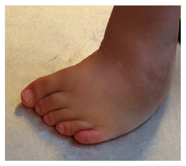
Clinical photograph of a human foot, anterior-oblique view, demonstrating a recurrent congenital talipes equinovarus (clubfoot) deformity following previous surgical revision. The image illustrates the 'CAVE' mnemonic components typical of residual clubfoot: cavus (high midfoot arch), forefoot adduction (inward deviation of the toes), and hindfoot varus (inward rotation of the heel). Soft tissue examination reveals significant swelling localized around the ankle joint and across the dorsal surface of the foot. Distal changes include callus formation on the plantar-lateral aspect and the tip of the first digit, indicating pathological weight-bearing and altered biomechanics. This visual serves as an educational example of incomplete surgical correction or disease recurrence in pediatric orthopedics, specifically within the context of Möbius syndrome management.
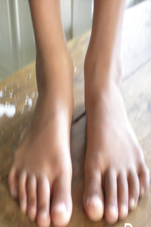
Clinical photograph showing an anterior (front) view of bilateral feet in a pediatric patient at a five-year follow-up for congenital clubfoot (talipes equinovarus) treatment. The image demonstrates the long-term anatomical outcome following corrective procedures, likely the Ponseti method. Residual findings include mild forefoot adduction bilaterally, where the forefoot curves inward toward the midline. The left foot exhibits a slightly more pronounced medial curvature of the lateral border compared to the right. Both feet display five well-formed digits without gross syndactyly or secondary skin lesions. The ankles appear slender with visible malleolar landmarks. The photograph serves as a clinical assessment of post-treatment alignment, focusing on the correction of the 'CAVE' components (cavus, adduction, varus, and equinus) associated with clubfoot deformity.
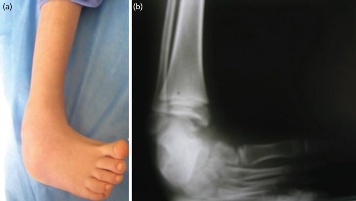
This composite educational resource consists of a clinical photograph and a corresponding diagnostic radiograph of a pediatric foot and ankle, illustrating congenital talipes equinovarus, commonly known as clubfoot. Panel (a) is a clinical photograph showing the characteristic morphology of the deformity, including severe equinus (plantarflexion), hindfoot varus (inversion of the heel), and forefoot adduction with the sole facing medially. Panel (b) is an anteroposterior X-ray demonstrating the underlying skeletal malalignment. Key radiological findings include the abnormal parallel relationship between the long axes of the talus and calcaneus (reduced talocalcaneal angle), marked plantarflexion of the talus relative to the tibia, and medial deviation of the metatarsals. These images serve as a teaching tool for orthopedic residents to recognize the 'CAVE' components of clubfoot: cavus, adductus, varus, and equinus. The materials are relevant for preoperative assessment in pediatric orthopedics and illustrate the degree of structural deviation requiring surgical or conservative corrective measures, such as the Ponseti method or Ilizarov external fixation.
CTEV: Congenital Talipes Equinovarus (Clubfoot)
- Cavus: high medial arch due to plantar-flexed first ray
- Adductus: forefoot deviated medially
- Varus: heel inverted
- Equinus: ankle plantar-flexed due to tight tendo-Achilles
Etiology
CTEV
 │
 ├─ Idiopathic / isolated clubfoot
 │    └─ Most common
 │       • Multifactorial inheritance
 │       • Genetic susceptibility + environmental influences
 │
 ├─ Intrinsic / neuromuscular causes
 │    ├─ Spina bifida / myelomeningocele
 │    ├─ Arthrogryposis
 │    ├─ Cerebral palsy
 │    └─ Peripheral nerve or muscle abnormalities
 │
 ├─ Syndromic causes
 │    ├─ Chromosomal disorders
 │    └─ Skeletal dysplasias / connective-tissue disorders
 │
 └─ Extrinsic / positional factors
      └─ Intrauterine constraint, oligohydramnios, uterine anomalies
Clinical features
NEWBORN WITH FOOT TURNED INWARD AND DOWNWARD
                  │
                  ▼
             Look for CAVE
                  │
    ┌─────────────┼──────────────┐
    ▼             ▼              ▼
 Cavus       Forefoot          Hindfoot
 High arch   adduction         varus
    │             │              │
    └──────────► Equinus ◄───────┘
              Ankle plantar-flexion
Examination findings
- Foot is small, rigid and difficult to correct passively.
- Sole faces medially and upward.
- Medial and posterior skin creases may be deep.
- Heel is small, inverted and poorly palpable.
- Calf on the affected side may be smaller, especially in unilateral CTEV.
- Can be unilateral or bilateral.
- Assess severity using Pirani score or Dimeglio classification.
- Examine the spine, hips, neurologic status, and the rest of the limbs to exclude associated neuromuscular or syndromic disease.
Investigations
- Usually a clinical diagnosis.
- X-rays are not routinely needed in a typical newborn with isolated CTEV.
- If obtained, radiographs may show a reduced talocalcaneal angle and persistent equinus. POSNA diagnostic notes
Management flow chart
DIAGNOSIS OF CTEV
        │
        ▼
Assess:
• Idiopathic vs syndromic/neuromuscular
• Severity: Pirani/Dimeglio score
• Associated anomalies
• Age and prior treatment
        │
        ▼
START EARLY, IDEALLY SOON AFTER BIRTH
        │
        ▼
PONSETI METHOD: FIRST-LINE TREATMENT
        │
        ▼
Weekly gentle manipulation + above-knee plaster cast
        │
        ▼
Correct deformities in this order: C → A → V → E
Cavus → Adductus → Varus → Equinus
        │
        ▼
After approximately 4-7 casts:
Is adequate ankle dorsiflexion achieved?
        │
   ┌────┴─────┐
   │          │
 YES          NO, residual equinus
   │          │
   ▼          ▼
Foot-abduction  Percutaneous Achilles tenotomy
brace           + final cast for about 3 weeks
   │                    │
   └────────────┬───────┘
                ▼
Foot-abduction brace
• 23 h/day for first 3 months
• Then during sleep and naps
• Continue until about 4-5 years of age
                │
                ▼
Regular follow-up for relapse
                │
    ┌───────────┴────────────┐
    ▼                        ▼
No relapse                Relapse/residual deformity
Continue brace            Reassess brace adherence
and surveillance                 │
                                 ▼
                       Repeat Ponseti casting
                                 │
                     ┌───────────┴───────────┐
                     ▼                       ▼
              Dynamic supination        Rigid/severe relapse
              in walking child                 │
                     │                       ▼
                     ▼             Limited surgery / tendon
       Tibialis anterior tendon     transfer / osteotomy /
       transfer, usually >2.5 yr    gradual correction in
                                    selected older children
Core principles of Ponseti treatment
- Do not correct equinus first. Correct in the sequence CAV(E).
- Use long-leg casts, changed weekly, to prevent tibial rotation.
- In most infants, residual equinus requires a percutaneous Achilles tenotomy.
- The correction is maintained with a foot-abduction brace. Non-adherence is the most important preventable cause of recurrence.
- Extensive posteromedial soft-tissue release is now reserved for resistant or recurrent rigid deformity because it can lead to a stiff, painful foot later. AAOS patient guidance
Exam-oriented summary
CTEV = CAVE
Cavus + Adductus + Varus + Equinus

Treatment = Ponseti
Manipulation + weekly long-leg casts
      ↓
Achilles tenotomy for residual equinus
      ↓
Foot-abduction brace until 4-5 years
      ↓
Relapse: repeat casts ± tibialis anterior tendon transfer
Clinical feature in detail
clubfoot clinical features
clubfoot clinical features examination CAVE associated conditions POSNA
Clinical features of CTEV in detail
1. CAVE deformities
| Component | Clinical appearance | Clinical examination |
|---|---|---|
| Cavus | High medial longitudinal arch | Plantar-flexion of the first metatarsal makes the forefoot supinated. The medial border appears high and curved. |
| Adductus | Forefoot deviates medially toward the midline | The lateral border of the foot is convex. The toes point inward. |
| Varus | Heel is inverted | On looking from behind, the calcaneum is turned inward. The sole faces medially rather than downward. |
| Equinus | Foot is plantar-flexed at ankle | Heel cannot be brought down to the ground. Passive ankle dorsiflexion is restricted because of a tight tendo-Achilles and posterior soft tissues. |
Important: The deformities occur together. It is not merely “in-toeing.” The hindfoot is fixed in varus and equinus.
Typical appearance on inspection
Anterior / medial view
        ↓
Toes point medially and the sole faces medially and upward
        ↓
Forefoot adduction + cavus
        ↓
Deep medial skin crease

Posterior view
        ↓
Heel is drawn upward and inward
        ↓
Hindfoot varus + equinus
        ↓
Deep posterior ankle crease
Other local signs
- Small foot on the affected side, particularly in unilateral CTEV.
- Small, underdeveloped calf muscles on the affected side. This may persist even after successful correction.
- Shortened Achilles tendon with a prominent heel cord.
- Heel appears small and “empty” because the calcaneus is pulled upward and lies in varus.
- Deep medial crease over the medial aspect of foot.
- Deep posterior crease above the heel.
- The lateral border is convex, while the medial border is concave.
- The talar head may be palpable prominently on the dorsolateral aspect of the foot.
- The foot is usually rigid, with limited passive correction.
Assessment of rigidity
Idiopathic structural CTEV
- Foot cannot be fully corrected by gentle passive manipulation.
- Hindfoot varus and equinus are fixed.
- Resistance is felt on attempting dorsiflexion and abduction.
- Requires treatment with serial manipulation and casting.
Positional talipes
- Foot may look like clubfoot but is flexible.
- Can usually be passively corrected to normal or near-normal position.
- There is no major fixed contracture, deep crease, or severe calf wasting.
- It commonly resolves with observation, stretching, or limited splintage.
Later clinical features in an untreated child
Untreated CTEV
      ↓
Child starts walking
      ↓
Weight borne on lateral border / dorsolateral surface of foot
      ↓
Callosities, abnormal gait, shoe-wear difficulty
      ↓
Painful and stiff foot in later childhood or adult life
- Walking occurs on the lateral border or dorsolateral surface of the foot rather than the sole.
- Callosities develop over the lateral border, dorsum, or head of talus.
- Shoe fitting is difficult.
- Gait is abnormal, often with in-toeing and limp.
- Progressive stiffness and pain may develop with age.
Later clinical features after treatment or in relapse
- Reappearance of adductus, varus, or equinus
- Reduced ankle dorsiflexion
- Heel rising early during gait
- Foot slipping repeatedly out of the brace
- Dynamic forefoot supination during swing phase or walking
- Weight bearing on the lateral border
Systemic examination and associated abnormalities
Examine for
- Spine: sacral dimple, tuft of hair, swelling, scoliosis, signs of spina bifida or tethered cord
- Neurologic function: spontaneous toe movement, foot sensation, muscle power and tone
- Hips: screen for developmental dysplasia of the hip
- Other limbs: arthrogryposis, limb deficiency, constriction bands
- Other congenital anomalies: facial, cardiac, renal, gastrointestinal, and chromosomal abnormalities where syndromic CTEV is suspected
Clinical severity assessment: Pirani score
| Midfoot signs | Hindfoot signs |
|---|---|
| Curved lateral border | Posterior crease |
| Medial crease | Empty heel |
| Lateral head of talus coverage | Rigid equinus |
- 0: normal
- 0.5: moderately abnormal
- 1: severely abnormal
- Total score ranges from 0 to 6.
- A higher score indicates a more severe, rigid deformity and often predicts the need for more casts and Achilles tenotomy.
One-line examination answer
CTEV presents at birth as a small, rigid foot turned downward and inward, with cavus, forefoot adduction, hindfoot varus and ankle equinus, deep medial and posterior creases, tight tendo-Achilles, a small heel, and calf wasting on the affected side.