
Which of the following endometrial lesions is associated with the highest risk of developing endometrial adenocarcinoma? • a. Chronic endometritis • b. Complex hyperplasia with atypia • c. Simple hyperplasia d. Squamous metaplasia
Answer: b. Complex hyperplasia with atypia
Breakdown of Each Option
| Lesion | Malignant Potential |
|---|---|
| Complex hyperplasia with atypia | ~29-40% risk of progression to adenocarcinoma |
| Simple hyperplasia (without atypia) | ~1% risk |
| Complex hyperplasia without atypia | ~3% risk |
| Chronic endometritis | No malignant potential (inflammatory condition) |
| Squamous metaplasia | No malignant potential (benign metaplastic change) |
Why Complex Hyperplasia with Atypia Carries Such High Risk
-
Nuclear atypia is the key driver - Atypical hyperplasia (now also called Endometrial Intraepithelial Neoplasia, or EIN) shows back-to-back complex glands with nuclear atypia: rounded nuclei, open (vesicular) chromatin, and conspicuous nucleoli.
-
Concurrent carcinoma is common - Up to 40-50% of women diagnosed with atypical hyperplasia are found to have concurrent adenocarcinoma when hysterectomy is performed. This means the biopsy diagnosis often understages the true pathology.
-
Genetic overlap with carcinoma - Atypical hyperplasia has considerable morphologic and genetic overlap with well-differentiated endometrioid adenocarcinoma (including shared PTEN mutations in 30-80% of endometrial carcinomas).
-
Management implication - Because of this risk, hysterectomy is the definitive treatment for atypical hyperplasia/EIN in postmenopausal women. Progestin therapy is reserved for young women wishing to preserve fertility, but carries a recurrence risk approaching 50%.
Why the Other Options Are Wrong
-
c. Simple hyperplasia - Caused by persistent estrogen stimulation, shows dilated glands with increased gland-to-stroma ratio but no cytologic atypia. Rarely progresses to carcinoma (~1%). Resolves with progestin therapy or withdrawal of estrogen stimulus.
-
a. Chronic endometritis - An inflammatory condition (often due to infection with organisms like Chlamydia, Mycoplasma, or following instrumentation). Associated with plasma cell infiltration of the stroma. It is not a premalignant lesion.
-
d. Squamous metaplasia - A benign, non-progressive change where squamous epithelium replaces glandular endometrial epithelium. No malignant potential on its own.
Sources: Robbins, Cotran & Kumar - Pathologic Basis of Disease, and Berek & Novak's Gynecology
Choose best option from endometrial polyps diagnostic methods? O a. X ray • b. CT • c. US • d. Pap test
Answer: c. Ultrasound (US)
Why Ultrasound is the Answer
"TVUS is the initial method of investigation, which reveals thickening of the echogenic endometrial stripe."
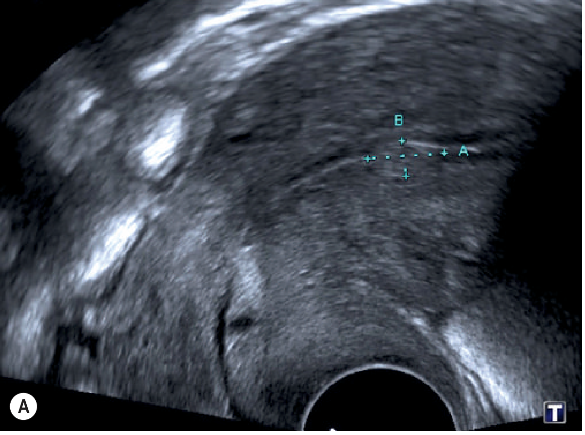
| Step | Modality | Role |
|---|---|---|
| 1st line | Transvaginal US (TVUS) | Detects endometrial stripe thickening; best initial test |
| Enhanced US | Saline infusion sonohysterography (SIS/SHG) | Instills saline to outline the polyp clearly, differentiates polyps from fibroids |
| Confirmatory | Hysteroscopy + biopsy | Gold standard - allows direct visualization and tissue sampling |
| Alternative imaging | MRI | Used when US is inconclusive; not first-line |
Why the Other Options Are Wrong
-
a. X-ray - Plain radiographs show bone and some soft tissue, but cannot visualize the uterine cavity or endometrium. No role in diagnosing endometrial polyps.
-
b. CT - CT has poor soft-tissue contrast resolution for intracavitary uterine lesions and exposes the patient to ionizing radiation. Not used to diagnose endometrial polyps.
-
d. Pap test (Papanicolaou smear) - Samples the cervix, not the endometrium. While endometrial cells can occasionally appear on a Pap smear (which may raise suspicion), the Pap test is not a diagnostic tool for endometrial polyps and has very poor sensitivity for endometrial pathology.
Key takeaway: When a patient presents with abnormal uterine bleeding or postmenopausal bleeding, transvaginal ultrasound is the first-line investigation. A threshold of ≥4 mm endometrial thickness on TVUS in postmenopausal women warrants further evaluation (hysteroscopy/biopsy). Saline infusion sonohysterography is the best ultrasound technique for definitively characterizing polyps.
Source: Grainger & Allison's Diagnostic Radiology; Pfenninger and Fowler's Procedures for Primary Care
Abnormal menstrual bleeding characterized by excessive bleeding at irregular intervals is best referred to as? • a. Dysmenorrhea • b. Menometrorrhagia ) c. Menorrhagia • d. Metrorrhagia
Answer: b. Menometrorrhagia
Definitions - Breaking Down the Terminology
| Term | Breakdown | Meaning |
|---|---|---|
| Menorrhagia | meno (menstrual) + rrhagia (excessive flow) | Heavy/prolonged bleeding at regular intervals - normal timing, abnormal amount (>80 mL or >7 days) |
| Metrorrhagia | metro (uterus) + rrhagia (flow) | Bleeding at irregular intervals between periods - abnormal timing, may be normal amount |
| Menometrorrhagia | meno + metro + rrhagia | Excessive bleeding at irregular intervals - both abnormal amount AND abnormal timing |
| Dysmenorrhea | dys (painful) + menorrhea | Painful menstruation - not about quantity or timing |
Why the Other Options Are Wrong
- a. Dysmenorrhea - Refers to painful periods, not abnormal bleeding quantity or timing. Has nothing to do with irregular intervals.
- c. Menorrhagia - Heavy bleeding but at regular intervals (normal cycle timing). Missing the "irregular intervals" component.
- d. Metrorrhagia - Irregular intermenstrual bleeding, but not necessarily excessive in amount. Missing the "excessive" component.
A Note on Modern Terminology
- "Menorrhagia" → now called Heavy Menstrual Bleeding (HMB)
- "Metrorrhagia" → now called Intermenstrual bleeding
- "Menometrorrhagia" → described as irregular + heavy menstrual bleeding
Sources: Tintinalli's Emergency Medicine; Pfenninger and Fowler's Procedures for Primary Care
Name most common malignancy of the stomach, Mostlly it occur in the gastric antrum; tumor with an intestinal morphology form bulky tumors and are composed of glandular structures while cancers with a diffuse infiltrative growth pattern are typically composed of signet-ring cells? • a. Chronic gastritis • b. Gastric Adenocarcinoma • c. Celiac disease • d. Acute pancreatitis • e. Acute Gastritis
Answer: b. Gastric Adenocarcinoma
Direct Textbook Confirmation
"Adenocarcinoma is the most common malignancy of the stomach, accounting for more than 90% of all gastric cancers."
"Gastric adenocarcinomas are classified according to their gross and histologic appearance into intestinal and diffuse types. Intestinal-type cancers tend to be bulky and are composed of glandular structures... Diffuse gastric cancers display an infiltrative growth pattern and are composed of dyscohesive cells with large mucin vacuoles that push the nucleus to the periphery, creating a signet ring cell appearance."
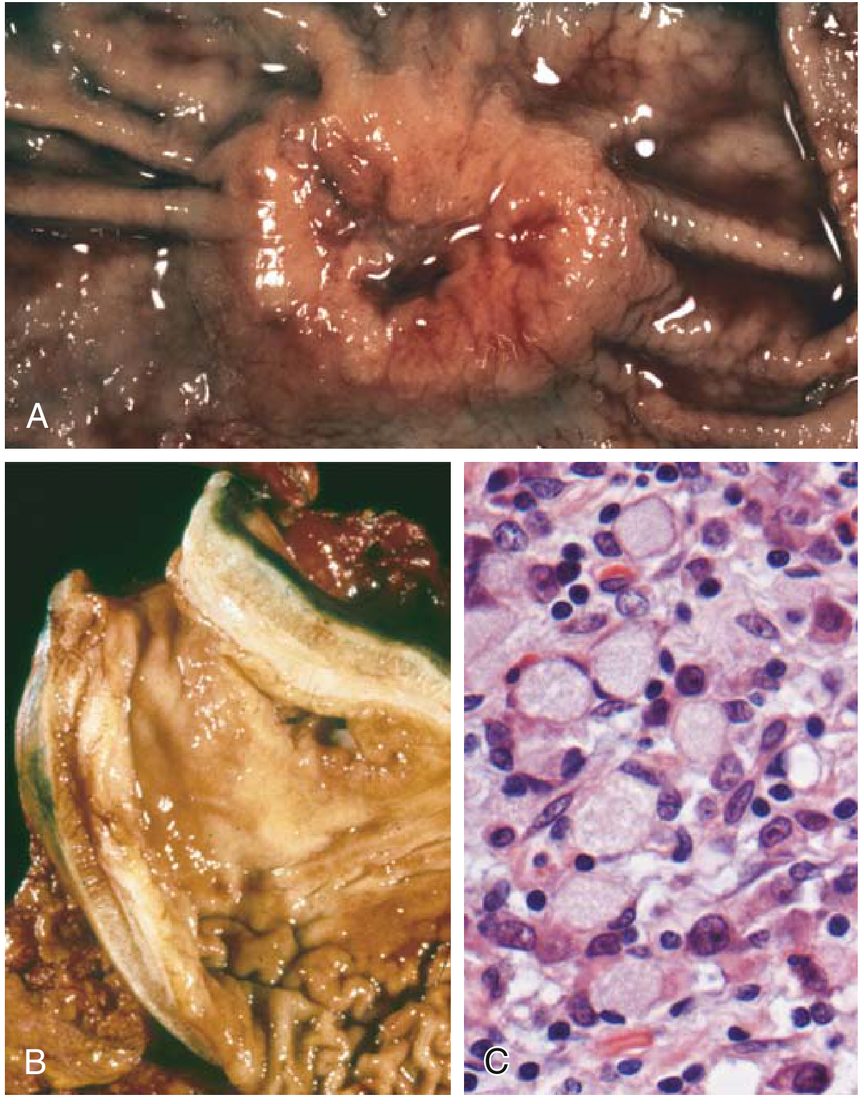
The Two Morphologic Subtypes - Key Features
| Feature | Intestinal Type | Diffuse Type |
|---|---|---|
| Growth pattern | Bulky mass / ulcer; cohesive growth | Infiltrative, dyscohesive |
| Histology | Malignant glandular structures | Signet ring cells |
| Mucin | Apical mucin vacuoles; mucin in gland lumina | Large mucin vacuoles push nucleus to periphery |
| Gross appearance | Exophytic or ulcerated tumor | Diffuse wall thickening - "linitis plastica" (leather bottle stomach) |
| Location | Antrum/lesser curvature | Entire stomach (diffuse) |
| Risk factors | H. pylori, atrophic gastritis, intestinal metaplasia, dietary carcinogens | CDH1 (e-cadherin) germline mutations; no clear precursor lesion |
| Epidemiology | More common in high-risk geographic areas (Japan, Chile) | Uniform worldwide distribution |
Why the Other Options Are Wrong
- a. Chronic gastritis - An inflammatory condition (not a malignancy); it is actually a risk factor for gastric adenocarcinoma, especially H. pylori-driven gastritis
- c. Celiac disease - An autoimmune enteropathy affecting the small intestine (not the stomach), triggered by gluten
- d. Acute pancreatitis - Inflammation of the pancreas; completely unrelated to gastric malignancy
- e. Acute gastritis - Acute mucosal inflammation of the stomach; not a malignancy and not associated with signet ring cells
Source: Robbins & Kumar Basic Pathology; Sabiston Textbook of Surgery
During which disorder biopsy specimens generally demonstrate H. pylori in infected individuals. Neutrophils infiltrate across the basement membrane and accumulate in the lumens of gastric glands, or pits, to create pit abscesses. When intense, inflammatory infiltrates may create thickened rugal folds, mimicking the endoscopic appearance of early cancers? • a. Chronic gastritis • b. Gastric Adenocarcinoma • c. Celiac disease • d. Acute pancreatitis • e. Acute Gastritis
Answer: a. Chronic Gastritis
Direct Textbook Confirmation
"Gastric biopsy specimens generally demonstrate H. pylori in infected individuals... Neutrophils infiltrate across the basement membrane and accumulate in the lumens of gastric glands, or pits, to create pit abscesses. When intense, inflammatory infiltrates may create thickened rugal folds, mimicking the endoscopic appearance of early cancers."
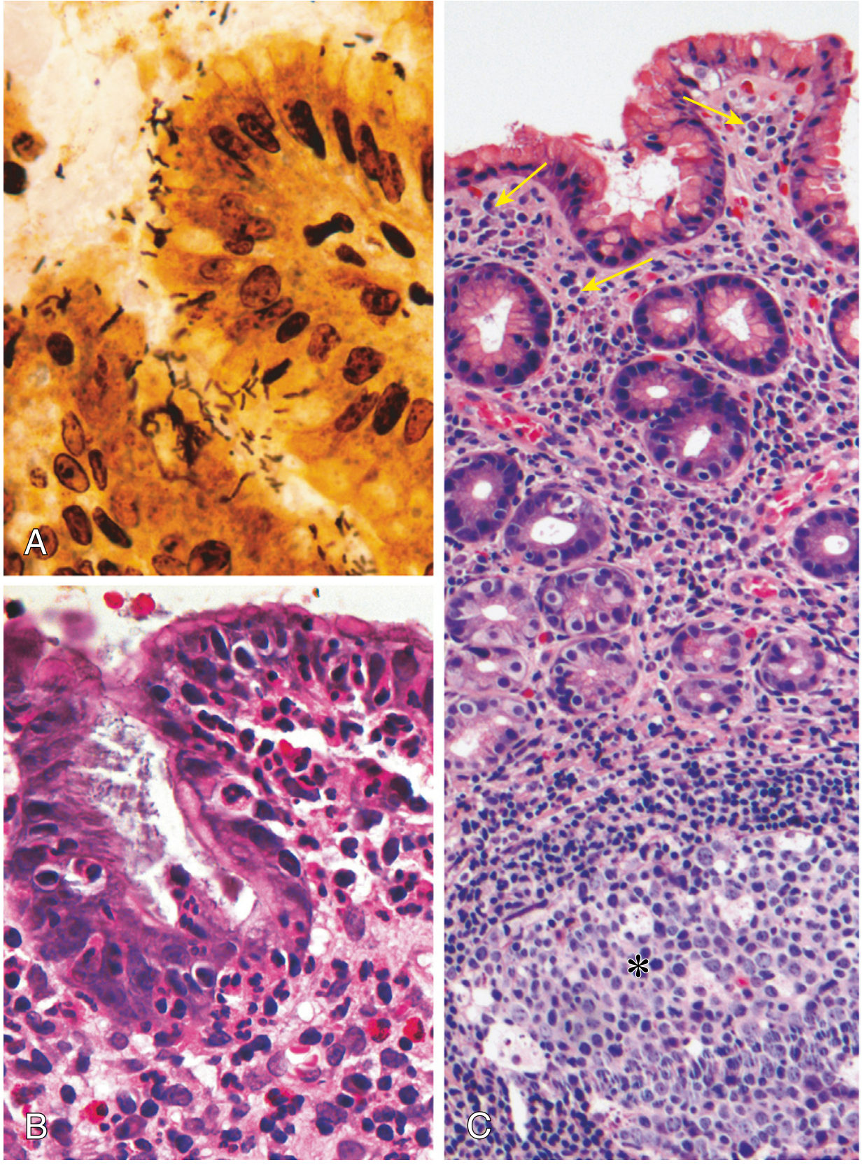
Key Pathological Features of H. pylori Chronic Gastritis
| Feature | Detail |
|---|---|
| Organism | Concentrated in superficial mucus overlying foveolar (surface) cells; best identified with Warthin-Starry silver stain or immunostain |
| Preferred biopsy site | Gastric antrum (most commonly infected) |
| Neutrophils | Cross the basement membrane → enter gland lumens → form pit abscesses |
| Chronic inflammatory cells | Large numbers of plasma cells + lymphocytes + macrophages in lamina propria |
| Rugal folds | Thickened by intense inflammation, mimicking early gastric cancer on endoscopy |
| Lymphoid tissue | Lymphoid aggregates with germinal centers = induced MALT (can transform to MALToma/lymphoma) |
| Long-term consequences | Intestinal metaplasia → gastric atrophy → increased risk of adenocarcinoma |
Why the Other Options Are Wrong
- b. Gastric Adenocarcinoma - A malignant tumor, not an inflammatory condition. Characterized by glandular (intestinal type) or signet ring cells (diffuse type), not pit abscesses or H. pylori infiltrates.
- c. Celiac disease - Autoimmune enteropathy of the small intestine; features villous atrophy and crypt hyperplasia in the duodenum/jejunum, not gastric pit abscesses.
- d. Acute pancreatitis - Inflammation of the pancreas (not stomach); features acinar cell necrosis, fat necrosis, and neutrophil infiltration of pancreatic parenchyma.
- e. Acute Gastritis - Acute gastritis is a transient mucosal injury (from NSAIDs, alcohol, stress) with superficial neutrophil infiltration and possible erosions, but does not produce the chronic inflammatory pattern with pit abscesses, plasma cells, lymphoid aggregates, and H. pylori colonization described in the question.
Key distinction: The presence of H. pylori + plasma cells + lymphoid aggregates + pit abscesses is the defining picture of chronic active gastritis (the "active" referring to the neutrophil component). Acute gastritis lacks plasma cells, lymphoid follicles, and H. pylori.
Source: Robbins, Cotran & Kumar Pathologic Basis of Disease; Robbins & Kumar Basic Pathology
Name most common Secondary amenorrhea etiology? Ovarian disease • b. Pituitary disease • c. Uterine disease • d. Hypothalamic dysfunction
Answer: d. Hypothalamic Dysfunction
Important Caveat First
"Pregnancy is the most common cause of secondary amenorrhea and must be excluded in all cases, regardless of patient history." - Harrison's Principles of Internal Medicine (22nd Ed.)
Why Hypothalamic Dysfunction is the Answer
"Functional hypothalamic amenorrhea (HA) is most commonly associated with conditions causing a mismatch between energy expenditure and energy intake and/or significant stress leading to increased corticotropin-releasing hormone (CRH), suppression of GnRH, and decreased TRH input."
Stress / Low body weight / Excessive exercise
↓
↑ CRH → Suppressed GnRH pulsatility
↓
↓ LH + FSH from pituitary
↓
No follicular development → No estrogen → No menses
Common Triggers of Functional Hypothalamic Amenorrhea
| Trigger | Example |
|---|---|
| Energy deficit | Anorexia nervosa, restrictive eating, rapid weight loss |
| Excessive exercise | Female athlete triad (amenorrhea + low energy + low bone density) |
| Psychosocial stress | Anxiety, depression, major life events |
| Chronic illness | Type 1 diabetes, celiac disease, hyperthyroidism |
Frequency Distribution of Secondary Amenorrhea Etiologies
| Etiology | Approximate Frequency |
|---|---|
| Hypothalamic dysfunction (FHA) | ~35-40% (most common non-pregnant cause) |
| Hyperprolactinemia (pituitary) | ~30% |
| Primary ovarian insufficiency (POI) | ~10% |
| Uterine/outflow tract (Asherman's) | ~5-7% |
| Thyroid / other systemic disease | ~5% |
Why the Other Options Are Less Common
-
a. Ovarian disease - Primary Ovarian Insufficiency (POI/premature ovarian failure) accounts for only ~10% of secondary amenorrhea. It presents with high FSH/LH (hypergonadotropic state) and hot flashes.
-
b. Pituitary disease - Hyperprolactinemia (most often from prolactinoma) is common but accounts for ~30%. Other pituitary causes include Sheehan syndrome (postpartum pituitary necrosis) and empty sella syndrome. Though pituitary causes are frequent, hypothalamic dysfunction is still more prevalent overall.
-
c. Uterine disease - Asherman syndrome (intrauterine adhesions from D&C or endometritis) is the most common outflow tract cause but accounts for only ~5-7% of total secondary amenorrhea cases. The patient is hormonally normal but cannot shed the endometrium.
Key clinical pearl: The initial workup after excluding pregnancy includes: serum β-hCG → TSH → prolactin → FSH. Low FSH points to hypothalamic/pituitary cause; high FSH points to ovarian failure.
Sources: Harrison's Principles of Internal Medicine 22nd Ed.; Berek & Novak's Gynecology; Tietz Textbook of Laboratory Medicine
Teratoma is? • a. cancer in which the tumor cells resemble stratified squamous epithelium; • b. tumor which made up of several different types of tissue, such as hair, muscle, bone • c. a lesion in which the neoplastic epithelial cells grow in glandular patterns. • d. papillary patterns that protrude into cystic spaces
Answer: b. Tumor made up of several different types of tissue, such as hair, muscle, bone
Definition - Confirmed from Textbooks
"Teratomas are tumors of disputed origin that often contain a variety of tissues, such as bone, hair, muscle, gut epithelia, and others. It is thought that these tumors arise from pluripotent stem cells that can differentiate into any of the three germ layers or their derivatives."
"If tumor cells arise from pluripotent stem cells, the tumor may contain cells that differentiate and resemble cells originating from all three germ layers. The result is the formation of a tumor that contains a variety of mature tissues arranged in an unorganized manner. Such masses are referred to as teratomas."
Histology of a Teratoma (Actual Microscopy)
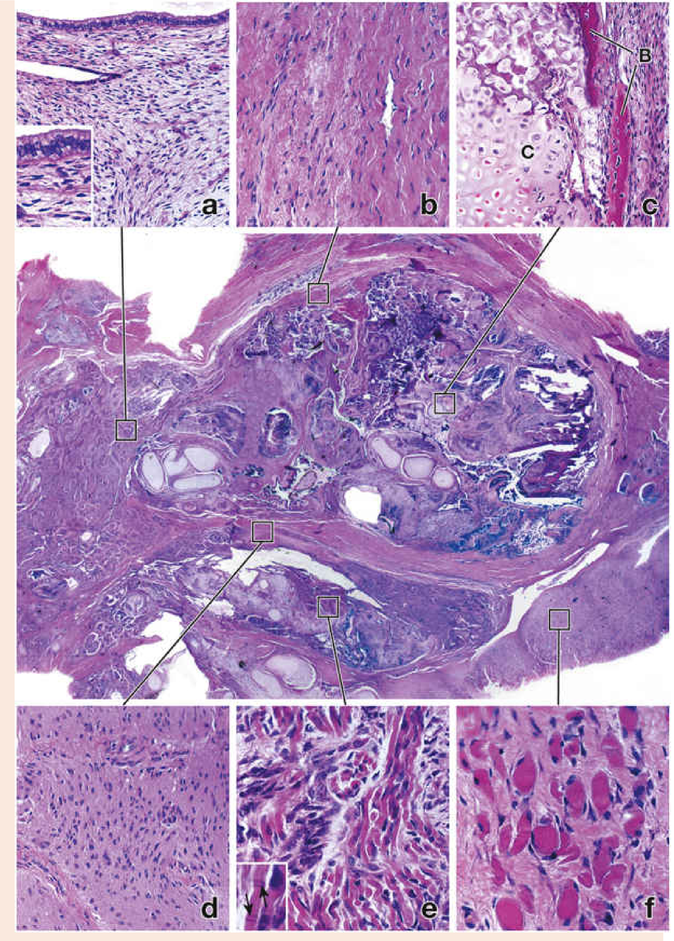
The Three Germ Layers in a Teratoma
| Germ Layer | Derived Tissues Found in Teratoma |
|---|---|
| Ectoderm | Skin, hair, sebaceous glands, teeth, neural tissue/brain |
| Mesoderm | Muscle (smooth, cardiac, skeletal), bone, cartilage, connective tissue |
| Endoderm | Gut epithelium, bowel segments, thyroid, respiratory epithelium |
Types of Teratoma
| Type | Features |
|---|---|
| Mature teratoma (Dermoid cyst) | All tissues well-differentiated; benign; most common in ovary; karyotype 46,XX |
| Immature teratoma | Contains primitive/embryonic tissue (esp. neuroepithelium); malignant potential |
| Monodermal teratoma | Composed predominantly of one tissue type (e.g., struma ovarii = thyroid tissue) |
Why the Other Options Are Wrong
- a. "Tumor cells resembling stratified squamous epithelium" = Squamous cell carcinoma - a carcinoma derived from squamous epithelium, not a teratoma.
- c. "Neoplastic epithelial cells growing in glandular patterns" = Adenocarcinoma - a glandular malignancy (e.g., gastric, colonic, endometrial adenocarcinoma).
- d. "Papillary patterns protruding into cystic spaces" = Papillary cystadenoma/cystadenocarcinoma - describes the architecture of tumors like serous ovarian tumors, not teratoma.
Sources: Langman's Medical Embryology; Histology: A Text and Atlas with Correlated Cell and Molecular Biology (Pawlina); Robbins, Cotran & Kumar Pathologic Basis of Disease