
Aesthetic posts for 15 marks with contents and references
aesthetic post and core classification fiber post ceramic post dentistry exam notes
"fiber post" AND esthetic AND "endodontically treated teeth"
fiber post ceramic post endodontically treated tooth restoration
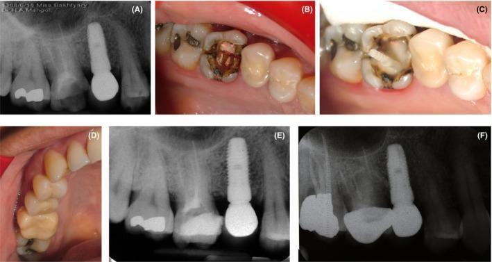
A multi-panel series documenting the diagnosis, restoration, and follow-up of a cracked maxillary right first molar. (A) Periapical radiograph showing an endodontically treated molar with a radiopaque root canal filling and existing restoration. (B) Intraoral clinical photograph following restoration removal, revealing a distinct mesiodistal crack line across the pulp chamber floor and secondary caries. (C) Intraoral view of the intermediate restoration stage featuring post and core fabrication with glass fiber-reinforced composite. (D) Clinical photograph of the final metal-ceramic crown restoration in situ, exhibiting restored occlusal anatomy. (E) Immediate postoperative periapical radiograph confirming proper crown seating and post placement. (F) Long-term follow-up periapical radiograph at 8 years, demonstrating sustained periapical health and structural stability of the prosthetic complex. The series illustrates the management of a symptomatic cracked tooth through adhesive sealing of the fracture line followed by full-coverage crown protection.
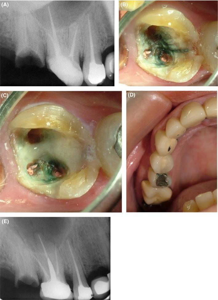
This composite figure illustrates the clinical management of a cracked maxillary right first molar that has previously undergone endodontic treatment. (A) Preoperative periapical radiograph showing an endodontically treated molar with a radiopaque amalgam restoration and no visible root pathology. (B) Intraoral photograph following removal of the previous restoration; transillumination reveals a dark, mesiodistal crack line running across the pulp chamber floor between the canal orifices, accompanied by green staining. (C) Clinical view showing the application of a resin-based cement (Panavia) over the identified crack line for stabilization. (D) Occlusal view of the final prosthetic metal-ceramic crown restoration in situ, demonstrating proper anatomical contour and integration within the dental arch. (E) Final follow-up periapical radiograph confirming the placement of a fiber post and core and the definitive crown restoration, showing stable periapical tissues and well-adapted margins. This series demonstrates a conservative endodontic-restorative approach to salvaging a tooth with a compromised pulpal floor.
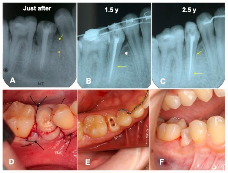
A series of dental images illustrating the 2.5-year progression of an autotransplanted premolar with porcine-derived demineralized dentin matrix (pDDM) grafting. (A-C) Periapical radiographs showing chronological healing: (A) Immediate post-operative state with radiopaque pDDM granules (arrows) and lack of a visible periodontal ligament (PDL) space; (B) At 1.5 years, showing remodeled bone-like radio-opacity (*) and initial PDL space formation (arrow) during orthodontic treatment; (C) At 2.5 years, demonstrating a continuous, healthy PDL space and lamina dura (arrows). (D-F) Matching intraoral clinical photographs: (D) Surgical site immediately post-transplantation with a mucoperiosteal flap secured by sutures; (E) Occlusal view during the treatment phase showing the endodontically treated root and orthodontic appliances; (F) Final clinical view following core build-up with resin composite and fiber post, ready for the definitive ceramic crown restoration. The sequence highlights successful tooth integration, bone remodeling around the graft, and orthodontic alignment.
Esthetic (Aesthetic) Posts — 15 Marks
1. Introduction
2. Definition
3. Need / Indications for a Post
- Extensive loss of coronal tooth structure (>2/3 of crown destroyed)
- Anterior/premolar teeth restored with all-ceramic crowns where esthetics is critical
- Insufficient ferrule and need for core retention
- Teeth requiring retention for a core prior to full-coverage restoration
4. Classification of Posts (Singh, Chandra, Pandit - 2015)
- Custom (cast) post and core
- Prefabricated post and core
- Metallic - Stainless steel, Titanium, Titanium alloy, Chrome-cobalt, Nickel-chromium, Gold alloy
- Non-metallic
- Non-esthetic: Carbon fiber post (black/dark grey)
- Esthetic posts:
- Fiber-reinforced composite posts - Glass fiber, Quartz fiber, Polyethylene (woven) fiber
- Ceramic posts - Zirconia (Zirconium oxide), Alumina
5. Ideal Requirements of an Esthetic Post
- Tooth-like color/translucency, no shine-through under ceramic restorations
- Modulus of elasticity close to dentin (to distribute stress evenly and reduce root fracture risk)
- High flexural and fatigue strength
- Good adhesion/bonding to resin cement and core material
- Biocompatible, corrosion resistant, radiopaque for radiographic visibility
- Easy retrievability if retreatment needed
6. Types of Esthetic Posts
A. Fiber-Reinforced Composite (FRC) Posts
| Type | Composition | Features |
|---|---|---|
| Carbon fiber post | Carbon fibers in epoxy resin | High strength but black color - esthetic limitation, mainly used posteriorly |
| Glass fiber post | E-glass/S-glass fibers in resin matrix | Most widely used esthetic post; tooth-colored, translucent, bonds well to resin cement |
| Quartz fiber post | Pure quartz fibers | Highly translucent, superior esthetics, high strength |
| Polyethylene fiber (woven) post | Braided/woven polyethylene ribbon | Flexible, used to reinforce direct composite core build-ups |
B. Ceramic Posts (Zirconia)
7. Clinical Technique (Cementation of Fiber Post - brief)
- Case selection and post space preparation (retain 4-5 mm apical gutta-percha for seal)
- Try-in of correctly sized post
- Etch and bond canal dentin (self-etch/total-etch adhesive)
- Silanate the fiber post surface
- Cement post with dual-cure resin cement
- Light cure/self-cure, build up composite core
- Tooth preparation and final crown fabrication
8. Recent Advances
- Anatomic/individualized fiber posts customized with flowable composite to match canal anatomy
- One-step post-core-buildup systems (e.g., translucent quartz posts with light-transmitting properties for deep cure)
- Silica/zirconia-nanoparticle coated fiber posts for improved bonding
- CAD/CAM milled fiber-reinforced or zirconia posts
9. Recent Evidence (Systematic Reviews)
- A 2021 meta-analysis found no significant difference in overall failure rates between fiber and metal posts, though failure modes differ (fiber posts fail more often by debonding, which is repairable, whereas metal/ceramic posts more often cause irreparable root fracture) (Martins et al., Journal of Dentistry, PMID: 34274439).
- A 2023 network meta-analysis similarly supports fiber posts as generally preferable due to more favorable (repairable) failure modes (Giok et al., J Prosthet Dent, PMID: 37827970).
- A 2020 meta-analysis confirmed intraradicular fiber posts improve fracture resistance of endodontically treated anterior teeth compared to no post, when adequate tooth structure/ferrule is considered (Jurema et al., PMID: 33546858).
10. Conclusion
References
- Singh S, Chandra A, Pandit RH. A New Classification of Post and Core. Indian Journal of Restorative Dentistry. Sept-Dec 2015;4(3):56-58.
- Sturdevant's Art and Science of Operative Dentistry, 6th ed. - Chapter on Restoration of Endodontically Treated Teeth.
- Shillingburg HT, et al. Fundamentals of Fixed Prosthodontics, 4th ed. - Post and Core sections.
- Martins MD, Junqueira RB, de Carvalho RF, et al. Is a fiber post better than a metal post for the restoration of endodontically treated teeth? A systematic review and meta-analysis. Journal of Dentistry. 2021. PMID: 34274439.
- Giok KC, Veettil SK, Menon RK. Comparative effectiveness of fiber and metal posts in the restoration of endodontically treated teeth: A systematic review with network meta-analysis. Journal of Prosthetic Dentistry. 2023. PMID: 37827970.
- Jurema ALB, Filgueiras AT, Santos KA, et al. Effect of intraradicular fiber post on the fracture resistance of endodontically treated and restored anterior teeth: A systematic review and meta-analysis. Journal of Prosthetic Dentistry. 2022. PMID: 33546858.
- Sarna-Boś K, et al. Dental Fiber-Post Systems: An In-Depth Review of Their Evolution and Current Status. PMC10215107.
But where are the contents
Contents
- Introduction - definition and background of esthetic posts
- Definition of esthetic (metal-free) post
- Need / Indications for using a post
- Contraindications
- Classification of Posts
- By fabrication method (cast vs prefabricated)
- By material (metallic vs non-metallic - esthetic vs non-esthetic)
- By configuration (parallel/tapered)
- By surface (smooth/serrated/self-threading)
- By fit (active/passive)
- Ideal Requirements of an esthetic post
- Types of Esthetic Posts
- A. Fiber-Reinforced Composite (FRC) Posts - Carbon, Glass, Quartz, Polyethylene
- B. Ceramic (Zirconia) Posts
- Advantages and Disadvantages of each
- Clinical Technique - cementation steps for a fiber post
- Recent Advances
- Recent Evidence (systematic reviews/meta-analyses)
- Conclusion
- Image - clinical example of fiber post/core build-up
- References (7 sources)