
Visceral leishmaniasis mocrobiology
visceral leishmaniasis Leishmania donovani amastigote macrophage
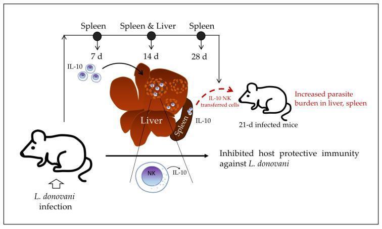
This pathophysiology diagram illustrates the immunoregulatory role of IL-10-producing natural killer (NK) cells during Visceral Leishmaniasis caused by Leishmania donovani infection in a murine model. The timeline tracks disease progression at 7, 14, and 28 days post-infection. At 7 days, IL-10 production begins in the spleen. By 14 days, the diagram shows a central representation of the liver and spleen where splenic NK cells migrate into liver granulomas, becoming a primary source of the immunosuppressive cytokine IL-10. Visual details include hepatic granulomas depicted as clusters of cells within the liver parenchyma. At 28 days, the model demonstrates an adoptive transfer experiment where IL-10-producing NK cells are transferred into 21-day infected recipient mice, resulting in an increased parasite burden in both the liver and spleen. The diagram concludes that these cells inhibit host protective immunity against L. donovani, shifting the immune response from pro-inflammatory to immunosuppressive during the late stages of infection.
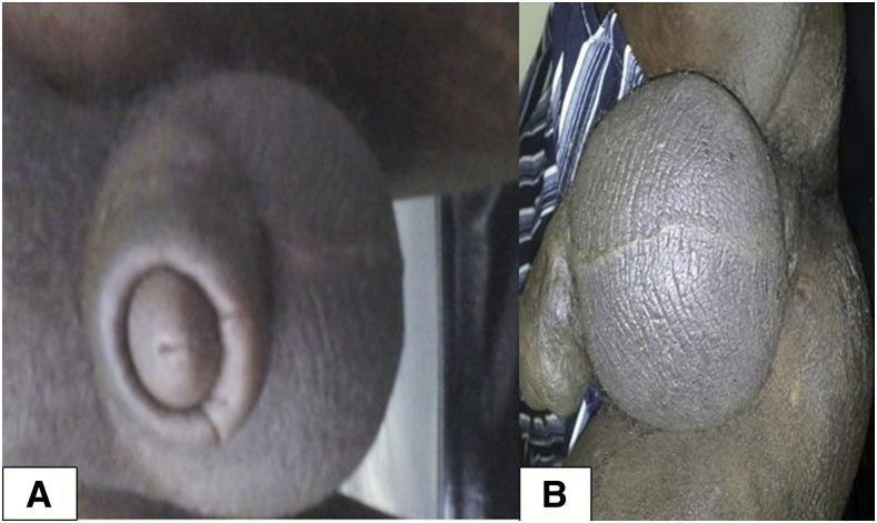
Two-panel clinical photograph (Figures 1A and 1B) illustrating atypical genital manifestations of visceral leishmaniasis in a patient with HIV coinfection. Panel A displays significant edema and swelling of the penile shaft. Panel B shows massive, diffuse scrotal swelling with a coarsely textured, thickened, and wrinkled skin surface. A focal nodular lesion is visible at the base of the scrotum. The visual findings represent cutaneous involvement in the context of systemic parasitic infection and immunodeficiency. This image serves as an educational reference for recognizing atypical extra-visceral presentations of Leishmania donovani infection, which may mimic other tropical or sexually transmitted diseases such as lymphogranuloma venereum. The presentation highlights the clinical significance of dermatological examination in HIV-positive patients living in or traveling from endemic regions (e.g., Ethiopia).
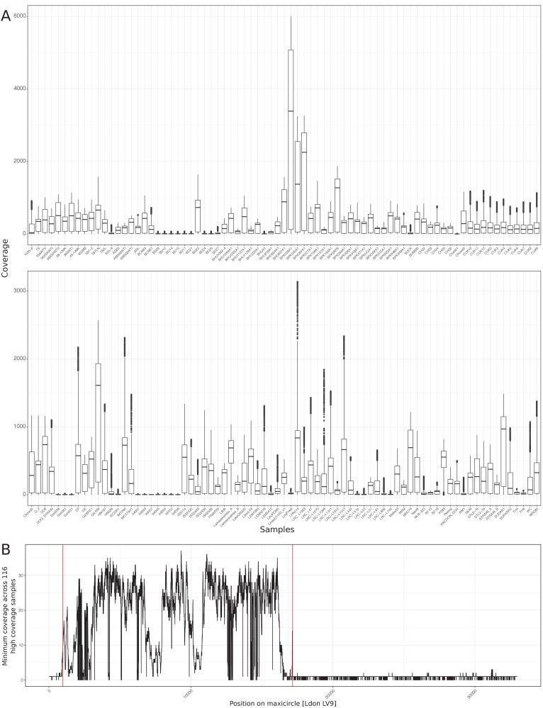
This figure presents genetic sequencing data analysis for maxicircle DNA (mitochondrial genome) coverage across 151 Leishmania isolates, supporting research into human visceral leishmaniasis. Panel A is a comparison chart featuring multiple boxplots. Each plot represents a specific sample (x-axis), showing the median, quartiles, and range of sequencing coverage (y-axis). This visualization highlights the significant variability in maxicircle DNA abundance and mapping success across different clinical isolates. Panel B is a line graph showing the minimum sequencing coverage across 116 high-coverage samples as a function of the nucleotide position on the L. donovani LV9 maxicircle reference (x-axis). The black line illustrates a fluctuating coverage profile, indicating regions of high and low mapping confidence along the mitochondrial genome. Two vertical red lines delineate a specific high-confidence region (approximately between positions 1,000 and 17,000) selected for subsequent phylogenetic reconstruction. This educational material demonstrates methods for qualifying genomic data and identifying conserved regions for species-level evolutionary analysis in parasitology and infectious diseases.
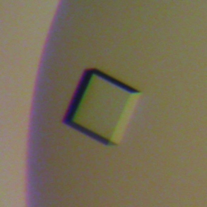
This diagnostic micrograph displays a protein crystal of Leishmania donovani pteridine reductase 1 (LdPTR1), a key enzyme in the parasite's folate metabolism. The visual demonstrates a single, well-defined orthorhombic plate crystal with approximate dimensions of 0.1 x 0.1 x 0.03 mm. The crystal is translucent with a smooth, planar surface and sharp geometric edges, though slight chromatic aberration is visible at the margins due to light refraction. This specimen was grown using the sitting-drop vapor diffusion method in a solution containing cofactor NADP+ and the inhibitor methotrexate. Such crystals are essential in structural biology and pharmacology for X-ray diffraction studies to determine the 3D molecular structure of protein targets, which is a critical step in rational drug design against visceral leishmaniasis. The image highlights the morphological characteristics required for successful crystallographic data collection in biomedical research.
"visceral leishmaniasis"[MeSH Terms] AND diagnosis
Visceral Leishmaniasis (VL) - Microbiology
The Organism
| Species | Transmission | Key Endemic Areas |
|---|---|---|
| L. donovani | Anthroponotic (human-to-human) | Indian subcontinent, East Africa |
| L. infantum (L. chagasi in the New World) | Zoonotic (dog reservoir) | Mediterranean, Middle East, Central Asia, Americas |
- Harrison's Principles of Internal Medicine 22E, p.1825
Morphology & Life Cycle
1. Promastigote (extracellular, in sandfly)
- Elongated, flagellated form (10-15 µm)
- Lives in the gut of the female sandfly vector
- Infective stage - inoculated into the human host during a blood meal
2. Amastigote (intracellular, in human)
- Small, ovoid, non-flagellated (2-4 µm)
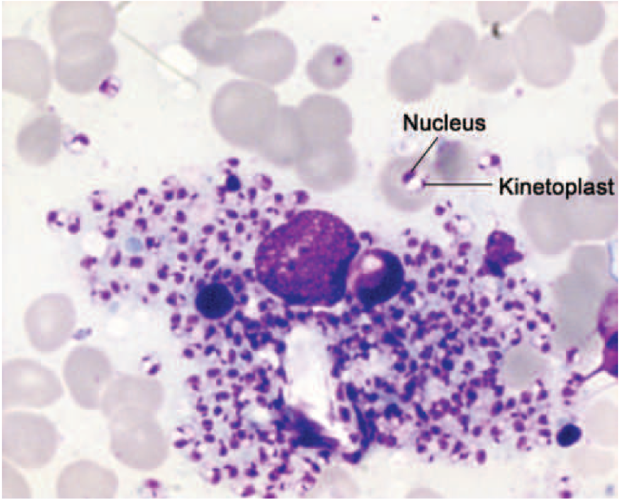
- Contains a nucleus and a characteristic kinetoplast (rod-like, contains multiple copies of mitochondrial DNA) - seen as a "dot" (nucleus) and "dash" (kinetoplast) on Giemsa stain
- Obligate intracellular parasite, residing within macrophages
Life Cycle Steps
- Female sandfly (Phlebotomus spp. in Old World; Lutzomyia spp. in New World) takes a blood meal and inoculates promastigotes
- Promastigotes are phagocytosed by macrophages/monocytes
- Inside the phagolysosome, promastigotes transform into amastigotes
- Amastigotes multiply by binary fission, fill the cell cytoplasm, and burst the cell
- Released amastigotes are phagocytosed by more macrophages - cycle repeats
- Parasite spreads via the reticuloendothelial system to spleen, liver, bone marrow, and lymph nodes
- Sandfly ingests amastigotes during feeding; amastigotes convert back to promastigotes in the sandfly gut
- Jawetz, Melnick & Adelberg's Medical Microbiology 28th Ed.
- Harrison's Principles of Internal Medicine 22E
Vectors
| Region | Vector |
|---|---|
| Indian subcontinent | Phlebotomus argentipes |
| East Africa (Sudan, Ethiopia) | P. orientalis, P. martini |
| Mediterranean/Middle East | P. perniciosus, P. ariasi |
| Americas | Lutzomyia longipalpis |
Epidemiology
-
East Africa now has the highest global incidence, followed by Brazil and the Indian subcontinent
-
WHO has declared elimination as a public health problem in Bangladesh and near-elimination in Nepal and India (98.7% decline)
-
In Mediterranean Europe, 70% of adult VL cases are HIV co-infected
-
IV drug users are at particular risk for co-infection
-
Children predominantly affected in immunocompetent endemic areas (Americas, Mediterranean); all ages affected in India and East Africa
-
Harrison's Principles of Internal Medicine 22E, p.1825
Immunopathogenesis
-
Protective response: IL-12 from antigen-presenting cells → IFN-γ, TNF-α from Th1 cells → activation of macrophages → nitric oxide production → parasitic killing
-
Disease promotion: Markedly elevated serum IL-10 in active VL
- IL-10 renders macrophages unresponsive to activation signals
- Downregulates TNF-α and nitric oxide production
- Suppresses antigen-presentation by DCs and macrophages
- Parasite survives and multiplies unopposed
-
Organs of the reticuloendothelial system (spleen, liver, lymph nodes, bone marrow) are predominantly affected
-
Polyclonal hypergammaglobulinemia occurs (antibodies are non-protective)
-
Patients with active disease have negative leishmanin skin tests (Montenegro test) - unlike immune individuals who show delayed-type hypersensitivity
-
Harrison's Principles of Internal Medicine 22E, p.1826
Clinical Features
- Incubation period: 2-3 months (up to 1 year)
- Fever: Intermittent, moderate-to-high grade with rigors/chills (classically biphasic - two daily spikes)
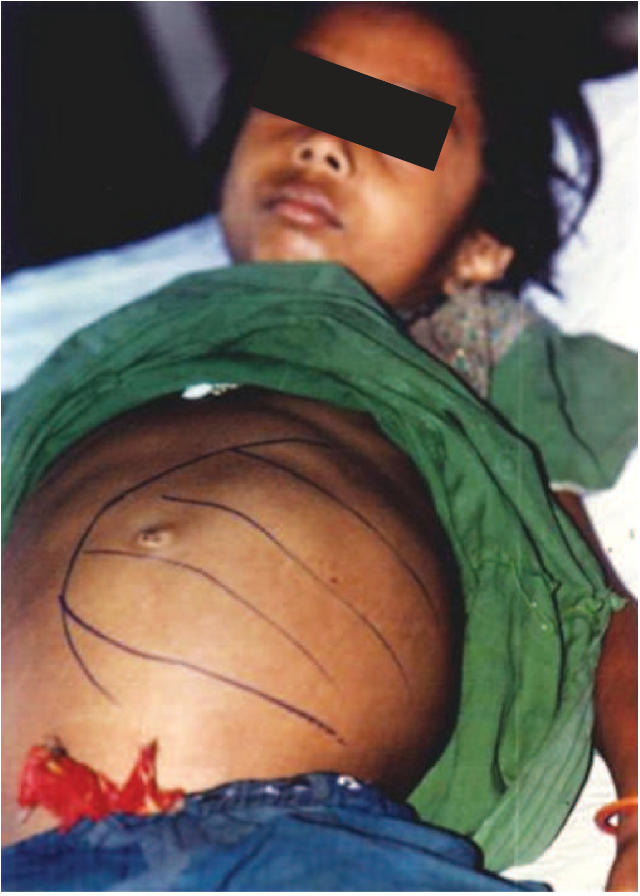
- Massive splenomegaly - hallmark finding (palpable by 2nd week, can be enormous)
- Hepatomegaly - usually moderate
- Lymphadenopathy - common everywhere except Indian subcontinent
- Weight loss, weakness
- Hyperpigmentation of skin (gives rise to the name kala-azar)
- Advanced disease: hypoalbuminemia → pedal edema and ascites
Labs
- Pancytopenia - leukopenia, anemia, thrombocytopenia (bone marrow involvement)
- Markedly elevated serum immunoglobulins (polyclonal)
- Raised hepatic aminotransferases
- Occasional hyperbilirubinemia
Diagnosis
Gold Standard
- Demonstration of amastigotes in tissue aspirate smears (Giemsa stain)
| Specimen | Sensitivity |
|---|---|
| Splenic aspirate | >95% (most sensitive but invasive) |
| Bone marrow aspirate | 60-85% |
| Lymph node aspirate | ~50% |
Serologic Tests
- ELISA and IFAT - used in sophisticated labs
- rK39 Rapid Diagnostic Test (RDT) - most widely used field test
- Detects antibodies to a recombinant antigen (39 amino acids from the kinesin region of L. infantum)
- Requires only a fingerprick drop of blood; result in 15 minutes
- Sensitivity ~98%, specificity ~90% in immunocompetent patients (lower in East Africa)
- Cannot be used to monitor cure or detect relapse (remains positive for years)
Molecular Tests
-
PCR - qualitative, highly sensitive, allows species identification
-
Real-time (quantitative) PCR - can monitor parasite load
-
LAMP (Loop-mediated isothermal amplification) - newer, performs better than microscopy using blood; simple visual readout, suitable for point-of-care in endemic areas
-
Harrison's Principles of Internal Medicine 22E, p.1827
Post-Kala-Azar Dermal Leishmaniasis (PKDL)
- A dermatosis that develops as a sequela after apparently successful treatment of VL
- Indian subcontinent variant: polymorphic lesions (macules, patches) coexisting with nodules
- Important reservoir of infection for maintaining anthroponotic transmission cycles
Treatment (Key Points)
- Liposomal amphotericin B - drug of choice in most settings (single-dose regimen in India)
- Miltefosine - oral agent; first-line in Indian subcontinent
- Antimonials (sodium stibogluconate, meglumine antimoniate) - historically first-line, now with widespread resistance in Bihar, India
- Amphotericin B deoxycholate - effective but toxic
- Paromomycin - aminoglycoside, used in combination regimens in East Africa
Summary Table
| Feature | Detail |
|---|---|
| Causative agents | L. donovani, L. infantum |
| Vector | Female sandfly (Phlebotomus / Lutzomyia) |
| Infective stage | Promastigote |
| Intracellular stage | Amastigote (in macrophages) |
| Key organelle | Kinetoplast (mitochondrial DNA) |
| Hallmark sign | Massive splenomegaly |
| Gold standard diagnosis | Splenic smear (>95% sensitivity) |
| Best field test | rK39 RDT |
| Immune evasion key | IL-10 - suppresses macrophage activation |
| Drug of choice | Liposomal amphotericin B / miltefosine |
Visceral leishmaniasis microbiology
Visceral Leishmaniasis (VL) - Microbiology
The Organism
| Species | Transmission | Key Endemic Areas |
|---|---|---|
| L. donovani | Anthroponotic (human-to-human) | Indian subcontinent, East Africa |
| L. infantum (L. chagasi in the New World) | Zoonotic (dog reservoir) | Mediterranean, Middle East, Central Asia, Americas |
Morphology & Life Cycle
1. Promastigote (in the sandfly - extracellular)
- Elongated, flagellated (10-15 µm)
- The infective stage - inoculated into the human host during a sandfly bite
2. Amastigote (in the human host - intracellular)
- Small, ovoid, non-flagellated (2-4 µm)
- Contains a nucleus and a distinctive kinetoplast (rod-like; packed mitochondrial DNA) - seen as a "dot and dash" on Giemsa stain
- Lives and multiplies inside macrophages
Life Cycle Steps
- Female sandfly inoculates promastigotes during a blood meal
- Promastigotes are phagocytosed by skin macrophages/monocytes
- Inside the phagolysosome, they transform into amastigotes
- Amastigotes multiply by binary fission, eventually burst the cell
- Released amastigotes are phagocytosed by more macrophages - cycle repeats
- Parasite disseminates via the reticuloendothelial system to spleen, liver, bone marrow, lymph nodes
- A new sandfly ingests amastigotes during feeding; these revert to promastigotes in the sandfly midgut
Vectors
| Region | Sandfly Vector |
|---|---|
| Indian subcontinent | Phlebotomus argentipes |
| East Africa | P. orientalis, P. martini |
| Mediterranean / Middle East | P. perniciosus, P. ariasi |
| Americas | Lutzomyia longipalpis |
Epidemiology
- East Africa carries the highest global burden, followed by Brazil and the Indian subcontinent
- WHO declared elimination as a public health problem in Bangladesh; India reported a 98.7% decline
- In Mediterranean Europe, 70% of adult VL cases are HIV co-infected
- L. infantum is the species in HIV/VL co-infection; IV drug users are a key at-risk group
- Reservoir for L. donovani: humans (anthroponotic); for L. infantum: dogs, foxes, jackals
Immunopathogenesis
- IL-12 (from APCs) → Th1 cells → IFN-γ + TNF-α → macrophage activation → nitric oxide → parasite killing
- These individuals test positive on the leishmanin (Montenegro) skin test (delayed-type hypersensitivity)
- Markedly elevated serum IL-10 suppresses macrophage activation
- IL-10 downregulates TNF-α and nitric oxide production
- Antigen-presentation by DCs and macrophages is suppressed
- Parasite survives and multiplies unchecked
- Polyclonal hypergammaglobulinemia develops (antibodies are non-protective)
- Patients have negative leishmanin skin tests during active disease
Clinical Features
- Incubation: 2-3 months (up to 1 year)
- Fever: Intermittent, moderate-to-high grade, with rigors/chills; classically biphasic (two daily spikes)
- Massive splenomegaly - the hallmark (palpable by week 2; can be enormous - see figure below)
- Moderate hepatomegaly
- Lymphadenopathy - present in most regions, rare on the Indian subcontinent
- Weight loss, weakness
- Skin hyperpigmentation - the origin of "kala-azar" (dark fever)
- Advanced: hypoalbuminemia → pedal edema, ascites
Laboratory Findings
| Finding | Cause |
|---|---|
| Pancytopenia (leukopenia, anemia, thrombocytopenia) | Bone marrow infiltration + hypersplenism |
| Polyclonal hypergammaglobulinemia | Non-specific B cell activation |
| Raised hepatic transaminases | Liver involvement |
| Hypoalbuminemia | Chronic disease |
Diagnosis
Gold Standard
| Specimen | Sensitivity |
|---|---|
| Splenic aspirate | >95% (most sensitive but invasive) |
| Bone marrow aspirate | 60-85% |
| Lymph node aspirate | ~50% |
Serologic Tests
- ELISA / IFAT - used in reference labs
- rK39 Rapid Diagnostic Test (RDT) - workhorse field test
- Detects antibodies to a recombinant antigen (39 amino acids from the kinesin region of L. infantum)
- Only a fingerprick of blood; result in 15 minutes
- Sensitivity ~98%, specificity ~90% in immunocompetent patients
- Sensitivity/specificity lower in East Africa
- Remains positive for years after cure - cannot be used to monitor treatment response
Molecular Diagnostics
- PCR - qualitative; highly sensitive; allows species identification
- Real-time PCR - quantitative; can monitor parasite load and relapse
- LAMP - performs better than microscopy on blood; minimal equipment needed; suitable for point-of-care use in endemic settings
Post-Kala-Azar Dermal Leishmaniasis (PKDL)
- Develops after apparently successful treatment of VL
- Indian subcontinent: polymorphic lesions (macules/patches + nodules)
- Important as a reservoir for continued anthroponotic transmission
Treatment Overview
| Drug | Notes |
|---|---|
| Liposomal amphotericin B | Drug of choice globally; single-dose regimen used in India |
| Miltefosine | Oral agent; first-line in Indian subcontinent |
| Sodium stibogluconate / Meglumine antimoniate | Historic first-line; widespread resistance in Bihar, India |
| Paromomycin | Aminoglycoside; used in combination in East Africa |
| Amphotericin B deoxycholate | Effective but nephrotoxic |
Quick Summary Table
| Feature | Detail |
|---|---|
| Causative agents | L. donovani, L. infantum |
| Vector | Female sandfly (Phlebotomus / Lutzomyia) |
| Infective stage (to human) | Promastigote |
| Intracellular stage | Amastigote (in macrophages) |
| Diagnostic organelle | Kinetoplast ("dot and dash") |
| Hallmark clinical sign | Massive splenomegaly |
| Gold standard diagnosis | Splenic smear (>95%) |
| Best field test | rK39 RDT (15 min, fingerprick) |
| Key immune evasion mechanism | IL-10 suppresses macrophage activation |
| Drug of choice | Liposomal amphotericin B / miltefosine |
Lymphatic filariasis
lymphatic filariasis elephantiasis microfilaria Wuchereria bancrofti
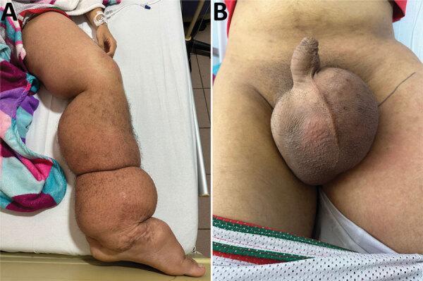
Two-panel clinical photograph illustrating the manifestations of lymphatic filariasis (elephantiasis) caused by Wuchereria bancrofti. Image A displays a side profile of the left lower limb exhibiting severe, generalized lymphedema from the foot to the groin. There is significant swelling and skin folds, erythematous patches on the thigh, and trophic skin changes including perimaleolar hypocromia and hypercromia. Image B provides a frontal view of the pelvic region, showing massive, bulbous scrotal edema (hydrocele) and induration. The skin of the scrotum appears thickened and tense. These findings are characteristic of chronic lymphatic obstruction leading to massive tissue enlargement and secondary dermatological changes. The images are highly relevant for tropical medicine and infectious disease specialties, demonstrating the late-stage physical sequelae of nematode-induced lymphatic damage.
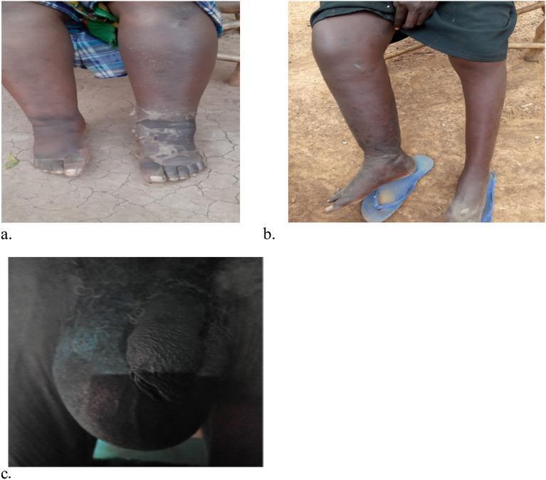
This set of clinical photographs illustrates chronic manifestations of lymphatic filariasis, a parasitic disease caused by Wuchereria bancrofti. Image (a) shows bilateral lymphedema of the lower extremities, characterized by significant swelling (elephantiasis) extending from the feet to the lower legs, with visible skin thickening, hyperkeratosis, and textural changes. Image (b) demonstrates unilateral lymphedema, where the right lower extremity exhibits marked enlargement and swelling compared to the relatively normal left leg. Image (c) displays severe scrotal swelling, indicative of a large hydrocele. The scrotal sac is massively distended, obscuring normal genital contours due to lymphatic fluid accumulation. These images highlight the progressive morbidity and physical deformities associated with untreated chronic lymphatic obstruction in endemic regions. The visuals serve as educational markers for late-stage morbidity management and disability prevention (MMDP) in tropical medicine.
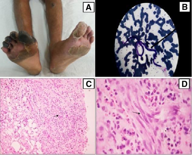
Educational multi-panel figure illustrating the clinical and pathological findings of lymphatic filariasis (Wuchereria bancrofti) masquerading as vasculitis. Panel A (Clinical Photograph): Displays the lower extremities of a patient with dry gangrene of the right hallux and lesser digits, alongside an ulcerated nodule on the left shin and diffuse skin excoriation, indicating severe peripheral vascular compromise. Panel B (Peripheral Blood Film): Giemsa-stained blood smear showing a characteristic microfilaria (approximately 240-250 um long) with identifiable micromorphological features. Panels C and D (Histopathology): Haematoxylin and Eosin (H&E) stained skin biopsy sections from the shin nodule. Low-power magnification (C, 100x) reveals dense inflammatory infiltrates spanning the dermis and subcutaneous fat with capillary proliferation. High-power magnification (D, 1000x) demonstrates a microfilaria (arrow) localized within a capillary lumen, surrounded by a mixed inflammatory cell population consisting of plasma cells, lymphocytes, and neutrophils. This visual set demonstrates the importance of parasitic evaluation in cases of atypical limb ischemia and nodular skin lesions.
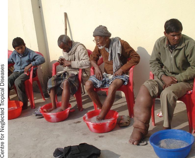
A clinical photograph depicting four male patients seated in a row, participating in foot hygiene practices for the management of lymphatic filariasis. The patients are seated in plastic chairs, each with their lower limbs positioned in basins containing soapy water. The individual on the far right exhibits advanced elephantiasis of the right lower extremity, characterized by massive non-pitting lymphedema, significant limb distortion, and skin thickening (pachydermia) with visible skin folds. Other patients show varying degrees of lower limb involvement or are engaged in preventive hygiene. The scene illustrates morbidity management and disability prevention (MMDP) protocols, specifically focusing on local skin care and hygiene to prevent secondary bacterial infections (dermatolymphangioadenitis) in patients with neglected tropical diseases (NTDs). The setting appears to be a community-based health intervention in a region endemic for Wuchereria bancrofti or Brugia malayi.
"lymphatic filariasis"[MeSH Terms] AND treatment
Lymphatic Filariasis
The Organisms
| Species | Proportion of cases | Key Endemic Areas |
|---|---|---|
| Wuchereria bancrofti | ~90% of all cases | Tropics/subtropics worldwide - Africa, South Asia, Southeast Asia, Pacific Islands, Americas |
| Brugia malayi | Most remaining cases | Eastern India, Indonesia, Malaysia, Philippines |
| Brugia timori | Minor | Islands of southeastern Indonesia only |
- Robbins & Cotran Pathologic Basis of Disease; Harrison's Internal Medicine 22E, p.1863
Morphology
Adult Worms
- Long, threadlike (males ~4 cm; females ~8-10 cm)
- Live in afferent lymphatics and lymph node sinuses
- May remain viable for >20 years
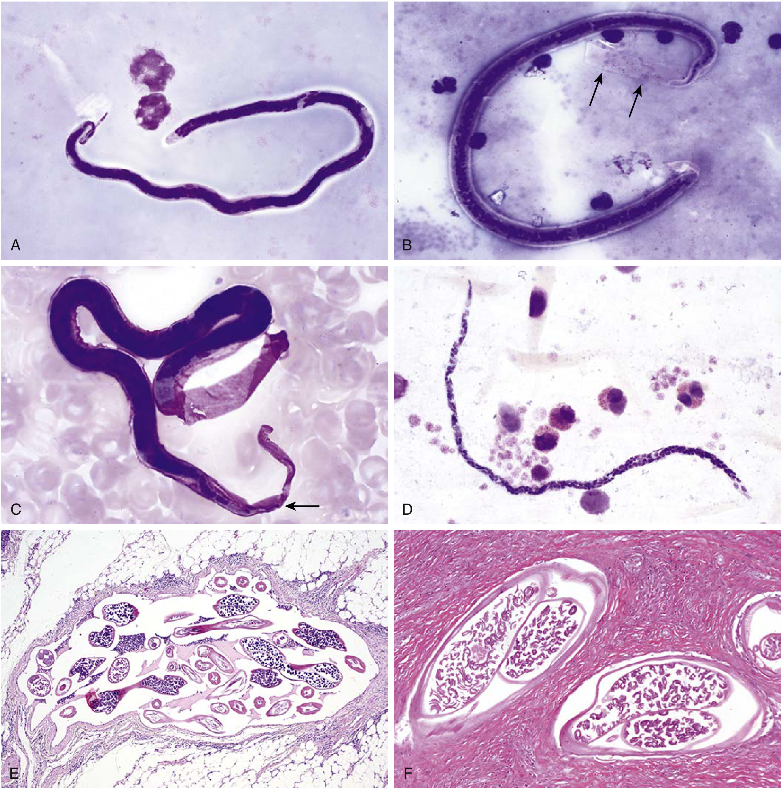
- W. bancrofti adults found in cross-section within lymphatics: associated lymphatic fibrosis (see panel E below)
Microfilariae (larval stage circulating in blood)
- Elongated larvae, ~220-300 µm
- Sheathed in all three lymphatic filarial species (the sheath is the remnant of the egg membrane)
- Key distinguishing features on Giemsa stain:
- W. bancrofti: tail tip has no nuclei; sheath does not readily stain with Giemsa
- B. malayi: two discrete nuclei at the tail tip; sheath stains deep pink with Giemsa
- Loa loa (for comparison): nuclei extending to the tail tip; diurnal periodicity
Life Cycle
- Infected mosquito takes a blood meal and deposits infective L3 larvae (filariform larvae) onto the skin; larvae enter through the bite wound
- L3 larvae migrate to lymphatic vessels and lymph nodes, where they develop through L4 into adult worms over 6-12 months
- Mated female adult worms release microfilariae into the bloodstream
- Microfilariae circulate in peripheral blood with nocturnal periodicity (peak at night, coinciding with mosquito feeding hours)
- A mosquito ingests microfilariae during a blood meal; microfilariae develop into infective L3 larvae in the mosquito's flight muscles over ~10-14 days
- Cycle repeats with the next bite
Vectors
| Region | Principal Mosquito Vector |
|---|---|
| Urban settings globally | Culex spp. (esp. C. quinquefasciatus) |
| Rural settings | Anopheles or Aedes spp. |
| B. malayi (nocturnal form) | Mansonia, Anopheles spp. |
| Pacific Islands (subperiodic W. bancrofti) | Aedes spp. |
Subperiodic forms: In the Pacific Islands (W. bancrofti) and forested areas (B. malayi), microfilariae circulate at all times, peaking in the afternoon - matching daytime biting vectors.
Epidemiology
- ~51 million people affected by W. bancrofti worldwide; total LF burden ~50-70 million
- Humans are the only definitive host for W. bancrofti
- B. malayi naturally also infects cats (zoonotic reservoir)
- A WHO-led global program for elimination (GPELF) using mass drug administration has significantly reduced case numbers
- Still endemic across 49 countries in Africa, Asia, Western Pacific, and the Americas
Pathogenesis & Immunology
Parasite Immune Evasion
- Elastases and trypsin-like proteases - facilitate tissue invasion
- Surface glycoproteins with antioxidant function - protect against reactive oxygen species
- Cystatin homologues - impair MHC class II antigen processing
- Serpins - inhibit neutrophil proteases (key inflammatory mediators)
- TGF-β and macrophage migration inhibitory factor homologues - dampen the host immune response
Role of Wolbachia
- Filarial nematodes harbor intracellular Wolbachia bacteria (endosymbionts)
- Wolbachia is required for nematode development and reproduction
- Antibiotics targeting Wolbachia (e.g., doxycycline) impair nematode survival and fertility
- Released Wolbachia antigens may contribute to inflammatory pathology
- Side effects of DEC/ivermectin treatment (fever, chills) may partly result from Wolbachia released from dying parasites
Pathology of Lymphatics
-
Adult worms (not microfilariae) cause the principal pathological damage
-
Adults cause lymphatic dilation, thickening of vessel walls, and infiltration of plasma cells, eosinophils, and macrophages
-
Repeated episodes lead to lymphangitis and lymphadenitis
-
Chronic infection results in fibrosis and obstruction of lymphatic vessels
-
Result: lymphedema and ultimately elephantiasis
-
Robbins & Cotran Pathologic Basis of Disease; Harrison's Internal Medicine 22E, p.1863
Clinical Stages
Stage 1: Asymptomatic Amicrofilaremia
- Exposed individual with no detectable microfilariae and no symptoms
- May reflect resistance to infection or undetectable infection level
Stage 2: Asymptomatic Microfilaremia
- Blood positive for microfilariae but no clinical disease
- Important reservoir in the community
- Subclinical lymphatic damage often present (hematuria, proteinuria on testing)
- Detected by night blood examination
Stage 3: Acute Manifestations
- Recurrent episodes of acute adenolymphangitis (ADL):
- Filarial fever
- Retrograde lymphangitis (travels from lymph node distally - distinguishes from bacterial ascending lymphangitis)
- Lymphadenitis
- Lymphedema of affected region
- Epididymo-orchitis in males
Stage 4: Chronic Obstructive Lesions (develops 10-15 years after first acute episode)
| Manifestation | Description |
|---|---|
| Elephantiasis | Massive, non-pitting lymphedema; most commonly legs, then scrotum, arms, penis, vulva, breasts |
| Hydrocele | Scrotal fluid accumulation (most common chronic manifestation of Bancroftian filariasis) |
| Chyluria | Milky urine due to leakage of chyle into the urinary tract |
| Tropical pulmonary eosinophilia (TPE) | Occult filariasis - hypersensitivity to microfilarial antigens; eosinophilia, nocturnal cough/wheezing, high IgE |
Brugian filariasis is generally milder than Bancroftian and rarely involves the genitalia (unless co-existing with Bancroftian infection).
Diagnosis
Direct Detection
| Method | Notes |
|---|---|
| Thick blood smear (Giemsa/Field stain) | Collected at night (10 PM - 2 AM) to match nocturnal periodicity; gold standard for microfilariae |
| Concentration techniques (membrane filtration, Knott's method) | More sensitive when microfilariae are sparse |
| Histopathology | Adult worms in cross-section within lymphatics |
Antigen Detection
- Circulating Filarial Antigen (CFA) ELISA or immunochromatographic card test - detects W. bancrofti adult worm antigen; highly sensitive and specific; can be performed during the day (no periodicity issue)
Antibody Tests
- ELISA/IFAT - less specific due to cross-reactivity with other helminths
Imaging
- Ultrasound ("filarial dance sign") - real-time movement of live adult worms in dilated scrotal lymphatics is pathognomonic
- Lymphoscintigraphy - reveals subclinical lymphatic dysfunction even in asymptomatic individuals
Molecular
- PCR - highly sensitive; used for species identification and epidemiological surveys
Treatment
Active Infection
| Drug | Dose / Regimen | Action |
|---|---|---|
| DEC (diethylcarbamazine) | 6 mg/kg/day × 12 days | Macro- and microfilaricidal; drug of choice |
| Albendazole | 400 mg twice daily × 21 days | Macrofilaricidal |
| Doxycycline | 4-6 week course (targets Wolbachia) | Significant macrofilaricidal activity; improves lymphedema |
| Ivermectin | 200 µg/kg single dose | Microfilaricidal (used in Africa where DEC contraindicated due to co-endemic Loa loa) |
Mass Drug Administration (MDA) Regimens
- Albendazole + DEC (single annual doses) - regions without Loa loa co-endemicity
- Albendazole + Ivermectin - Africa (DEC avoided due to Loa loa risk)
- Triple therapy (Albendazole + DEC + Ivermectin) - single-dose regimen shown to sustain microfilarial clearance for ≥2 years; accelerates elimination programs
Caution: In Loa loa co-endemic areas, both DEC and ivermectin can precipitate severe encephalopathy in patients with high Loa loa microfilaremia.
Chronic Manifestations
- Lymphedema management: skin hygiene, limb elevation, exercise (complex decongestive physiotherapy), prevention of secondary bacterial infections
- Hydrocele: surgical correction
- Doxycycline (6 weeks) can improve filarial lymphedema even without confirmed active infection
- DEC has no benefit for irreversible elephantiasis
Side Effects of DEC/Ivermectin
-
Fever, chills, arthralgias, headache, nausea, vomiting
-
Severity proportional to microfilarial load
-
Mediated by antigens/Wolbachia released from dying parasites (Mazzotti-like reaction)
-
Harrison's Internal Medicine 22E, p.1865
Summary Table
| Feature | W. bancrofti | B. malayi | B. timori |
|---|---|---|---|
| Global burden | ~90% | Most remainder | Minor |
| Distribution | Worldwide tropics | East/SE Asia | SE Indonesia only |
| Vector | Culex, Anopheles, Aedes | Mansonia, Anopheles | Anopheles |
| Periodicity | Nocturnal (subperiodic in Pacific) | Nocturnal / Subperiodic | Subperiodic |
| Microfilariae sheath | Present (weak Giemsa) | Present (deep pink Giemsa) | Present |
| Tail tip nuclei | Absent | 2 discrete nuclei | Present |
| Genital involvement | Common (hydrocele, scrotal elephantiasis) | Rare | Rare |
| Zoonotic reservoir | None (humans only) | Cats | None known |
| Drug of choice | DEC | DEC | DEC |
Quick Concept Map
Mosquito bite → L3 larvae → Lymphatics → Adult worms (20+ yr lifespan)
↓
Microfilariae → Bloodstream (nocturnal)
↓
Lymphatic inflammation + obstruction
↓
Acute: fever, lymphangitis, ADL → Chronic: elephantiasis, hydrocele, chyluria
↓
Occult: Tropical Pulmonary Eosinophilia
Anti epileptics
antiepileptic drug mechanism of action sodium channel GABA epilepsy
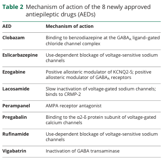
Table 2 Mechanism of action of the 8 newly approved antiepileptic drugs (AEDs) <table><thead><tr><th>AED</th><th>Mechanism of action</th></tr></thead><tbody><tr><td>Clobazam</td><td>Binding to benzodiazepine at the GABA<sub>A</sub> ligand-gated chloride channel complex</td></tr><tr><td>Eslicarbazepine</td><td>Use-dependent blockage of voltage-sensitive sodium channels</td></tr><tr><td>Ezogabine</td><td>Positive allosteric modulator of KCNQ2-5; positive allosteric modulator of GABA<sub>A</sub> receptors</td></tr><tr><td>Lacosamide</td><td>Slow inactivation of voltage-gated sodium channels; binds to CRMP-2</td></tr><tr><td>Perampanel</td><td>AMPA receptor antagonist</td></tr><tr><td>Pregabalin</td><td>Binding to the α2-δ protein subunit of voltage-gated calcium channels</td></tr><tr><td>Rufinamide</td><td>Use-dependent blockage of voltage-sensitive sodium channels</td></tr><tr><td>Vigabatrin</td><td>Inactivation of GABA transaminase</td></tr></tbody></table>
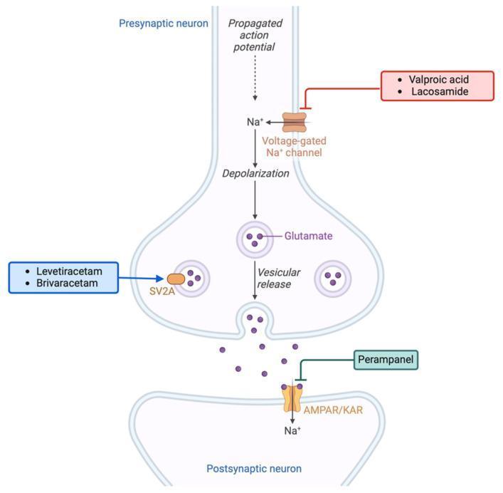
This pathophysiology diagram illustrates the excitatory glutamatergic synapse and the mechanism of action of various antiseizure medications (ASMs). The presynaptic neuron shows a propagated action potential leading to sodium (Na+) influx via voltage-gated Na+ channels and subsequent depolarization. This triggers the vesicular release of glutamate into the synaptic cleft. On the postsynaptic neuron, glutamate binds to AMPA/kainate receptors (AMPA/KAR), resulting in further Na+ influx. The diagram highlights three pharmacological intervention points: (1) Valproic acid and Lacosamide inhibiting voltage-gated Na+ channels; (2) Levetiracetam and Brivaracetam targeting the Synaptic Vesicle Protein 2A (SV2A) to modulate vesicular release; and (3) Perampanel acting as an antagonist at the postsynaptic AMPA/KAR receptors. This visual summary explains how these medications regulate neuronal hyperexcitability, which is clinically relevant in both epilepsy management and the treatment of peritumoral hyperexcitability in glioma patients.
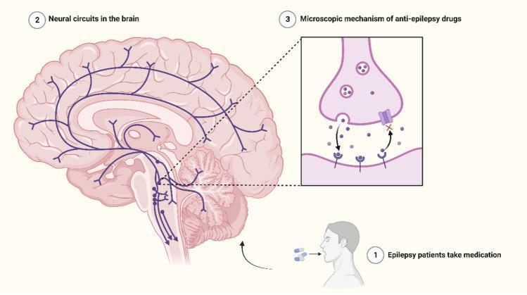
This medical infographic illustrates the pharmacological management of epilepsy at both the macro-anatomical and microscopic levels. The main panel presents a mid-sagittal and lateral perspective of the human brain, highlighting 'Neural circuits in the brain' with blue schematic lines. These pathways demonstrate connectivity between the brainstem, thalamus, and various cortical regions, representing the networks involved in seizure activity and regulation. A primary inset focuses on the 'Microscopic mechanism of anti-epilepsy drugs' at a synapse. It depicts a pre-synaptic terminal containing neurotransmitter vesicles, the synaptic cleft, and a post-synaptic membrane. The diagram shows neurotransmitters being released and moving toward receptors; however, a drug molecule is shown binding to a specific receptor or channel, effectively blocking synaptic transmission or modulating signal intensity. A supplementary icon shows a patient taking oral medication, linking systemic drug administration to these localized neural circuit interventions. This visual is designed for neurology and pharmacology education, emphasizing how targeted therapy suppresses epileptic seizures by regulating neuronal excitation and inhibition within specific cerebral axes.
Antiepileptic Drugs (AEDs)
Overview & Goals of Treatment
-
~70% of epilepsy patients achieve complete or near-complete seizure control with medications
-
~50% respond to the first drug tried as monotherapy; a further ~15% respond to a second agent
-
Remaining cases (~30%) are considered treatment-resistant epilepsy
-
Start with the lowest effective dose, using once- or twice-daily dosing when possible
-
Monotherapy is always preferred initially - polytherapy is rarely additive and multiplies side effects
-
Adams & Victor's Principles of Neurology 12th Ed.
Mechanisms of Action
1. Sodium Channel Blockade (Use-Dependent)
- Drugs: Phenytoin, Carbamazepine, Oxcarbazepine, Eslicarbazepine, Lamotrigine, Lacosamide (slow inactivation), Rufinamide, Valproate (partial)
2. GABA Enhancement
- Benzodiazepines - bind to GABA-A receptor (benzodiazepine site) → increased frequency of Cl⁻ channel opening
- Barbiturates (Phenobarbital) - bind to GABA-A receptor (barbiturate site) → increased duration of Cl⁻ channel opening
- Vigabatrin - irreversible inhibitor of GABA transaminase → increases synaptic GABA levels
- Tiagabine - inhibits GABA reuptake transporter (GAT-1)
- Clobazam - binds benzodiazepine site on GABA-A receptor
3. Calcium Channel Blockade
- Ethosuximide, Valproate - block low-threshold T-type Ca²⁺ channels in thalamic neurons (key mechanism in absence seizures)
- Gabapentin, Pregabalin - bind to α₂-δ subunit of voltage-gated Ca²⁺ channels → reduce glutamate/GABA release
4. Glutamate (Excitatory) Antagonism
- Perampanel - selective, non-competitive AMPA receptor antagonist
- Felbamate - blocks NMDA receptor (glycine site)
- Topiramate - blocks AMPA/kainate receptors (among multiple mechanisms)
5. Synaptic Vesicle Protein 2A (SV2A) Modulation
- Levetiracetam, Brivaracetam - bind SV2A on presynaptic vesicles → modulate neurotransmitter release; unique mechanism with broad-spectrum efficacy
6. Other / Multiple Mechanisms
- Valproate - multiple mechanisms: Na⁺ channel blockade, T-type Ca²⁺ channel blockade, increased GABA synthesis/decreased GABA degradation
- Topiramate - Na⁺ channel blockade + AMPA antagonism + GABA-A enhancement + carbonic anhydrase inhibition
- Ezogabine (Retigabine) - positive allosteric modulator of KCNQ2-5 potassium channels → membrane hyperpolarization
Classification: Generations of AEDs
First-Generation (Classical) AEDs
| Drug | Trade Name | Mechanism | Primary Indications | Key Adverse Effects |
|---|---|---|---|---|
| Phenytoin | Dilantin | Na⁺ channel blockade | Focal, GTC, status epilepticus | Nystagmus, ataxia, gingival hyperplasia, hirsutism, coarsening of facies, osteoporosis, teratogenic (fetal hydantoin syndrome), zero-order kinetics (narrow TI) |
| Carbamazepine | Tegretol | Na⁺ channel blockade | Focal, GTC; also trigeminal neuralgia | Diplopia, ataxia, hyponatremia (SIADH), leukopenia, SJS/TEN (esp. HLA-B*1502 in Asians), autoinduction, hepatotoxicity |
| Valproate | Depakote/Epilim | Na⁺ + Ca²⁺ block, ↑GABA | Broad-spectrum: GTC, absence, myoclonic, focal | Hepatotoxicity (fatal in <2 yr), teratogenic (neural tube defects - NTDs; avoid in pregnancy), weight gain, tremor, hair loss, pancreatitis, thrombocytopenia |
| Phenobarbital | Luminal | GABA-A (↑Cl⁻ duration) | GTC, focal, neonatal seizures | Sedation, cognitive dulling, tolerance, dependence, enzyme induction (CYP), teratogenic |
| Ethosuximide | Zarontin | T-type Ca²⁺ blockade | Absence seizures only | GI upset, hiccups, drowsiness; no effect on other seizure types |
| Primidone | Mysoline | GABA-A (metabolized to phenobarbital) | GTC, focal | Same as phenobarbital; also essential tremor |
Second-Generation AEDs
| Drug | Trade Name | Mechanism | Primary Indications | Key Adverse Effects |
|---|---|---|---|---|
| Lamotrigine | Lamictal | Na⁺ channel blockade | Focal, GTC, absence, Lennox-Gastaut; safe in pregnancy | SJS (especially with rapid titration or valproate co-use), dizziness, diplopia, rash |
| Oxcarbazepine | Trileptal | Na⁺ channel blockade | Focal, GTC | Hyponatremia, dizziness, diplopia; less enzyme induction than CBZ |
| Levetiracetam | Keppra | SV2A modulation | Broad-spectrum: focal, GTC, myoclonic, JME | Irritability, behavioral changes (most common), somnolence; no drug interactions, safe in pregnancy |
| Gabapentin | Neurontin | α₂-δ Ca²⁺ channel | Focal seizures; neuropathic pain, postherpetic neuralgia | Somnolence, ataxia, weight gain, edema |
| Pregabalin | Lyrica | α₂-δ Ca²⁺ channel | Focal seizures; neuropathic pain, fibromyalgia | Weight gain, edema, somnolence |
| Topiramate | Topamax | Na⁺ block + AMPA antagonist + GABA enhancement + CA inhibition | Focal, GTC, Lennox-Gastaut; migraine prophylaxis | Cognitive impairment ("Dope-amax"), kidney stones, weight loss, metabolic acidosis, glaucoma, oligohidrosis, teratogenic (oral clefts) |
| Tiagabine | Gabitril | GABA reuptake inhibitor | Adjunctive for focal seizures | Dizziness, tremor, GI; may worsen absence/myoclonic seizures |
| Vigabatrin | Sabril | GABA transaminase inhibitor | Infantile spasms (West syndrome), focal (adjunct) | Permanent visual field defects (retinal toxicity - requires regular ophthalmological monitoring) |
| Felbamate | Felbatol | NMDA antagonism | Lennox-Gastaut (reserved) | Aplastic anemia, hepatic failure (black box warnings - restricted use) |
| Zonisamide | Zonegran | Na⁺ + T-Ca²⁺ block, CA inhibition | Focal, GTC, myoclonic | Kidney stones, oligohidrosis, metabolic acidosis, weight loss |
Third-Generation (Newer) AEDs
| Drug | Trade Name | Mechanism | Primary Indications | Key Adverse Effects |
|---|---|---|---|---|
| Lacosamide | Vimpat | Slow inactivation of Na⁺ channels; CRMP-2 binding | Focal seizures (adjunct/monotherapy) | Dizziness, diplopia, headache, PR interval prolongation |
| Perampanel | Fycompa | AMPA receptor antagonist | Focal, GTC (adjunct) | Dizziness, somnolence, aggression/psychiatric effects |
| Brivaracetam | Briviact | SV2A (higher affinity than levetiracetam) | Focal seizures | Somnolence, dizziness; less behavioral side effects than levetiracetam |
| Eslicarbazepine | Aptiom | Na⁺ channel blockade (active metabolite of oxcarbazepine) | Focal seizures (monotherapy/adjunct) | Dizziness, hyponatremia, diplopia |
| Cenobamate | Xcopri | Na⁺ channel blockade + GABA-A positive modulator | Focal seizures | Somnolence, dizziness, drug interactions; Drug Reaction with Eosinophilia and Systemic Symptoms (DRESS) risk |
Pharmacokinetic Data (Key Values)
| Drug | Adult Dose (mg/day) | Half-life (h) | Therapeutic Level (µg/mL) |
|---|---|---|---|
| Valproic acid | 1,000-3,000 | 6-15 | 50-100 |
| Phenytoin | 300-400 | 12-36 | 10-20 |
| Carbamazepine | 600-1,200 | 14-25 | 4-12 |
| Phenobarbital | 90-200 | 40-120 | 15-40 |
| Lamotrigine | 300-500 | 15-60 | 2-7 |
| Levetiracetam | 500-3,000 | ~6-8 | - |
| Ethosuximide | 750-1,500 | 40-60 | 40-100 |
| Oxcarbazepine | 900-2,400 | 1-5 (prodrug) | - |
Long half-lives (phenytoin, phenobarbital, ethosuximide) → once-daily dosing (preferably at bedtime) Shorter half-lives (valproate, carbamazepine) → spaced dosing throughout the day
- Adams & Victor's Principles of Neurology 12th Ed., Table 15-6
Drug Choice by Seizure Type
Adults
| Seizure Type | First Choice | Second Choice | Notes |
|---|---|---|---|
| Focal (partial) | Carbamazepine, Levetiracetam, Lamotrigine | Oxcarbazepine, Valproate | Carbamazepine is classic first-line |
| Generalized Tonic-Clonic (GTC) | Valproate, Lamotrigine, Levetiracetam | Carbamazepine, Topiramate | Avoid CBZ/PHT in IGE (may worsen) |
| Absence | Ethosuximide (pure absence), Valproate | Lamotrigine | Ethosuximide for pure absence; valproate if GTC also present |
| Myoclonic | Valproate, Levetiracetam | Clonazepam, Lamotrigine | Avoid carbamazepine (worsens myoclonus) |
| Juvenile Myoclonic Epilepsy (JME) | Valproate, Levetiracetam | Lamotrigine, Topiramate | Lifetime treatment often required |
Children (Adams & Victor's Table 15-7)
| Seizure Type | First Choice | Second | Third |
|---|---|---|---|
| GTC | Valproate, Carbamazepine | Lamotrigine, Oxcarbazepine | Phenytoin |
| Myoclonic | Valproate, Levetiracetam | Lamotrigine | Phenobarbital, Clobazam |
| Absence | Valproate | Topiramate, Levetiracetam, Ethosuximide | Lamotrigine |
| Focal | Carbamazepine, Phenytoin | Valproate, Levetiracetam | Lamotrigine, Vigabatrin, Topiramate |
| Infantile Spasms (West) | ACTH, Vigabatrin | Valproate | Lamotrigine |
| Lennox-Gastaut | Valproate | Topiramate, Lamotrigine | Levetiracetam |
Drug Interactions
| Interaction | Effect |
|---|---|
| Valproate + Lamotrigine | Valproate inhibits lamotrigine metabolism → 2-fold increase in lamotrigine levels → SJS risk; must halve lamotrigine dose |
| Valproate + Phenobarbital | Both on CYP pathway → toxicity buildup |
| Carbamazepine epoxide + Valproate | VPA inhibits epoxide hydrolase → accumulation of toxic CBZ-epoxide → toxicity at "normal" CBZ levels |
| Carbamazepine (inducer) + OCPs | Reduces contraceptive efficacy |
| Phenytoin (inducer) | Induces CYP1A2, 2C9, 3A4 → reduces levels of warfarin, statins, OCPs, cyclosporine |
| Phenobarbital | Broad CYP induction similar to phenytoin |
| Levetiracetam | No hepatic metabolism, no protein binding → essentially no significant drug interactions |
Special Populations
Pregnancy
- Safest options: Lamotrigine, Levetiracetam (least teratogenic data)
- Most teratogenic: Valproate (neural tube defects, facial clefts, cognitive impairment in child - avoid if possible), Phenytoin (fetal hydantoin syndrome), Phenobarbital, Topiramate (oral clefts)
- All women of childbearing age on AEDs should take folic acid 5 mg/day
- Use the lowest effective dose; avoid polytherapy in pregnancy
Status Epilepticus (Acute Management)
- Benzodiazepines (lorazepam IV, diazepam IV/rectal, midazolam IM) - first line
- Fosphenytoin or Valproate IV - second line
- Levetiracetam IV - increasingly used as second line
- Phenobarbital IV - second line if above fail
- Anesthetic agents (propofol, midazolam infusion, thiopental) - refractory status
Important Side Effects at a Glance
| Side Effect | Drug(s) |
|---|---|
| Stevens-Johnson Syndrome / TEN | Carbamazepine (esp. HLA-B*1502), Lamotrigine, Phenytoin |
| Aplastic anemia + Liver failure | Felbamate (black box) |
| Visual field defects (permanent) | Vigabatrin |
| Gingival hyperplasia + hirsutism | Phenytoin |
| Hepatotoxicity (fatal, <2 yr) | Valproate |
| Neural tube defects | Valproate (highest risk), Carbamazepine |
| Hyponatremia (SIADH) | Carbamazepine, Oxcarbazepine, Eslicarbazepine |
| Weight gain | Valproate, Gabapentin, Pregabalin |
| Weight loss + kidney stones | Topiramate, Zonisamide |
| Cognitive impairment | Topiramate (most), Phenobarbital, Phenytoin |
| Behavioral/psychiatric effects | Levetiracetam (irritability), Perampanel (aggression) |
| Enzyme induction (CYP) | Phenytoin, Carbamazepine, Phenobarbital, Oxcarbazepine |
| Zero-order (saturation) kinetics | Phenytoin (narrow therapeutic index - small dose changes → large level changes) |
Mnemonic Summary
Anti cancer drugs for pharma
anticancer drug mechanism cell cycle chemotherapy classification
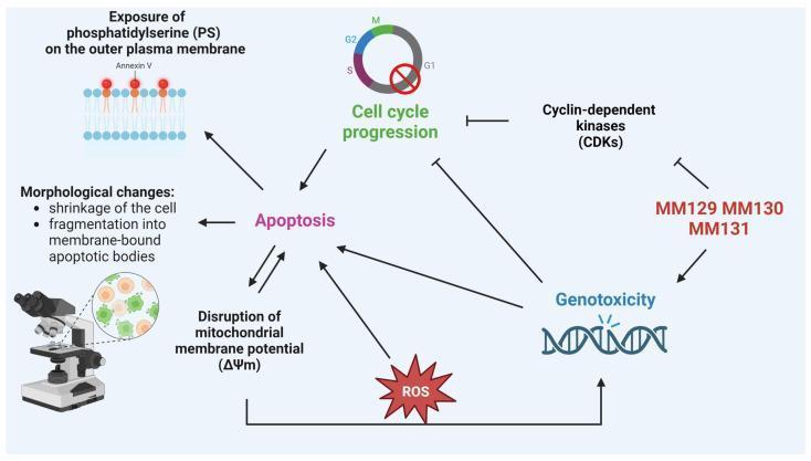
This pathophysiology diagram illustrates the anticancer mechanism of action for sulfonamide derivatives MM129, MM130, and MM131. The pathway depicts these compounds inhibiting cyclin-dependent kinases (CDKs) and inducing genotoxicity, represented by DNA strand breaks. These actions lead to a blockade of cell cycle progression (arrested at G1, S, G2, or M phases) and the direct induction of apoptosis. The diagram highlights several hallmark features of programmed cell death: the exposure of phosphatidylserine (PS) on the outer plasma membrane (detectable by Annexin V), disruption of mitochondrial membrane potential (ΔΨm), and characteristic morphological changes including cell shrinkage and fragmentation into membrane-bound apoptotic bodies. A positive feedback loop is shown where the loss of mitochondrial potential leads to the generation of reactive oxygen species (ROS), which in turn exacerbates genotoxicity and further promotes apoptosis. This illustration is designed for advanced biomedical education regarding signal transduction, oncology pharmacology, and cell death mechanisms.
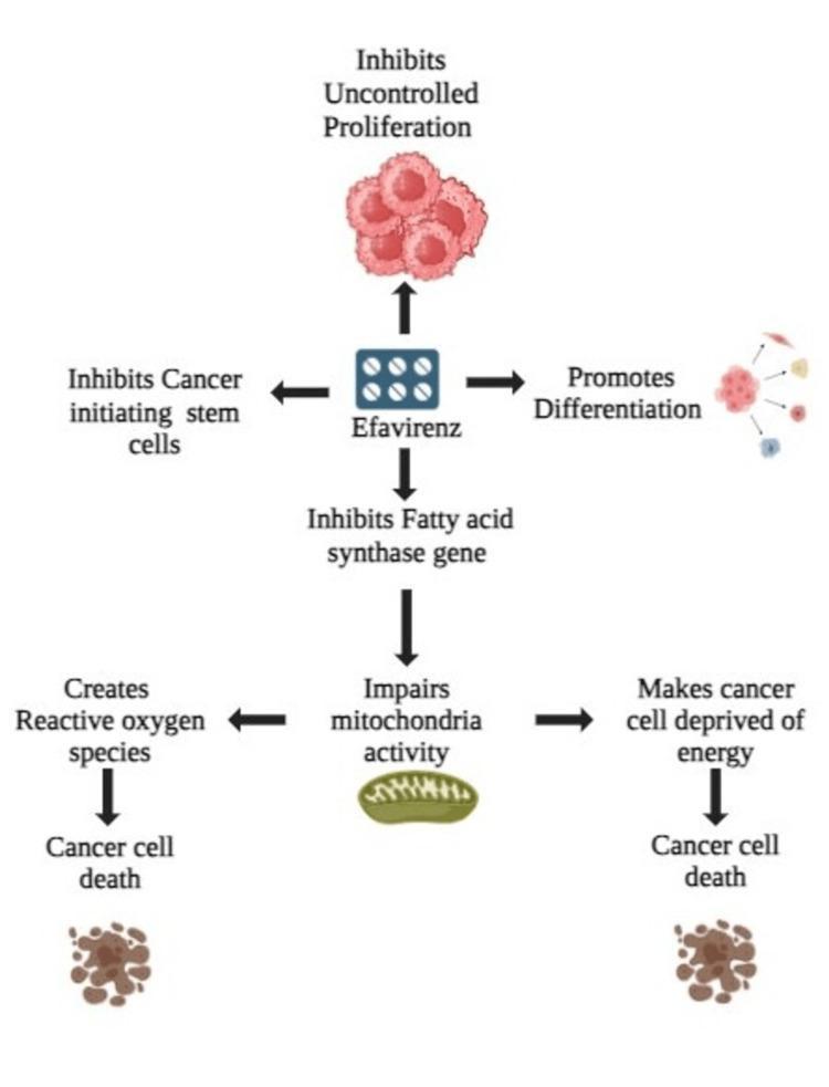
This pathophysiology diagram illustrates the multi-targeted anticancer mechanism of Efavirenz. The central element of the flow indicates the drug's impact on four primary biological processes: inhibition of uncontrolled proliferation (depicted as a dense red cell cluster), inhibition of cancer-initiating stem cells, promotion of cellular differentiation (visualized as a cell cluster diversifying into distinct types), and inhibition of the fatty acid synthase gene. The pathway then tracks the downstream consequences of fatty acid synthase inhibition, which leads to impaired mitochondrial activity, represented by an illustration of a mitochondrion. This mitochondrial dysfunction bifurcates into two detrimental cellular states: the creation of reactive oxygen species (ROS) and cellular energy deprivation. Both branches culminate in cancer cell death, represented visually by fragmented, necrotic brown cell clusters. The diagram serves as an educational summary of how an antiretroviral medication can be repurposed for oncology by disrupting metabolic and proliferative pathways essential for tumor survival and stemness.
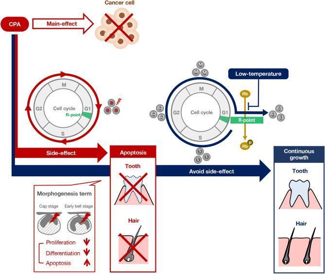
A pathophysiology diagram illustrating the mechanisms by which cyclophosphamide (CPA) induces dental and integumentary side effects and the mitigating role of a low-temperature approach. The diagram contrasts two pathways: one at physiological temperature (red) and one at low temperature (blue). In the standard pathway, CPA exerts its primary anticancer effect but simultaneously causes side effects by disrupting morphogenesis during cap and early bell stages. This leads to reduced proliferation/differentiation and increased apoptosis in tooth germs and hair follicles, visually depicted by crossed-out anatomical icons. A corresponding cell cycle diagram shows unrestricted progression through the G1/S/G2/M phases. In the low-temperature model, a protective mechanism is shown where cooling induces Rb protein-mediated inhibition at the G1 R-point (restriction point), arresting the cell cycle. This temporary stagnation prevents CPA from damaging actively dividing dental and hair cells, resulting in 'Continuous growth' and normal morphology of the tooth and hair structures. This summarizes the potential of cryotherapy to prevent chemotherapy-induced ectodermal organ damage.
Anticancer Drugs - Pharmacology
Classification Overview
- Alkylating Agents (and Platinum Complexes)
- Antimetabolites
- Antitumor Antibiotics
- Plant-Derived Agents (Vinca alkaloids, Taxanes, Topoisomerase inhibitors)
- Hormonal Agents (SERMs, Aromatase inhibitors, GnRH analogs, Androgens/Antiandrogens)
- Targeted / Biologic Agents (Tyrosine kinase inhibitors, Monoclonal antibodies, mTOR inhibitors)
- Miscellaneous (Asparaginase, Hydroxyurea, Procarbazine)
Cell Cycle Specificity
| Category | Cell Cycle Phase | Examples |
|---|---|---|
| Cell Cycle Non-Specific (CCNS) | Any phase | Alkylating agents, Nitrosoureas, Anthracyclines, Cisplatin, Dacarbazine |
| S-phase specific | DNA synthesis | Methotrexate, 5-FU, Cytarabine, Gemcitabine, 6-MP |
| M-phase specific | Mitosis (metaphase) | Vinca alkaloids (block polymerization), Taxanes (stabilize microtubules) |
| G2/M specific | G2 and M | Bleomycin, Etoposide, Taxanes |
| G1 specific | Gap 1 | Asparaginase, Steroids |
I. Alkylating Agents
- Myelosuppression and immunosuppression (dose-limiting)
- Toxicity to dividing mucosal cells and hair follicles (mucositis, alopecia)
- Delayed pulmonary fibrosis
- Reproductive toxicity (premature menopause, sterility)
- Leukemogenesis (secondary acute leukemia - up to 5% risk; highest with mechlorethamine, procarbazine, thiotepa)
A. Nitrogen Mustards
| Drug | Key Uses | Unique Toxicity / Notes |
|---|---|---|
| Mechlorethamine | Hodgkin lymphoma (MOPP), topical for cutaneous lymphoma | Vesicant - severe vascular damage on injection |
| Cyclophosphamide | NHL, Hodgkin, multiple myeloma, breast, ovarian, neuroblastoma, Wilms tumor, sarcoma, autoimmune diseases | Hemorrhagic cystitis (acrolein metabolite) - prevent with MESNA + hydration; activated by hepatic CYP450 |
| Ifosfamide | Germ cell testicular cancer, sarcomas | Hemorrhagic cystitis + neurotoxicity (chloroacetaldehyde metabolite); requires MESNA |
| Melphalan | Multiple myeloma, high-dose with BMT | Similar to cyclophosphamide; CNS seizures (neurotoxicity) |
| Chlorambucil | CLL, NHL, Waldenström's macroglobulinemia | Oral; least toxic nitrogen mustard |
| Bendamustine | CLL, NHL | Hybrid: nitrogen mustard + benzimidazole; lacks cross-resistance with classical alkylators |
B. Alkyl Sulfonates
| Drug | Key Uses | Unique Toxicity |
|---|---|---|
| Busulfan | CML, pre-BMT conditioning | Pulmonary fibrosis ("busulfan lung"), prolonged pancytopenia, skin hyperpigmentation, sterility |
C. Nitrosoureas
| Drug | Key Uses | Unique Toxicity |
|---|---|---|
| Carmustine (BCNU) | Malignant gliomas, Hodgkin lymphoma, BMT conditioning | Pulmonary toxicity, hepatotoxicity; also available as implantable wafer (Gliadel) for brain tumors |
| Lomustine (CCNU) | Brain tumors, Hodgkin lymphoma | Oral; prolonged myelosuppression (nadir 4-6 weeks) |
| Streptozocin | Pancreatic islet cell tumors | Diabetogenic (selectively toxic to β-cells); nephrotoxic |
D. Platinum Coordination Complexes
| Drug | Key Uses | Unique Toxicity / Notes |
|---|---|---|
| Cisplatin | Testicular (gold standard), ovarian, bladder, lung, head & neck | Nephrotoxicity (dose-limiting - distal tubule); Ototoxicity (high-frequency hearing loss); severe nausea/vomiting; peripheral neuropathy; requires aggressive pre- and post-hydration |
| Carboplatin | Ovarian, lung; alternative when cisplatin not tolerated | Myelosuppression (dose-limiting); less nausea, nephro-, neuro-, ototoxicity; dose by AUC (Calvert formula) |
| Oxaliplatin | Colorectal cancer (FOLFOX regimen) | Cold-induced peripheral neuropathy (acute, distinctive); cumulative peripheral neuropathy; mild myelosuppression |
- Lippincott Illustrated Reviews: Pharmacology
II. Antimetabolites
A. Folate Antagonists
| Drug | Mechanism | Key Uses | Adverse Effects |
|---|---|---|---|
| Methotrexate (MTX) | Inhibits dihydrofolate reductase (DHFR) → blocks conversion of DHF to THF → depletes tetrahydrofolate → blocks thymidylate and purine synthesis | ALL (maintenance), osteosarcoma, choriocarcinoma, lymphoma, breast cancer, RA, psoriasis (non-oncologic) | Myelosuppression, mucositis, hepatotoxicity, renal toxicity (high dose); Leucovorin rescue required with high-dose MTX |
| Pemetrexed | Inhibits multiple folate-dependent enzymes (DHFR, thymidylate synthase, GARFT) | Non-small cell lung cancer, mesothelioma | Myelosuppression, mucositis; supplement with folic acid + vitamin B12 to reduce toxicity; pretreat with corticosteroids |
B. Purine Analogs
| Drug | Mechanism | Key Uses | Notes |
|---|---|---|---|
| 6-Mercaptopurine (6-MP) | Thiol analog of hypoxanthine; incorporated as false metabolite into DNA/RNA; inhibits purine synthesis | ALL maintenance, Crohn disease | Prodrug; metabolized by xanthine oxidase → reduce dose with allopurinol (XO inhibitor) |
| 6-Thioguanine | Similar to 6-MP | AML | Also metabolized by XO |
| Fludarabine | Phosphate prodrug; triphosphate incorporated into DNA/RNA; inhibits DNA polymerase | CLL, low-grade NHL, hairy cell leukemia | S-phase specific; resistance via ↓ uptake or ↓ deoxycytidine kinase |
| Cladribine (2-CDA) | Purine analog resistant to adenosine deaminase | Hairy cell leukemia (drug of choice), CLL |
C. Pyrimidine Analogs
| Drug | Mechanism | Key Uses | Unique Points |
|---|---|---|---|
| 5-Fluorouracil (5-FU) | Fluorine at position 5 of uracil → converted to 5-FdUMP → inhibits thymidylate synthase → depletes thymidine (no DNA synthesis) | Colorectal, breast, gastric, pancreatic, ovarian cancers; topical for basal cell carcinoma | IV only (severe GI toxicity orally); enhanced by leucovorin (increases 5-FdUMP-TS binding) |
| Capecitabine | Oral prodrug of 5-FU; activated in tumor by thymidine phosphorylase → tumor-selective | Colorectal, metastatic breast cancer | Oral; unique toxicity: hand-foot syndrome (palmar-plantar erythrodysesthesia); less nausea/stomatitis than 5-FU |
| Cytarabine (Ara-C) | Pyrimidine antagonist; phosphorylated to ara-CTP → inhibits DNA polymerase; incorporated into DNA → chain termination | AML (drug of choice), ALL, CML blast crisis | S-phase specific; not oral (deaminated to inactive ara-U in gut); high-dose causes cerebellar toxicity |
| Gemcitabine | Nucleoside analog; triphosphate inhibits DNA polymerase and ribonucleotide reductase; incorporated into DNA | Pancreatic cancer, NSCLC, bladder, ovarian cancer | IV; "self-potentiating" (increases own active metabolite) |
III. Antitumor Antibiotics
| Drug | Mechanism | Key Uses | Unique Toxicity |
|---|---|---|---|
| Doxorubicin (Adriamycin) | DNA intercalation + Topoisomerase II inhibition + free radical generation | Breast, lung, ovarian, sarcoma, lymphoma, leukemia | Cumulative cardiotoxicity (dilated cardiomyopathy) at doses >450 mg/m²; red urine; myelosuppression; vesicant (severe extravasation injury) |
| Daunorubicin | Same as doxorubicin | AML, ALL | Cardiotoxicity limit: >550 mg/m²; vesicant |
| Epirubicin | Same as anthracyclines | Breast cancer | Cardiotoxicity limit: >900 mg/m² (less cardiotoxic); vesicant |
| Idarubicin | Same | AML | Limit: >150 mg/m²; risk of tumor lysis syndrome; vesicant |
| Bleomycin | Oxidative DNA strand breaks (bleomycin + Fe²⁺ → free radicals) | Testicular cancer (BEP), Hodgkin lymphoma | Pulmonary fibrosis ("bleomycin lung") - dose-limiting; skin hyperpigmentation; minimal myelosuppression (unique); active in G2/M phase |
| Dactinomycin (Actinomycin D) | DNA intercalation; blocks RNA polymerase | Wilms tumor, rhabdomyosarcoma, choriocarcinoma, Ewing sarcoma | Potent myelosuppression, mucositis, vesicant |
| Mitomycin C | Prodrug → alkylates DNA (behaves like alkylating agent) | Gastric, bladder (intravesical), colon | Myelosuppression (delayed), pulmonary toxicity, hemolytic uremic syndrome |
Cardiotoxicity prevention: Dexrazoxane (iron chelator) reduces anthracycline cardiotoxicity; liposomal doxorubicin has less cardiotoxicity.
IV. Plant-Derived Agents
A. Vinca Alkaloids (from Catharanthus roseus - periwinkle plant)
| Drug | Key Uses | Key Toxicity |
|---|---|---|
| Vincristine | ALL (children), Wilms tumor, Hodgkin/NHL, rhabdomyosarcoma, Ewing sarcoma; "O" in R-CHOP | Peripheral neuropathy (dose-limiting: paresthesias, areflexia, foot drop, constipation); minimal myelosuppression |
| Vinblastine | Testicular cancer (BEP/VBP), Hodgkin lymphoma, Kaposi sarcoma | Myelosuppression (dose-limiting); less neuropathy than vincristine |
| Vinorelbine | NSCLC, breast cancer | Myelosuppression, mild neuropathy |
CRITICAL: Vinca alkaloids cause DEATH if given intrathecally - never administer IT. Fatal neurotoxicity.
B. Taxanes (from Taxus - yew tree)
| Drug | Key Uses | Key Toxicity |
|---|---|---|
| Paclitaxel | Ovarian, breast, NSCLC, Kaposi sarcoma | Neutropenia (dose-limiting); peripheral neuropathy; hypersensitivity reactions (premedicate with dexamethasone + diphenhydramine + H2-blocker); alopecia |
| Docetaxel | Breast, NSCLC, prostate, gastric | Neutropenia; fluid retention/edema (pretreat with dexamethasone); peripheral neuropathy |
| Nab-paclitaxel (Abraxane) | Breast, pancreatic, NSCLC | Albumin-bound formulation - no Cremophor vehicle - fewer hypersensitivity reactions |
C. Topoisomerase Inhibitors
| Drug | Type | Mechanism | Key Uses | Toxicity |
|---|---|---|---|---|
| Etoposide (VP-16) | Epipodophyllotoxin | Inhibits Topoisomerase II → DNA strand breaks | Testicular, SCLC, lymphoma, AML | Myelosuppression, nausea, alopecia; secondary leukemia risk |
| Teniposide | Epipodophyllotoxin | Same as etoposide | ALL, brain tumors | Similar to etoposide |
| Irinotecan (CPT-11) | Camptothecin | Inhibits Topoisomerase I | Colorectal cancer (FOLFIRI), SCLC | Diarrhea (dose-limiting, early and late); myelosuppression; atropine for early cholinergic diarrhea |
| Topotecan | Camptothecin | Inhibits Topoisomerase I | Ovarian, SCLC | Myelosuppression |
V. Hormonal Agents
A. Selective Estrogen Receptor Modulators (SERMs)
| Drug | Mechanism | Uses | Adverse Effects |
|---|---|---|---|
| Tamoxifen | Estrogen antagonist in breast tissue; agonist in bone and endometrium | ER+ breast cancer (first-line), breast cancer prevention in high-risk women | Hot flashes, vaginal bleeding, thromboembolism; endometrial cancer (due to uterine agonism); visual changes |
| Raloxifene | Antagonist in breast AND uterus; agonist in bone | Prevention of ER+ breast cancer in postmenopausal women; osteoporosis | Hot flashes; no endometrial cancer risk (unlike tamoxifen) |
| Fulvestrant | Pure estrogen receptor downregulator/antagonist | Hormone receptor-positive metastatic breast cancer | Hot flashes, injection site reactions, elevated LFTs |
B. Aromatase Inhibitors (Postmenopausal women only)
| Drug | Type | Uses | Adverse Effects |
|---|---|---|---|
| Anastrozole | Non-steroidal | First-line breast cancer in postmenopausal women | Hot flashes, joint pain, osteoporosis, cardiovascular events; no endometrial cancer |
| Letrozole | Non-steroidal | Breast cancer, anovulatory infertility | Similar to anastrozole |
| Exemestane | Steroidal (irreversible) | Breast cancer | Similar; androgenic side effects |
C. GnRH Analogs (for Prostate Cancer)
| Drug | Mechanism | Use |
|---|---|---|
| Leuprolide, Goserelin | GnRH agonist - initially stimulates, then downregulates pituitary GnRH receptors → ↓ LH/FSH → ↓ testosterone ("medical castration") | Advanced prostate cancer |
Testosterone flare: Initial increase in testosterone before suppression. Use antiandrogen (flutamide) concurrently to block this.
D. Antiandrogens
| Drug | Mechanism | Use |
|---|---|---|
| Flutamide, Bicalutamide | Competitive AR antagonists | Prostate cancer |
| Enzalutamide | AR antagonist (also blocks nuclear translocation) | Castration-resistant prostate cancer |
VI. Targeted Agents (Key Examples)
| Drug | Target | Use | Key Toxicity |
|---|---|---|---|
| Imatinib (Gleevec) | BCR-ABL tyrosine kinase inhibitor | CML (revolutionary), GIST (c-KIT) | Fluid retention, myelosuppression, GI effects |
| Trastuzumab (Herceptin) | Anti-HER2/neu monoclonal Ab | HER2+ breast cancer, gastric cancer | Cardiotoxicity (synergistic with anthracyclines - avoid combination) |
| Rituximab | Anti-CD20 monoclonal Ab | B-cell lymphoma, CLL, RA | Infusion reactions, reactivation of HBV/TB |
| Bevacizumab (Avastin) | Anti-VEGF monoclonal Ab | Colorectal, lung, glioblastoma | Hypertension, impaired wound healing, thrombosis, GI perforation |
| Cetuximab | Anti-EGFR monoclonal Ab | Colorectal (KRAS wild-type), head & neck | Acneiform rash (biomarker of response), hypomagnesemia |
| Erlotinib, Gefitinib | EGFR tyrosine kinase inhibitor | NSCLC (EGFR mutation +) | Rash, diarrhea, interstitial lung disease |
| Vemurafenib, Dabrafenib | BRAF V600E inhibitor | Melanoma (BRAF mutant) | Paradoxical skin tumor induction, photosensitivity |
| Pembrolizumab, Nivolumab | Anti-PD-1 (checkpoint inhibitors) | Melanoma, NSCLC, many others | Immune-related adverse events (colitis, pneumonitis, hepatitis, endocrinopathies) |
VII. Miscellaneous Agents
| Drug | Mechanism | Key Use | Toxicity |
|---|---|---|---|
| Asparaginase | Depletes serum asparagine (tumor cells lack asparagine synthetase) | ALL | Anaphylaxis, pancreatitis, coagulation abnormalities, hepatotoxicity; no myelosuppression |
| Hydroxyurea | Inhibits ribonucleotide reductase → blocks deoxyribonucleotide synthesis (S-phase specific) | CML, sickle cell disease, polycythemia vera | Myelosuppression, mucositis, leg ulcers |
| Procarbazine | Methylates DNA; generates free radicals; MAO inhibitor | Hodgkin lymphoma (MOPP/BEACOPP) | Myelosuppression; disulfiram-like reaction with alcohol; MAO inhibition - avoid tyramine-rich foods; leukemogenesis |
| Thalidomide / Lenalidomide | Anti-angiogenic + immunomodulatory | Multiple myeloma | DVT/PE (use with aspirin/LMWH prophylaxis); teratogenic; peripheral neuropathy (thalidomide) |
Important Combination Regimens
| Regimen | Components | Used For |
|---|---|---|
| CHOP | Cyclophosphamide, Doxorubicin (H), Vincristine (O), Prednisone | NHL |
| R-CHOP | Rituximab + CHOP | B-cell NHL |
| ABVD | Doxorubicin (A), Bleomycin, Vinblastine, Dacarbazine | Hodgkin lymphoma |
| BEP | Bleomycin, Etoposide, Cisplatin (P) | Testicular cancer |
| FOLFOX | 5-FU + Leucovorin + Oxaliplatin | Colorectal cancer |
| FOLFIRI | 5-FU + Leucovorin + Irinotecan | Colorectal cancer |
| CMF | Cyclophosphamide, Methotrexate, 5-FU | Breast cancer |
| MOPP | Mechlorethamine, Vincristine (O), Procarbazine, Prednisone | Classic Hodgkin (historical) |
High-Yield Adverse Effects Summary
| Toxicity | Drug(s) |
|---|---|
| Cardiotoxicity (cardiomyopathy) | Doxorubicin, Daunorubicin, Trastuzumab (synergistic) |
| Pulmonary fibrosis | Bleomycin, Busulfan, Carmustine, Methotrexate, Cyclophosphamide |
| Hemorrhagic cystitis | Cyclophosphamide, Ifosfamide → prevent with MESNA |
| Peripheral neuropathy | Vincristine (severe), Cisplatin, Paclitaxel, Oxaliplatin |
| Nephrotoxicity | Cisplatin (prevent with hydration), Methotrexate, Ifosfamide |
| Ototoxicity | Cisplatin |
| Secondary leukemia (leukemogenesis) | Alkylating agents, Etoposide, Procarbazine |
| Endometrial cancer | Tamoxifen |
| Hand-foot syndrome | Capecitabine (most common), 5-FU, Sorafenib |
| Cold-induced neuropathy | Oxaliplatin |
| Minimal myelosuppression | Bleomycin, Asparaginase, Vincristine |
| Methotrexate toxicity rescue | Leucovorin (folinic acid) |
| 6-MP: reduce dose with allopurinol | 6-MP (allopurinol inhibits xanthine oxidase → ↑ 6-MP levels) |