
Explain structure and function of Long bone
Reading File
Now I'll search the medical library for long bone structure and function content, running parallel searches:
Finding Sources
Finding Sources
Good. Now let me read the primary histology content and fetch the diagram image:
Reading File
Reading File
Finding Sources
Reading File
Reading File
Reading File
Excellent content retrieved. Now let me fetch the key diagrams and search for osteon/Haversian canal details:
Loading Image
Loading Image
Finding Sources
Reading File
Finding Sources
Finding Sources
Loading Image
I now have comprehensive content from multiple authoritative textbooks along with excellent diagrams. Let me compile the full answer.
Structure and Function of Long Bone
What is a Long Bone?
Long bones are longer in one dimension than in any other, consisting of a shaft and two ends. Examples include the femur, tibia, fibula, humerus, radius, ulna, metacarpals, and phalanges.
Gross (Macroscopic) Structure
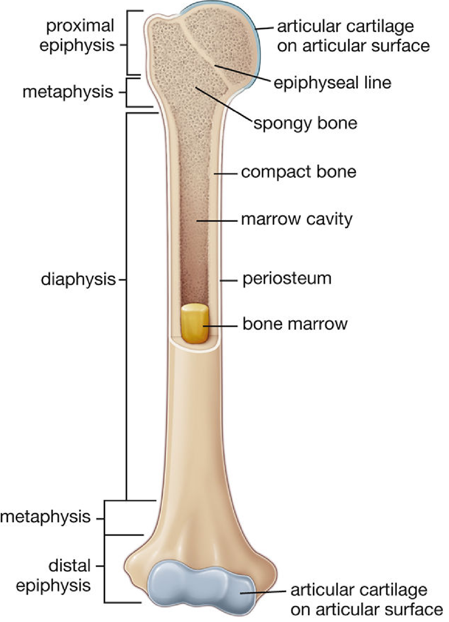
The diagram below, from Histology: A Text and Atlas with Correlated Cell and Molecular Biology, shows all major regions:
1. Diaphysis (Shaft)
The cylindrical midportion of the bone. In the adult, it is formed almost entirely of compact (cortical) bone, enclosing a large central medullary (marrow) cavity containing yellow bone marrow (mostly fat). A thin inner layer of spongy bone faces the medullary cavity. The diaphysis is the strongest region and withstands compressive and torsional forces.
2. Epiphyses (Ends)
The expanded proximal and distal ends of the bone. Each epiphysis consists chiefly of spongy (cancellous/trabecular) bone with only a thin outer shell of compact bone. The articular surfaces of the epiphyses are covered by smooth hyaline (articular) cartilage that reduces friction and absorbs shock at joints.
3. Metaphysis
The flared region between the diaphysis and the epiphysis. It contains a mix of compact and cancellous bone. In growing individuals, it is adjacent to the epiphyseal (growth) plate - a cartilaginous plate that is the site of longitudinal bone growth. After growth ceases, this plate ossifies into the epiphyseal line. The metaphysis has greater vascularity than the diaphysis, which is why it heals faster after fracture.
4. Epiphyseal (Growth) Plate
A cartilaginous plate present at the junction of the epiphysis and metaphysis in growing bones. This is the zone of endochondral ossification responsible for lengthening the bone. It is weaker than surrounding bone and susceptible to Salter-Harris fractures in children.
Coverings and Linings
5. Periosteum
The outer fibrocellular sheath covering all bone surfaces except articular cartilage. It consists of:
- An outer fibrous layer - dense connective tissue with collagen fibers and fibroblasts
- An inner osteogenic (cambium) layer - contains osteoprogenitor cells and osteoblasts, responsible for appositional (width) growth and fracture repair
The periosteum is anchored to the bone cortex by Sharpey's fibers - bundles of collagen that project from the periosteum into the outer layers of bone tissue. It is richly supplied with sensory nerves (responsible for bone pain) and blood vessels.
6. Endosteum
A thin cellular layer lining:
- The medullary cavity
- All trabecular surfaces of cancellous bone
- Haversian and Volkmann canals
It consists of osteogenic (progenitor) cells and thin reticular fibers, and is the primary site of bone resorption and remodeling.
Bone Tissue Types
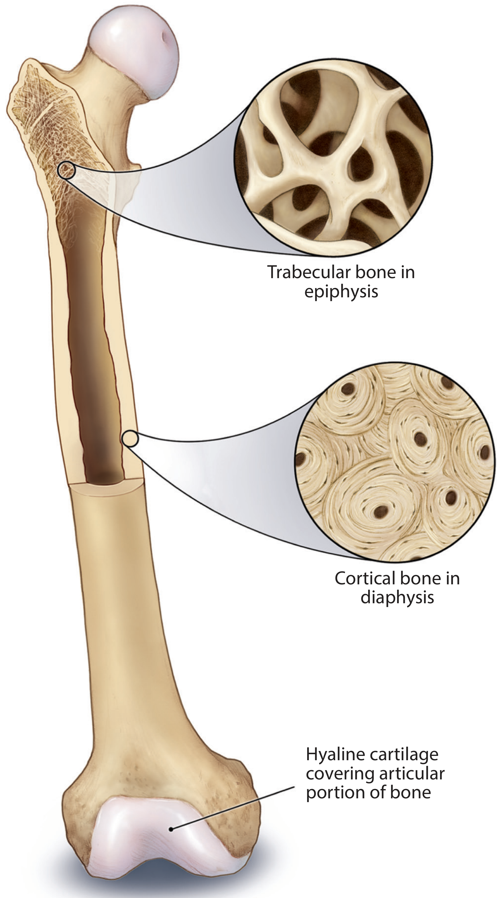
The diagram below shows the contrast between trabecular bone in the epiphysis and cortical bone in the diaphysis:
Compact (Cortical) Bone (~80% of skeletal mass)
- Forms the outer cortex; most abundant in the diaphysis shaft
- Organized into functional units called osteons (Haversian systems)
Osteon structure:
- A central Haversian canal containing blood vessels (capillaries), lymphatics, and nerves, running parallel to the long axis of the bone
- Surrounded by 5-20 concentric lamellae (rings of mineralized collagen fibers)
- Each lamella contains osteocytes (mature bone cells) sitting in spaces called lacunae
- Osteocytes extend cytoplasmic processes through tiny tunnels called canaliculi, connecting adjacent lacunae - this creates a communication network for nutrient/waste exchange via gap junctions
- The outer boundary of each osteon is the cement line (rich in noncollagen proteins and mineralized collagen)
Connecting canals:
- Volkmann's (perforating) canals - transverse channels connecting adjacent Haversian canals to each other and to the periosteum and medullary cavity; they carry blood vessels but lack concentric lamellae
Interstitial lamellae: Irregular remnants of previously remodeled osteons, filling spaces between intact osteons.
Cancellous (Spongy/Trabecular) Bone (~20% of skeletal mass)
- Forms the interior of the epiphyses and lines the inner shaft wall
- Consists of a 3D lattice of trabeculae (thin bony spicules/struts, usually <200 µm thick) separated by interconnecting marrow spaces filled with red marrow
- Trabeculae are oriented along lines of mechanical stress, providing maximum strength with minimal weight
- All trabecular surfaces are covered by endosteum
- No Haversian canals - osteocytes receive nutrients by diffusion directly from marrow spaces
| Feature | Compact Bone | Cancellous Bone |
|---|---|---|
| Also known as | Cortical bone | Spongy/trabecular bone |
| Location | Outer cortex, diaphysis | Interior of epiphyses, inner shaft |
| Organization | Osteons with Haversian canals | Trabeculae with marrow spaces |
| Proportion | ~80% of bone mass | ~20% of bone mass |
| Main function | Mechanical strength | Load distribution, hematopoiesis |
Bone Cells
| Cell | Origin | Function |
|---|---|---|
| Osteoprogenitor cells | Mesenchymal stem cells | Precursor to osteoblasts; activated during growth and repair |
| Osteoblasts | Osteoprogenitor cells | Secrete osteoid (organic bone matrix); once surrounded by matrix, become osteocytes |
| Osteocytes | Osteoblasts | Maintain bone viability; mechanosensing; regulate remodeling via gap junctions and canalicular network |
| Bone-lining cells | Quiescent osteoblasts | Remain on surfaces after deposition stops; initiate remodeling in response to mechanical stress |
| Osteoclasts | Hematopoietic progenitors (monocyte lineage) | Resorb bone matrix; found in Howship's lacunae; responsible for remodeling and calcium homeostasis |
Bone Matrix Composition
- Organic component (35%): Type I collagen (90%) + non-collagen proteins (BMPs, osteocalcin, osteopontin, etc.) - provides tensile strength and flexibility
- Inorganic component (65%): Hydroxyapatite crystals [Ca₁₀(PO₄)₆(OH)₂] - provides compressive strength and hardness; also stores calcium and phosphate
Functions of Long Bones
| Function | Mechanism |
|---|---|
| Mechanical support | Rigid framework that bears body weight and resists deformation |
| Movement | Serve as levers for skeletal muscle action; joints between bones allow locomotion |
| Protection | Encases and protects vital structures (e.g., bone marrow) |
| Mineral reservoir | Stores 99% of body calcium and 85% of body phosphate; released on demand via osteoclast activity to maintain serum calcium homeostasis |
| Hematopoiesis | Red marrow in cancellous bone (and in some diaphyses in children) produces all blood cell lineages |
| Fat storage | Yellow marrow in the adult diaphyseal cavity stores triglycerides as an energy reserve |
| Endocrine | Osteocalcin (secreted by osteoblasts) acts as a hormone regulating insulin secretion and energy metabolism; FGF-23 regulates phosphate balance |
Blood Supply
Long bones receive blood via three main routes:
- Nutrient artery - enters through the nutrient foramen in the diaphysis, supplies the medullary cavity and inner 2/3 of the cortex
- Metaphyseal and epiphyseal arteries - supply the ends of the bone; especially important for the growth plate
- Periosteal arteries - supply the outer 1/3 of the cortical bone
Sources: Histology: A Text and Atlas with Correlated Cell and Molecular Biology, pp. 582-584 | Junqueira's Basic Histology, pp. 365-371 | Forensic Anthropology: A Comprehensive Introduction, pp. 103-104 | Textbook of Family Medicine 9e, p. 810
This is a shared conversation. Sign in to Orris to start your own chat.