

7190.jpg
Answer for this question
Reading File
The image shows two essay questions. I'll answer both:
Question 2 (15M, 2025): Toxoplasmosis in pregnancy (cat litter box, fetal hydrocephalus, cervical lymphadenopathy)
Question 1 (15M, 2025): Typhoid fever (step-ladder fever, coated tongue, relative bradycardia, hepatosplenomegaly)
Searching sources...
Finding Sources
Finding Sources
Finding Sources
Finding Sources
Reading File
Reading File
Finding Sources
Finding Sources
Finding Sources
Finding Sources
Reading File
Reading File
Reading File
Reading File
I now have excellent source material. Let me also fetch the life cycle diagram for Toxoplasma and read the Typhoid diagnosis section.
Loading Image
Reading File
I now have all the information needed for both comprehensive answers. Let me compile them.
Question 2 (15M, 2025): Toxoplasmosis in Pregnancy
Clinical Scenario: 28-year-old pregnant woman (2nd trimester), mild fever, fatigue, cervical lymphadenopathy, cat litter box exposure, fetal hydrocephalus on anomaly scan.
a) Probable Clinical Condition
Congenital Toxoplasmosis (maternal toxoplasmosis with vertical transmission to the fetus).
The classic triad pointing to this diagnosis:
- Exposure to cat feces (litter box)
- Maternal lymphadenopathy and flu-like illness
- Fetal hydrocephalus (ventriculomegaly) on anomaly scan
b) Causative Organism, Mode of Transmission, and Life Cycle
Causative Organism: Toxoplasma gondii - an obligate intracellular protozoan parasite.
Mode of Transmission:
- Ingestion of oocysts from cat feces (contaminated soil, water, or litter box) - most relevant here
- Ingestion of bradyzoite-containing tissue cysts in raw/undercooked meat (pork, lamb, venison)
- Transplacental (vertical) transmission from infected mother to fetus
- Organ transplantation or blood transfusion from an infected donor
Life Cycle (see diagram below):
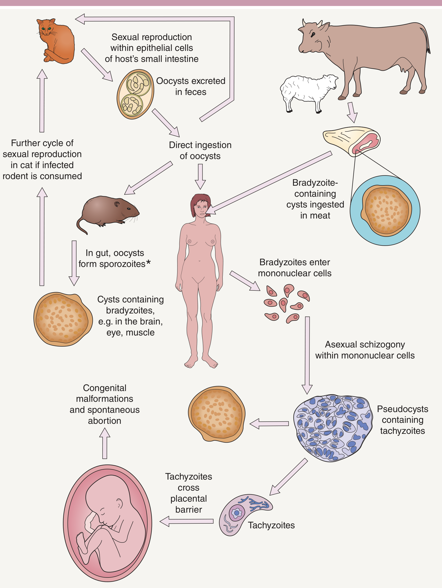
- Definitive host: Cats and wild felines (sexual reproduction occurs only in the intestinal epithelium of cats)
- In the cat's small intestine, sexual reproduction produces oocysts, which are excreted in feces
- Oocysts sporulate in the environment and become infective
- When ingested by an intermediate host (humans, rodents, livestock): oocysts release sporozoites in the gut → invade mononuclear cells → asexual schizogony → form pseudocysts containing tachyzoites (rapidly multiplying, acute phase)
- Tachyzoites disseminate hematogenously and can cross the placental barrier → congenital malformations
- Over time, tachyzoites encyst as bradyzoites in brain, eyes, and muscle (latent/chronic phase - tissue cysts)
- If a cat eats an infected intermediate host (e.g., rodent with bradyzoite cysts), the sexual cycle restarts in the cat intestine
(Sources: Dermatology 2-Volume Set 5e, Fig. 83.20; Sherris & Ryan's Medical Microbiology, 8th Ed.)
c) Risk Factors and Diagnostic Methods
Risk Factors:
- Owning/handling cats (especially indoor/outdoor cats that eat raw meat)
- Cleaning cat litter boxes (oocysts sporulate and become infective within 1-5 days)
- Eating raw or undercooked meat (pork, lamb, venison)
- Gardening without gloves (oocysts in soil)
- Immunosuppression (HIV/AIDS, transplant recipients) - risk of reactivation
- First-time infection during pregnancy (>90% of pregnant individuals have no prior immunity)
- Third trimester infection = highest risk of fetal transmission (~65%)
- First trimester infection = lower transmission rate but most severe fetal disease
Diagnostic Methods:
| Method | Notes |
|---|---|
| Serological testing (IgM/IgG) | IgM rises in acute infection; IgG appears 1-2 weeks later. IgM can persist >1 year - limits utility for timing |
| Avidity testing | High avidity IgG = infection >3-4 months ago (rules out recent acute infection) |
| PCR (amniotic fluid) | Gold standard for confirming fetal infection after 18 weeks; detects T. gondii DNA |
| Amniocentesis + PCR | To confirm whether the fetus is infected; guides treatment decisions |
| Fetal ultrasound | Hydrocephalus, periventricular calcifications, hepatosplenomegaly, ascites, microcephaly |
| Reference lab confirmation | Required before treating (high false-positive rate with standard serology) |
(Source: Creasy & Resnik's Maternal-Fetal Medicine)
d) Preventive Measures, Especially in Pregnancy
For all pregnant women (per Creasy & Resnik's Maternal-Fetal Medicine, and Sherris & Ryan's Medical Microbiology):
- Avoid cat litter boxes if at all possible. If unavoidable, change litter daily (oocysts take 1-5 days to sporulate and become infective), wear gloves, and wash hands thoroughly afterward
- Avoid raw/undercooked meat - cook meat until juices run clear (internal temperature >66°C / 150°F); freezing meat also destroys cysts
- Wash hands thoroughly after handling raw meat, gardening, or contact with soil
- Wash fruits and vegetables carefully to remove possible oocyst contamination
- Avoid unpasteurized milk and dairy products
- Keep pet cats indoors and feed them only commercial/cooked food (reduces their risk of infection)
- Wear gloves when gardening and avoid contact with soil potentially contaminated by cat feces
- Serological screening - routine screening not recommended in the US, but indicated in immunocompromised/HIV-positive pregnant women
If maternal infection is confirmed:
- Spiramycin (1 g PO three times daily) - reduces late sequelae; continue throughout gestation unless fetal infection confirmed
- If fetal infection confirmed by PCR: Switch to pyrimethamine + sulfadiazine + folic acid
- Neonates with congenital toxoplasmosis: Treat with pyrimethamine + sulfadiazine + leucovorin for 1 year
Question 1 (15M, 2025): Typhoid Fever
Clinical Scenario: 22-year-old, step-ladder fever ×7 days, abdominal pain, malaise, constipation, coated tongue, mild hepatosplenomegaly, relative bradycardia (pulse-temperature dissociation). Blood sent for culture and serology.
a) Probable Clinical Condition
Enteric (Typhoid) Fever, caused most likely by Salmonella enterica serovar Typhi.
Key diagnostic clues:
- Step-ladder fever (gradually rising fever over days)
- Relative bradycardia (slow pulse despite high fever - pathognomonic)
- Constipation (not diarrhea, in the early phase)
- Coated tongue (thick white coating - "typhoid tongue")
- Hepatosplenomegaly (involvement of RES)
- Young adult, likely in an endemic area
b) Mode of Transmission and Laboratory Diagnosis
Mode of Transmission:
Salmonella Typhi is a strictly human pathogen; chronic carriers are the primary reservoir.
- Fecal-oral route - ingestion of food or water contaminated with feces from infected individuals or carriers
- Infecting dose: ~10⁵ to 10⁶ bacteria
- Chronic carriers (especially those with gallstones) shed the organism in bile into feces for years
- Contaminated water supplies in endemic areas are a major vehicle
Pathogenesis (for context): After ingestion → S. Typhi passes through intestinal epithelium via M cells overlying Peyer's patches → enters lymphatics → bloodstream → spreads to liver, spleen, bone marrow (RES) → multiplies in macrophages (inhibits oxidative burst) → seeds bloodstream continuously → LPS endotoxin triggers step-wise fever → biliary tree reinfects intestine (~2-week cycle).
Laboratory Diagnosis:
| Test | Timing & Notes |
|---|---|
| Blood culture | Positive in weeks 1-2 (first 7-10 days); most sensitive method (~80% sensitivity in 1st week). Best early test. |
| Bone marrow culture | Most sensitive (~90%), positive even after antibiotics started; gold standard |
| Stool culture | Positive from 2nd week onward; earlier cultures usually negative |
| Urine culture | Positive from 2nd-3rd week |
| Widal test (serology) | Detects agglutinating antibodies to O (somatic) and H (flagellar) antigens of S. Typhi. O agglutinins appear by end of 1st week; rising titers in paired samples (4-fold rise) are significant. Limitations: low specificity, cross-reactions with other Salmonellae, prior vaccination can give false positives |
| Typhidot / Tubex | Rapid serological tests detecting anti-LPS IgM; faster than Widal |
| PCR | Detects S. Typhi DNA in blood; highly specific but not widely available |
| CBC | Characteristically shows leukopenia with relative lymphocytosis (unlike most bacterial infections); may also see mild anemia and thrombocytopenia |
(Sources: Jawetz, Melnick & Adelberg's Medical Microbiology 28e; Sherris & Ryan's Medical Microbiology 8e; Harrison's Principles of Internal Medicine 22e)
c) (Truncated in image - likely: "Write a note on treatment/complications/prevention")
Treatment:
- First-line: Fluoroquinolones (ciprofloxacin, ofloxacin) for 10-14 days - effective for susceptible strains
- For resistant strains (MDR typhoid): Azithromycin (oral) or Ceftriaxone/Cefixime (3rd gen cephalosporins)
- Dexamethasone for severe/complicated typhoid (encephalopathy, high fever)
Complications (if untreated):
- Intestinal hemorrhage (from Peyer's patch necrosis)
- Intestinal perforation (week 2-3; most feared)
- Myocarditis, encephalopathy, disseminated intravascular coagulation (DIC)
- Carrier state (~1-3% become chronic carriers)
Prevention:
- Safe water supply and proper sewage disposal
- Food hygiene and handwashing
- Identification and treatment of chronic carriers
- Vaccination: Two licensed vaccines available:
- Oral live attenuated Ty21a vaccine (3-4 doses)
- Vi capsular polysaccharide (ViCPS) parenteral vaccine (single dose)
This is a shared conversation. Sign in to Orris to start your own chat.