
Gerd
"gastroesophageal reflux"[MeSH Terms]
gastroesophageal reflux disease endoscopy esophagitis Barrett esophagus
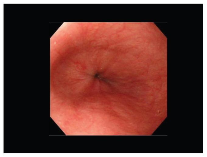
An endoluminal photograph captured during an upper gastrointestinal endoscopy, showing the distal esophagus and the lower esophageal sphincter (LES) region. The esophageal mucosa is smooth, glistening, and pinkish-red, with a normal vascular pattern and no evidence of mucosal breaks, erosions, or ulcerations. The LES appears as a central, closed punctate opening without evidence of widening or sliding hiatal hernia. There is no visible evidence of Barrett’s esophagus, esophagitis, or leukoplakia (whitish mucosa). This clinical image represents a normal endoscopic finding of the esophagus in a patient presenting with symptoms of gastroesophageal reflux disease (GERD), serving as a baseline for differentiating between non-erosive reflux disease (NERD) and erosive esophagitis.
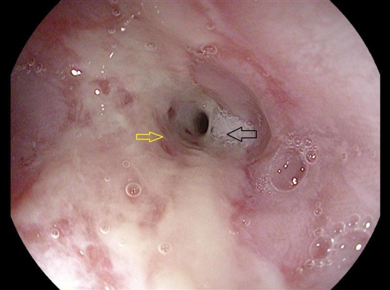
This diagnostic endoscopic image captures the luminal view of the esophagus, demonstrating classic findings of chronic gastroesophageal reflux disease (GERD) complications. A yellow arrow indicates areas of erosive esophagitis, characterized by erythematous, irregular mucosal lesions and visible friability of the esophageal lining. Distal to these inflammatory changes, a black arrow highlights a significant circumferential esophageal stricture. The stricture is visible as a fixed, concentric narrowing of the lumen, which reduces the internal diameter and restricts the passage of the endoscope. The surrounding mucosa appears thickened and pale pinkish-white, with scattered small air bubbles (saliva) and evidence of chronic tissue remodeling. This visual representation serves as a key educational example of peptic stricture formation resulting from untreated erosive esophagitis and is highly relevant for gastroenterology training in diagnostic endoscopy and the assessment of Barrett's esophagus.
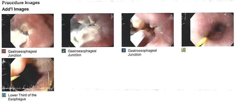
This set of five diagnostic endoscopic images documents an upper digestive endoscopy focused on the esophagus and gastroesophageal junction (GEJ). Images 1, 2, and 3 specifically illustrate the GEJ, showing significant mucosal irregularities including whitish, plaque-like areas or exudates overlaying erythematous tissue. These findings are clinically consistent with inflammatory changes, such as esophagitis or potentially post-ablative scarring in a patient with a history of Barrett’s esophagus. Images 4 and 5 display the lower third of the esophagus, maintaining a patent lumen with pink mucosa. A thin, yellow medical instrument or probe is visible within the esophageal lumen in the lower right and bottom images, likely used for measurement or guidance during the diagnostic procedure. The compilation serves to teach endoscopic identification of GEJ pathology and mucosal surface evaluation in the context of chronic gastroesophageal reflux disease (GERD) and its complications.
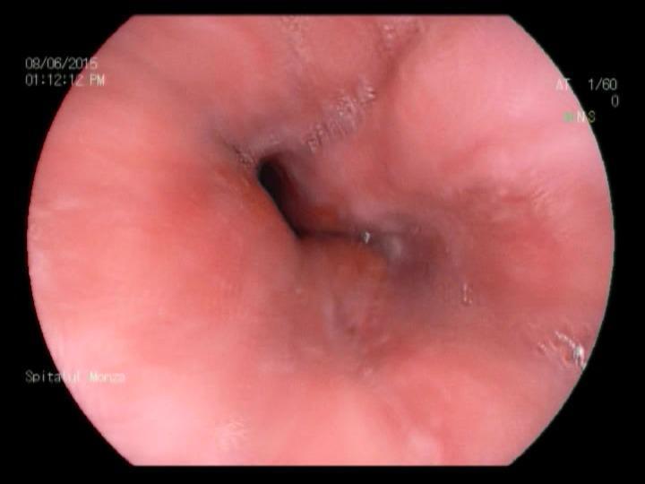
This diagnostic image is an upper gastrointestinal endoscopy (EGD) photograph of the human esophagus under white light imaging. The image displays the esophageal lumen and the surrounding mucosa. The mucosa is predominantly pink but exhibits subtle signs of minimal esophagitis, characterized by mild erythema (reddish hue) and slight blurring of the normal vascular pattern. The texture is relatively smooth, though there are fine, non-confluent linear markings and slight longitudinal ridges along the walls, indicating early mucosal irritation. The central lumen is patent but appears irregularly shaped due to mucosal redundancy and minor fold thickening. There are no macroscopic signs of deep ulcerations, strictures, or Barrett's esophagus visible in this view. This clinical image is representative of Grade A esophagitis according to the Los Angeles Classification, demonstrating minimal mucosal breaks. It serves as an educational tool for gastroenterology and internal medicine to distinguish between normal esophageal lining and early inflammatory changes associated with gastroesophageal reflux disease (GERD).
Gastroesophageal reflux disease (GERD)
Pathophysiology
- Lower esophageal sphincter (LES): Low LES pressure, transient LES relaxations, and a shortened intra-abdominal LES length all promote reflux. A hiatal hernia worsens this.
- Obesity: Obesity raises intra-abdominal pressure, lowers LES pressure, increases gastric acid production, and impairs esophageal motility. Higher estrogen levels may add to acid exposure. The association is stronger in women and in Caucasians. - Yamada's Textbook of Gastroenterology
- Obesity raises the odds of GERD (OR 2.15), erosive esophagitis (OR 1.87), and Barrett's esophagus (OR about 4.0). It also raises the risk of esophageal adenocarcinoma.
- In another meta-analysis, GERD odds were 1.43 for overweight and 1.94 for BMI above 30. - Sleisenger and Fordtran's GI and Liver Disease
- Weight loss improves symptoms, and this was shown in an intervention trial.
- Other contributors: Delayed gastric emptying, smoking, alcohol, pregnancy, and some drugs (anticholinergics, nitrates, calcium channel blockers).
Clinical features
- Typical: Heartburn and regurgitation.
- Extra-esophageal: Chronic cough, hoarseness, laryngitis, asthma, dental erosion, and globus.
- Alarm features (need endoscopy): Dysphagia, odynophagia, weight loss, GI bleeding or anemia, persistent vomiting, and age over about 50 with new symptoms.
Complications
Diagnosis
- Patients with typical symptoms and no alarm features can have an empiric PPI trial.
- Upper endoscopy is used for alarm features, refractory symptoms, or long-standing disease. Esophagitis is graded by the Los Angeles classification (A to D), and biopsies are taken if Barrett's esophagus is suspected.
- Ambulatory pH or pH-impedance monitoring is used when the diagnosis is uncertain, especially in non-erosive disease or when a patient is on no therapy and is being considered for surgery.
- Manometry is done before antireflux surgery to exclude achalasia and other motility disorders.
Management
- Lifestyle: Lose weight, raise the head of the bed, avoid late meals, and avoid personal trigger foods, smoking, and alcohol.
- Medical therapy (Katzung's Basic and Clinical Pharmacology, 16th ed.):
- Infrequent heartburn (fewer than 3 times a week): Antacids or intermittent H2 blockers. Antacids work fast but last only 1 to 2 hours. H2 blockers last 6 to 10 hours and can be taken before meals.
- Frequent heartburn: Twice-daily H2 blockers or PPIs.
- Erosive esophagitis: PPIs are preferred. H2 blockers heal fewer than 50% of cases.
- A standard PPI course is once daily, 30 to 60 minutes before a meal, for 8 weeks. Use the lowest effective dose for maintenance.
- Surgical or endoscopic options: Laparoscopic fundoplication, magnetic sphincter augmentation, or bariatric surgery (Roux-en-Y gastric bypass) in obese patients. These suit patients with PPI-refractory regurgitation, objective evidence of GERD, or those who do not want long-term drugs.
- Barrett's esophagus: Treat with PPI and endoscopic surveillance. Dysplasia is treated with endoscopic ablation or resection.
Recent evidence check
- Antireflux surgery for reflux-related cough (PMID 39206849)
- Laryngopharyngeal reflux, GERD, and dental erosion (PMID 39826062)
- GERD after esophageal atresia surgery (PMID 40775173)
- PPI dosing and long-term safety
- Barrett's esophagus surveillance
- Surgical options
- Pediatric GERD
- GERD in pregnancy
- Exam-style notes