
Histology diagrams topic for GIT, urinary system
Reading File
Now let me search the library for histology diagrams on GIT and urinary system simultaneously.
Finding Sources
Finding Sources
Finding Sources
Finding Sources
Finding Sources
Finding Sources
Two histology textbooks are available. Let me search them for GIT and urinary system histology sections.
Finding Sources
Finding Sources
Excellent! Now let me read these key sections from both textbooks in parallel.
Reading File
Reading File
Reading File
Reading File
I have the content. Now let me fetch the key diagrams from both sections.
Reading File
Reading File
I have the diagrams and content. Let me now fetch the key images in parallel.
Loading Image
Loading Image
Loading Image
Let me get the complete kidney image URL and more GIT diagrams.
Reading File
Reading File
Now let me fetch the key diagrams - kidney cross section, nephron, and additional GIT images.
Loading Image
Loading Image
Reading File
Excellent! Now let me fetch more key diagrams - renal corpuscle and GIT stomach/small intestine diagrams.
Loading Image
Reading File
Now let me get the stomach and small intestine diagrams from the GIT chapter.
Reading File
Reading File
Loading Image
Reading File
I now have extensive content and diagrams. Let me fetch one more important diagram - the jejunum plicae/villi and then compile the comprehensive answer.
Loading Image
I now have all the diagrams and content needed. Here is the full comprehensive histology reference for GIT and Urinary System:
Histology Diagrams: GIT & Urinary System
Source: Junqueira's Basic Histology, 17e & Histology: A Text and Atlas (Pawlina & Ross)
PART 1 - GASTROINTESTINAL TRACT (GIT)
Overview: The Digestive System
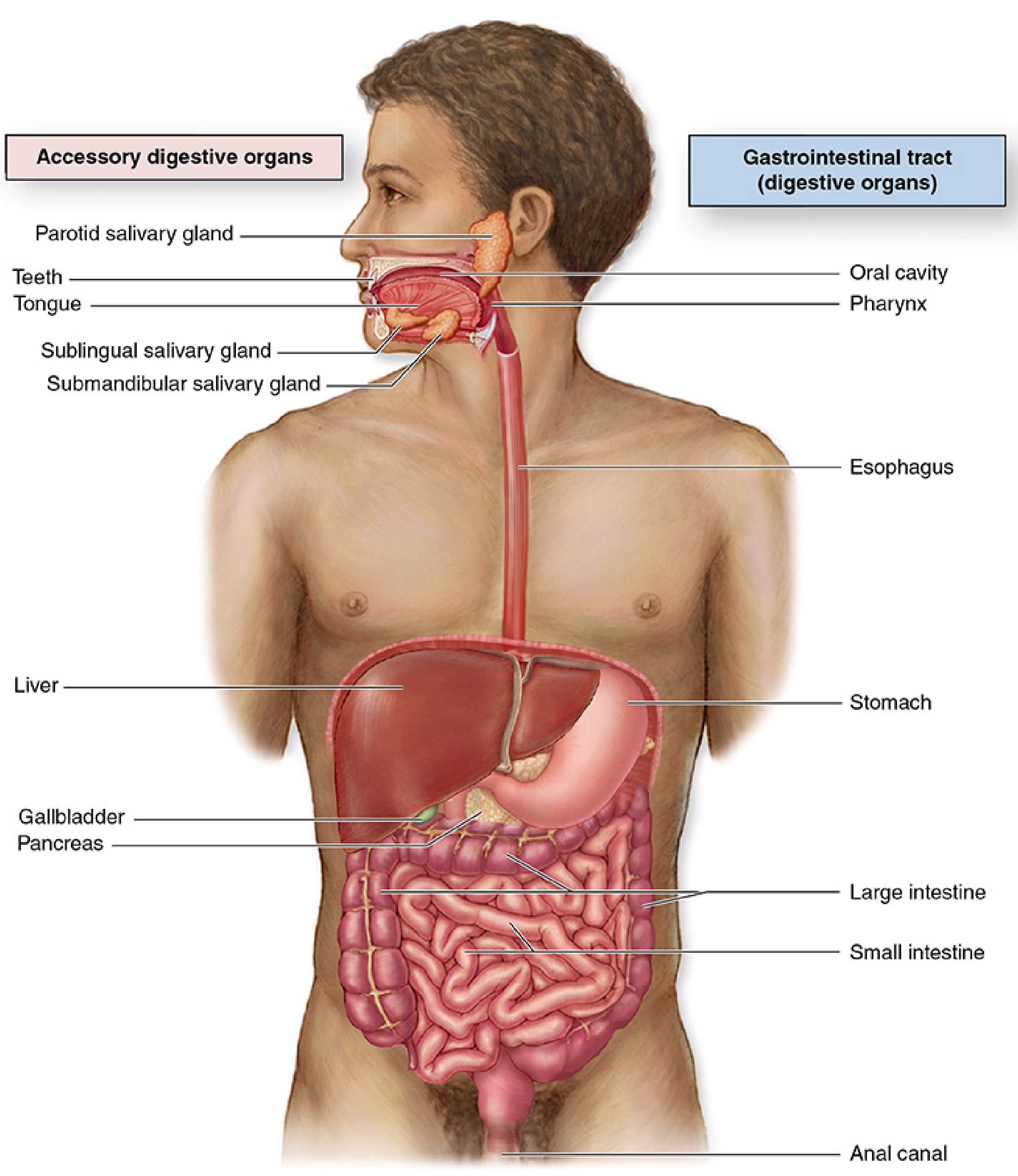
The GI tract (alimentary canal) runs from the oral cavity to the anus. The accessory digestive organs include the salivary glands, liver, gallbladder, and pancreas. The tract itself includes: oral cavity, pharynx, esophagus, stomach, small intestine (duodenum, jejunum, ileum), large intestine, rectum, and anal canal.
Diagram 1 - General Layers of the GI Tract Wall
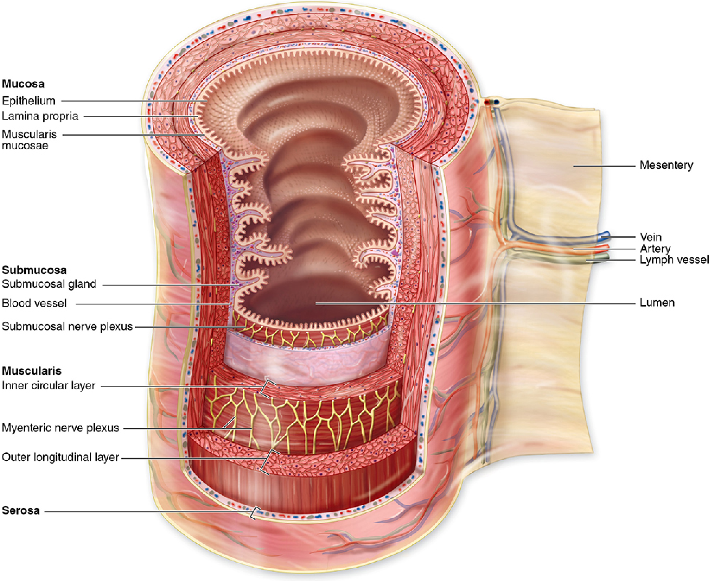
From the esophagus to the rectum, the wall of the GI tract has four main layers:
| Layer | Components | Key Points |
|---|---|---|
| Mucosa | Epithelium + Lamina propria + Muscularis mucosae | Varies regionally; contains glands and lymphoid tissue |
| Submucosa | Dense CT + Meissner's (submucosal) nerve plexus | May contain glands (Brunner's in duodenum) and Peyer's patches (ileum) |
| Muscularis (externa) | Inner circular + Outer longitudinal smooth muscle; Auerbach's (myenteric) plexus between them | Striated in upper esophagus; generates peristalsis |
| Serosa/Adventitia | Simple squamous mesothelium (serosa) or loose CT (adventitia) | Serosa where peritoneum covers; adventitia elsewhere (e.g. esophagus) |
Diagram 2 - Stomach Wall (H&E, Low Power)
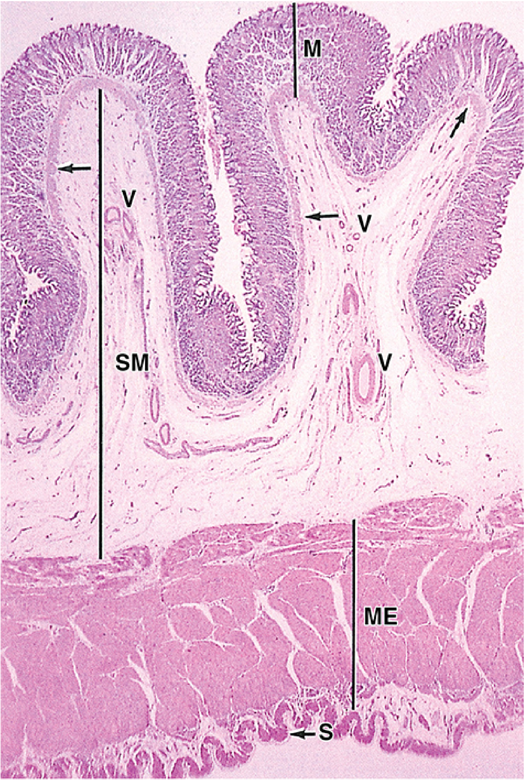
The image shows the fundic stomach in cross section. Key features:
- M (Mucosa): Packed with branching gastric glands; muscularis mucosae (arrows) visible at its base
- SM (Submucosa): Loose connective tissue with blood vessels (V)
- ME (Muscularis externa): Thick smooth muscle (inner oblique + middle circular + outer longitudinal in stomach)
- S (Serosa): Thin outermost layer
Key cellular components of gastric glands (fundus/body):
| Cell Type | Location | Function |
|---|---|---|
| Surface mucous cells | Lumen + gastric pits | Secrete thick mucus + HCO₃⁻ (protective) |
| Mucous neck cells | Neck of gland | Secrete less alkaline mucus; stem cell precursors |
| Parietal cells | Upper/middle gland | Secrete HCl + intrinsic factor; packed with mitochondria |
| Chief (zymogenic) cells | Lower gland | Secrete pepsinogen |
| Enteroendocrine cells | Scattered | Peptide hormones (gastrin, somatostatin, etc.) |
The cardiac and pyloric regions have only mucous cells in their glands - no parietal or chief cells.
Diagram 3 - Small Intestine: Plicae Circulares & Villi (Jejunum, Masson Trichrome)
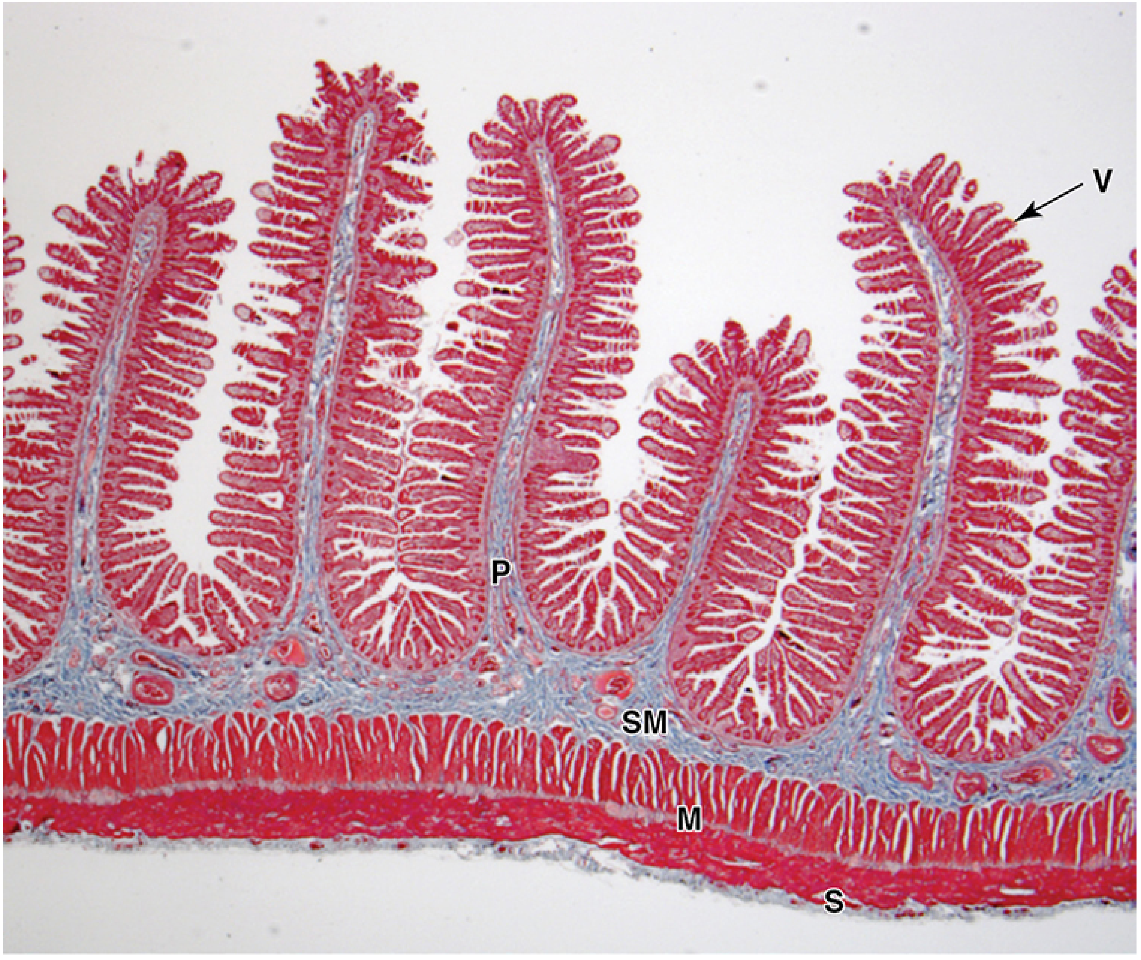
The small intestine has three surface-amplifying structures that multiply the absorptive area ~600-fold total:
- Plicae circulares (P) - folds of mucosa + submucosa; most developed in jejunum
- Villi (V) - finger-like projections of mucosa; increase area 10x
- Microvilli (brush border) - on enterocyte apical surface; increase area 20x
Cells of the small intestinal epithelium:
- Enterocytes - tall absorptive cells; prominent brush border; absorb sugars, amino acids, fatty acids
- Goblet cells - secrete mucus
- Paneth cells - deep in gland crypts; secrete defensins (antimicrobial)
- Enteroendocrine cells - hormone-secreting
- M cells - overlie Peyer's patches; sample luminal antigens
Regional differences in small intestine:
| Region | Distinguishing Feature |
|---|---|
| Duodenum | Brunner's glands in submucosa (mucus + HCO₃⁻ to neutralize acid) |
| Jejunum | Tallest villi, most plicae; highest absorption rate |
| Ileum | Peyer's patches (lymphoid nodules in mucosa/submucosa) |
Large Intestine - Key Histological Features
- No villi - flat mucosal surface
- Abundant goblet cells in simple tubular glands (crypts of Lieberkühn)
- No Paneth cells, no Brunner's glands
- Functions: water/electrolyte absorption, mucus production
- Appendix has prominent lymphoid aggregates in mucosa/submucosa
Esophagus - Summary
- Epithelium: Non-keratinized stratified squamous throughout most of its length
- Muscularis: Upper third = striated; middle = mixed; lower third = smooth muscle
- Outer layer: Adventitia (no serosa, as it lies in mediastinum)
- Abrupt change at esophagogastric junction to simple columnar epithelium (Z-line)
PART 2 - URINARY SYSTEM
Overview
The urinary system consists of: paired kidneys → ureters → urinary bladder → urethra.
Functions of the kidneys include:
- Regulation of water, electrolytes, and acid-base balance
- Excretion of metabolic wastes
- Secretion of renin, erythropoietin
- Activation of vitamin D to calcitriol
- Gluconeogenesis during prolonged fasting
Diagram 4 - Kidney: Cross-Section (Gross Histology)
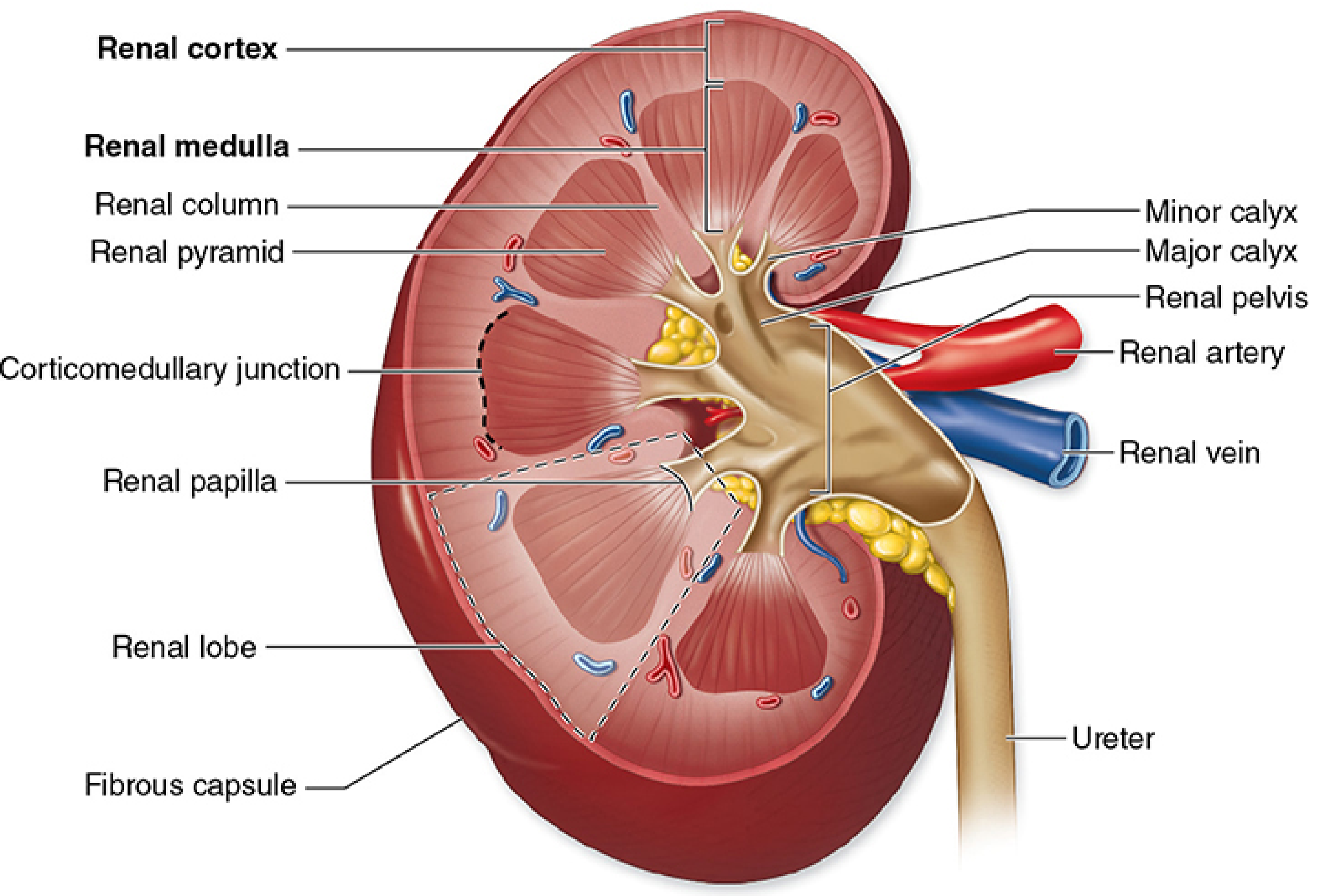
| Structure | Description |
|---|---|
| Renal cortex | Outer region; contains glomeruli, PCT, DCT - appears granular |
| Renal medulla | Inner region; contains loops of Henle, collecting ducts - appears striated |
| Renal pyramids | Conical medullary units; base at corticomedullary junction, apex = renal papilla |
| Renal columns | Extensions of cortex between pyramids |
| Renal papilla | Apex of each pyramid; drains urine into minor calyx |
| Minor/Major calyces | Collect urine from papillae; merge to form renal pelvis |
| Renal pelvis | Upper expanded part of ureter within hilum |
| Fibrous capsule | Thin outer covering |
Diagram 5 - The Nephron and Its Parts
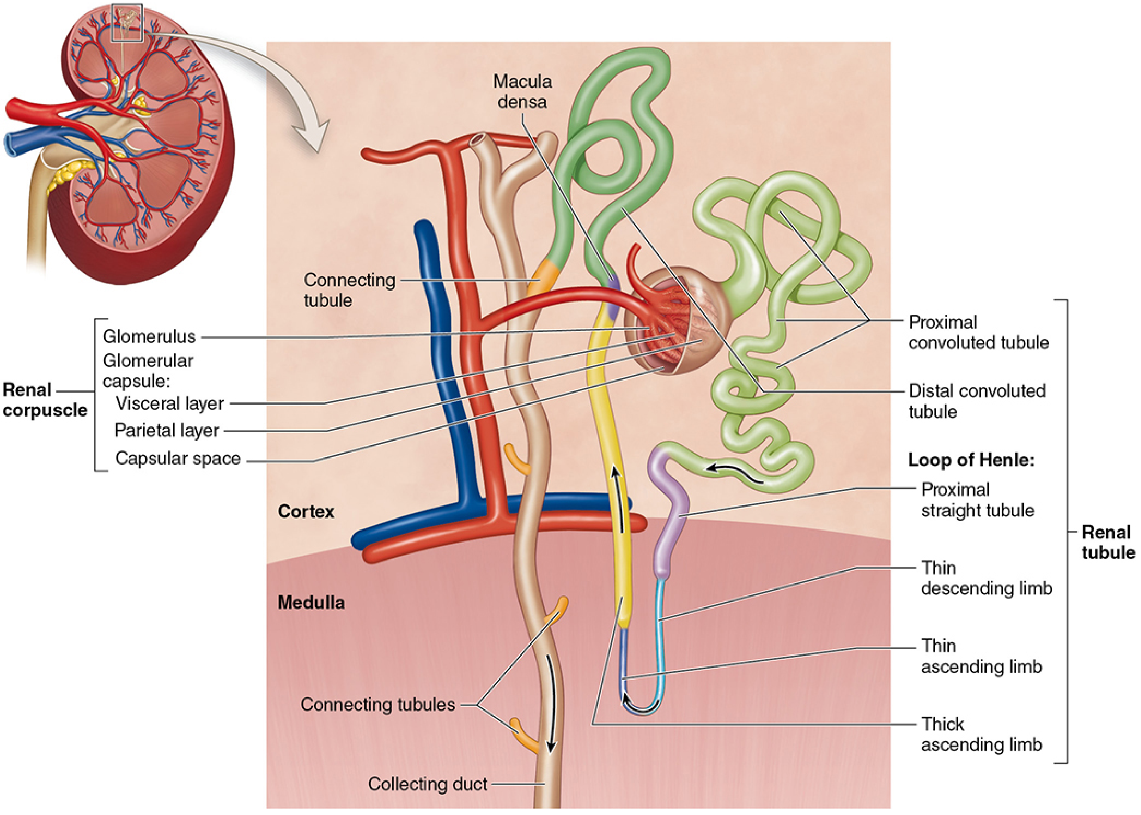
Each kidney contains ~1.25 million nephrons. Each nephron consists of:
| Segment | Location | Key Histology / Function |
|---|---|---|
| Renal corpuscle | Cortex | Glomerulus + Bowman's capsule; blood filtration |
| Proximal convoluted tubule (PCT) | Cortex | Cuboidal cells, prominent brush border, acidophilic; reabsorbs ~65% of filtrate |
| Proximal straight tubule | Outer medulla | Continuation of PCT |
| Thin descending limb | Medulla | Flat squamous cells; permeable to water |
| Thin ascending limb | Medulla | Flat squamous cells; impermeable to water |
| Thick ascending limb (TAL) | Medulla/cortex | Cuboidal cells; actively pumps NaCl; impermeable to water |
| Distal convoluted tubule (DCT) | Cortex | Cuboidal cells, NO brush border; contains macula densa at JGA |
| Connecting tubule | Cortex | Links DCT to collecting duct |
| Collecting duct | Cortex → medulla | Principal cells (water reabsorption via ADH) + intercalated cells (acid-base) |
Diagram 6 - Renal Corpuscle (Detail)
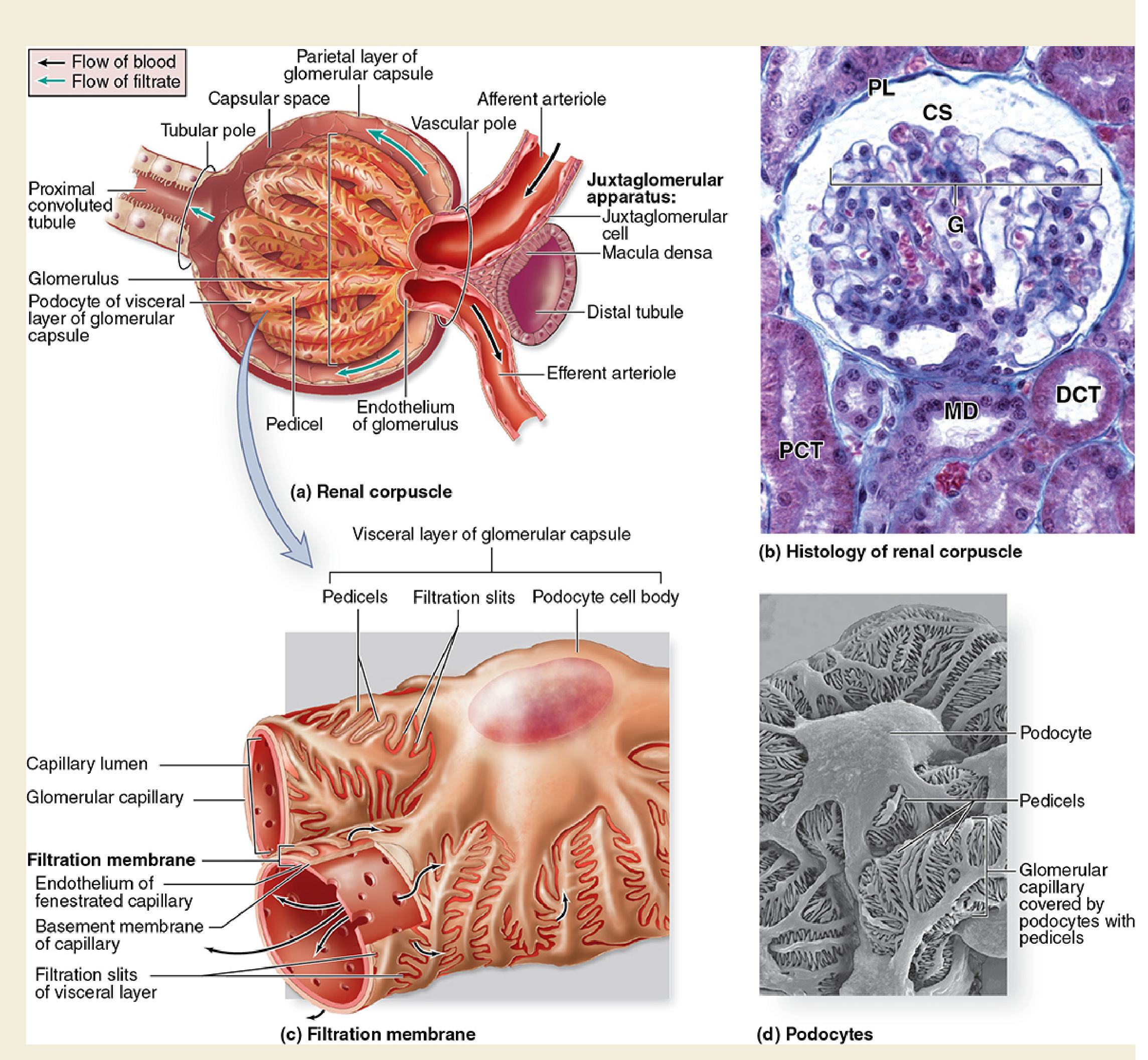
This composite figure shows:
(a) Renal Corpuscle diagram - Labeled components:
- Glomerulus - tuft of fenestrated capillaries
- Visceral layer (podocytes) - complex epithelial cells with pedicels covering the capillaries
- Parietal layer - simple squamous epithelium forming outer wall of Bowman's capsule
- Capsular space - receives the ultrafiltrate
- Afferent arteriole (enters) → efferent arteriole (exits) at the vascular pole
- Tubular pole - where PCT begins
- Juxtaglomerular (JG) cells - in the wall of the afferent arteriole; secrete renin
- Macula densa - thickened DCT cells; sense NaCl concentration; regulate renin release
(b) H&E micrograph showing G (glomerulus), CS (capsular space), PL (parietal layer), PCT, DCT, and MD (macula densa)
(c) Filtration membrane - three layers for ultrafiltration:
- Fenestrated endothelium of glomerular capillary
- Glomerular basement membrane (GBM)
- Podocyte pedicels with filtration slits
(d) SEM of podocytes - showing the interdigitating pedicels covering the glomerular capillary surface
Ureters, Urinary Bladder & Urethra
| Structure | Epithelium | Notes |
|---|---|---|
| Ureters | Transitional epithelium (urothelium) - 5-7 cell layers | Muscularis: inner longitudinal + outer circular smooth muscle; adventitia |
| Urinary bladder | Transitional epithelium (urothelium) - 3 cell layers when distended | Superficial cells = large "umbrella cells" (fusiform vesicles in cytoplasm); muscularis = detrusor (3 interwoven smooth muscle layers) |
| Urethra | Variable: proximal = transitional → stratified squamous distally | Male urethra lined by pseudostratified in prostatic/membranous portions |
Transitional epithelium (urothelium) - unique feature: cells change shape with bladder filling. When empty, cells are cuboidal/columnar; when distended, cells flatten. Superficial "umbrella cells" have special plaques (uroplakins) that form an impermeable barrier against urine.
Juxtaglomerular Apparatus (JGA) - Key Diagram Point
The JGA is located where the DCT returns to contact its own glomerulus. It consists of:
- Juxtaglomerular (granular) cells - modified smooth muscle of afferent arteriole; store and secrete renin
- Macula densa - tall, closely packed DCT cells that sense tubular Na⁺/Cl⁻ concentration
- Extraglomerular mesangial cells (Lacis cells) - signaling role
This apparatus is the effector of the renin-angiotensin-aldosterone system (RAAS) for blood pressure regulation.
Quick Comparison Table: Tubule Identification in Histology
| Tubule | Epithelium | Brush border | Lumen | Tip |
|---|---|---|---|---|
| PCT | Large cuboidal, granular/acidophilic | Prominent | Indistinct (brush border fills it) | Most abundant in cortex |
| DCT | Smaller cuboidal, less acidophilic | Absent | Clear/wide | Contacts glomerulus (macula densa) |
| Thick ascending limb | Cuboidal | Absent | Clear | In outer medulla + cortex |
| Thin limb (loop) | Flat squamous | None | Small | Inner medulla |
| Collecting duct | Cuboidal, pale cells; distinct borders | None | Clear, prominent | Cortex to papilla |
Sources: Junqueira's Basic Histology - Text and Atlas, 17e (pp. 730-822, 948-1000) | Histology: A Text and Atlas with Correlated Cell and Molecular Biology (Pawlina & Ross)
This is a shared conversation. Sign in to Orris to start your own chat.