
Varicocele in detail simple language for surgery university exam
Reading File
Now let me search the medical library for varicocele content across relevant books simultaneously.
Finding Sources
Finding Sources
Excellent. Now let me read the most relevant textbook sections in parallel - Bailey & Love (surgery exam gold standard), Campbell-Walsh Urology (comprehensive), Gray's Anatomy, and Smith & Tanagho.
Reading File
Reading File
Reading File
Reading File
Excellent content from Bailey & Love and other textbooks. Now let me read the rest of the Bailey & Love section (treatment/surgery) and the Campbell-Walsh key points section.
Reading File
Reading File
Reading File
Now let me get the treatment/surgery details from Bailey & Love and fetch the key clinical image.
Reading File
Loading Image
I now have comprehensive information from multiple authoritative textbooks. Let me compile the complete answer.
Varicocele - Complete Surgery Exam Notes
Sources: Bailey & Love's Short Practice of Surgery 28e, Smith & Tanagho's General Urology 19e, Campbell-Walsh-Wein Urology, Gray's Anatomy for Students
Definition
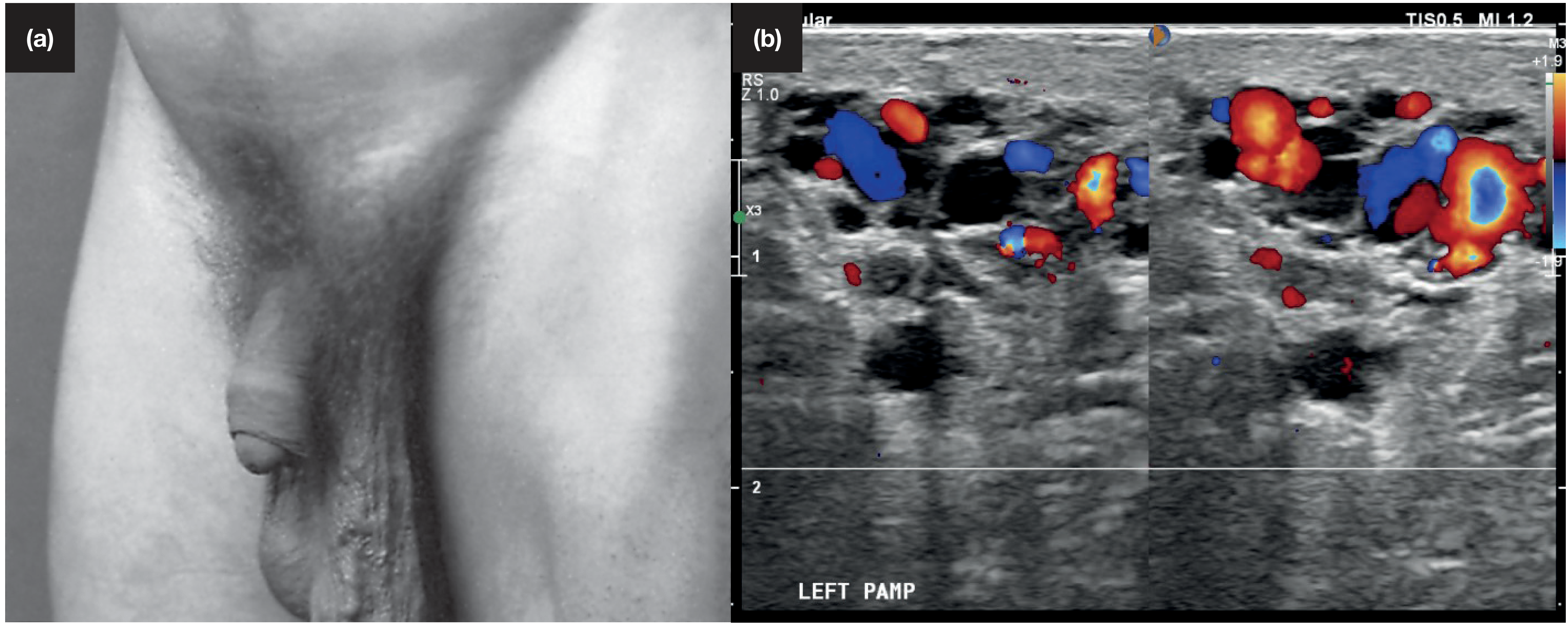
A varicocele is an abnormal dilatation and tortuosity of the veins of the pampiniform plexus - the network of veins draining the testis within the spermatic cord. Think of it as "varicose veins of the scrotum."
Incidence (Numbers to Remember)
| Population | Prevalence |
|---|---|
| General adult males | 10-20% |
| Infertile males | ~40% |
| Bilateral in healthy men | <10% |
| Bilateral in infertile men | up to 20% |
| Left-sided | 90% |
- Develops during puberty (typically after age 10, peak at Tanner stage 3)
- Rarely seen in boys under 10
- Does NOT spontaneously regress
Why Mostly Left-Sided? (Anatomy - Exam Favourite)
Three key anatomical reasons:
- Left testicular vein drains into the left renal vein at a RIGHT ANGLE - higher resistance, higher back-pressure. The right spermatic vein drains obliquely into the IVC - lower pressure.
- Left testicular vein is LONGER than the right - higher hydrostatic column of blood.
- Nutcracker effect - the left renal vein gets compressed between the Superior Mesenteric Artery (SMA) and the Aorta, raising pressure in the left renal vein and transmitted back into the testicular vein.
Result: Blood refluxes backwards through incompetent valves down the testicular vein into the pampiniform plexus, causing dilatation.
Right-sided varicocele alone is RARE - always investigate to exclude a retroperitoneal mass (e.g. renal tumour) compressing the right testicular vein.
Pathophysiology - Three Theories
- Absent/incompetent valves at the junction of testicular vein with renal vein (left) or IVC (right) - allows retrograde reflux
- Nutcracker phenomenon - SMA compresses left renal vein against the aorta, obstructing testicular venous outflow
- Angulation at the junction of the left testicular vein and left renal vein
How Does It Cause Infertility?
The most widely accepted theory:
- Reflux of warm blood from the abdomen raises the intratesticular temperature
- Normal spermatogenesis requires the testes to be 2-3°C cooler than body temperature
- The pampiniform plexus normally acts as a countercurrent heat exchanger - cool arterial blood is cooled further by venous blood flowing past it
- A varicocele disrupts this exchange → elevated temperature → impaired spermatogenesis
Other theories:
- Reflux of renal/adrenal metabolites (e.g. cortisol, catecholamines) down the spermatic vein
- Increased hydrostatic pressure in testicular vasculature
- Pituitary-gonadal hormonal dysfunction
Semen effects: Abnormalities in concentration, motility (most profound), and morphology. Also reduces sperm DNA integrity.
Clinical Features
Symptoms:
- Most are asymptomatic (found incidentally)
- "Dragging" or "heavy" discomfort in the scrotum - worse at end of day, worse on standing
- Typically presents in adolescence or early adulthood
Signs (examine standing, then lying):
| Sign | Detail |
|---|---|
| Affected side hangs lower | Left scrotum lower than right |
| "Bag of worms" | Classic feel on palpation - soft, compressible tortuous veins above the testis |
| Cough impulse | Present in larger varicoceles |
| Veins decompress on lying | Gravity empties the dilated veins - allows you to palpate the testis properly |
| Testicular atrophy | In longstanding cases - testis smaller and softer |
Key exam point: If a varicocele does NOT decompress on lying down → suspect secondary cause (renal tumour, retroperitoneal mass).
Grading (WHO/Clinical Classification)
| Grade | Description |
|---|---|
| Subclinical | Not palpable or visible; detected only by Doppler USS or venography |
| Grade 1 | Palpable only during Valsalva manoeuvre; not visible |
| Grade 2 | Palpable at rest; NOT visible |
| Grade 3 | Visible AND palpable at rest |
Investigations
- Doppler Ultrasound - gold standard; confirms diagnosis in doubtful cases; vein diameter >3 mm with retrograde flow on Valsalva
- Semen analysis - assess fertility impact (sperm count, motility, morphology)
- Renal USS - mandatory if right-sided varicocele or if varicocele does not decompress supine (to exclude renal cell carcinoma)
- Venography - for percutaneous embolization planning
Treatment
Indications for Treatment
- Infertility - clinical varicocele + abnormal semen analysis + otherwise unexplained infertility
- Pain/discomfort - significant symptoms
- Testicular atrophy - especially in adolescents (to halt progressive damage)
- Adolescent varicocele with documented growth arrest of testis (careful - risk of overtreatment)
Varicocele repair is NOT indicated for: subclinical varicocele alone, or normal semen parameters - no proven benefit on pregnancy rates.
Treatment Options
1. Percutaneous (Radiological) Embolization
- First-line treatment at most centres (especially when discomfort is the main complaint)
- Catheter inserted via right femoral vein → right common iliac vein → IVC → left renal vein → left testicular vein → pampiniform plexus
- Metal coils or sclerosants injected to occlude the vessels
- Advantages: Day procedure, minimal pain, rapid return to work (1 day)
- Disadvantages: 10-15% technical failure rate; 0-10% recurrence; not available everywhere
2. Surgical Ligation (Varicocelectomy)
Three surgical approaches - remember these for the exam:
| Approach | Level of Ligation | Notes |
|---|---|---|
| Retroperitoneal (Palomo) | High ligation at level of internal inguinal ring / retroperitoneum | Fewest vessels to ligate; highest recurrence (misses collaterals) |
| Inguinal (Ivanissevich) | At inguinal canal level | Most common traditional approach; good access |
| Subinguinal (Microsurgical) | Below external inguinal ring | Gold standard surgical technique; lowest recurrence, fewest complications |
| Laparoscopic | Retroperitoneal (similar to Palomo) | Higher recurrence 5-25%; useful if bilateral |
Microsurgical varicocelectomy (subinguinal, using operating microscope) is the most effective surgical method - lowest recurrence (0-15%), best semen improvement (66%), fewer complications.
3. Comparison of Outcomes (Smith & Tanagho)
| Outcome | Incisional | Laparoscopic | Radiological |
|---|---|---|---|
| Semen improvement | 66% | 50-70% | 60% |
| Pregnancy rate | 35% | 12-32% | 10-50% |
| Recurrence | 0-15% | 5-25% | 0-10% |
| Technical failure | Negligible | Small | 10-15% |
| Days to return to work | 5 | 5.3 | 1 |
4. Watchful Waiting
- Spontaneous pregnancy rate ~16% - a valid option if semen not severely abnormal
Complications of Varicocelectomy
- Recurrence (~20% after embolization; lower after microsurgical repair)
- Hydrocele - most common surgical complication; due to lymphatic damage (reduced with microsurgical technique)
- Testicular artery injury - leads to testicular atrophy (avoided by microscopic identification)
- Wound infection / haematoma
Varicocele and Fertility - Key Points
- Most common surgically correctable cause of male subfertility
- Varicocele present in ~40% of infertile men vs 15% of general population
- Cochrane review evidence: treatment improves chance of spontaneous pregnancy in men with clinical varicocele + abnormal semen + unexplained infertility
- Varicocelectomy may also improve outcomes with assisted reproduction (IVF/ICSI)
- About 85% of men with varicocele have fathered children - so not all varicoceles are clinically significant
Summary Box (Bailey & Love)
- Varicocele is common; 90% are left sided
- Associated with progressive testicular damage from adolescence onward and reduced fertility
- Varicocele repair is effective in men with: clinical varicocele + low sperm count + otherwise unexplained infertility
- Microsurgical varicocelectomy is the most effective surgical technique
- First-line intervention is usually percutaneous embolization; if it fails or recurs, surgical ligation is appropriate
Quick Mnemonics
Left-sided predominance: "LEFT RENAL = RIGHT ANGLE + LONG VEIN + NUTCRACKER"
Grading: "Sub-1-2-3"
- Sub = subclinical (ultrasound only)
- 1 = Valsalva only
- 2 = palpable at rest
- 3 = visible + palpable
Bag of WORMS = Varicocele (classic description)
This is a shared conversation. Sign in to Orris to start your own chat.