

1000445890.jpg
As per tjis format write examnation of recurrent patella dislocation
Here is the Examination of Recurrent Patella Dislocation written in the Knee Examination Proforma format shown in your image:
Knee Examination Proforma – Recurrent Patella Dislocation
1. Gait, Foot Progression Angle
- Antalgic gait or avoidance gait due to fear of giving way
- Toe-out gait (increased foot progression angle) suggesting external tibial torsion
- Trendelenburg gait if associated hip abductor weakness
2. Attitude
- Knee held in slight flexion to avoid quadriceps contraction
- Leg may be externally rotated
- If currently dislocated: patella visibly displaced laterally, knee held in slight flexion
3. Inspection
-
General findings: Overlying skin - bruising/ecchymosis after acute episode, swelling (hemarthrosis), old scar if previously operated, sinus if infected
-
Specific findings of inspection:a. From front:
- Deformity: Genu valgum (most common predisposing factor), increased Q angle
- Muscle wasting: Vastus medialis obliquus (VMO) wasting prominently
- Position of patella: Patella alta (high-riding patella), lateral tilt of patella, "grasshopper eye" appearance (bilateral lateral displacement)
- Limb length discrepancy
- Supra/parapatellar swelling (hemarthrosis or effusion)
- Pelvis position and ankle-foot: Pes planus, pronated foot, increased femoral anteversion
b. From side:- Sagittal plane deformity: Patella alta visible as high-riding patella
- Flexion deformity if acute episode
- Genu recurvatum (associated with ligamentous laxity)
c. From back:- Popliteal fossa - any swelling
- Spine - scoliosis if associated
- Calf muscle wasting
- Ankle-foot deformity (pes planus)
4. Palpation
- Local rise in temperature (if acute/recent dislocation)
- Tenderness:
- Medial retinaculum tenderness (torn MPFL - medial patellofemoral ligament)
- Lateral retinaculum tenderness
- Medial femoral condyle tenderness (MPFL femoral attachment)
- Lateral patellar facet tenderness
- Anterior knee tenderness
- Joint line tenderness (associated chondral damage)
- Patellar tap - effusion/hemarthrosis
- Crepitus on patellar movement (chondromalacia patellae)
- Synovial hypertrophy
- Retropatellar tenderness (patella-facet grinding)
- Hyperlaxity of joints (generalised ligamentous laxity - Beighton score)
- Confirmation of palpatory characteristics of swelling, scar, sinus, and deformities
5. Movements: Active and passive
- Flexion-extension range: usually full unless acute episode
- Extensor lag (if VMO weak or acute injury)
- Flexion deformity
- Patellar tracking during active flexion-extension: J-sign (patella jumps laterally at terminal extension - pathognomonic of recurrent dislocation)
- Crepitus, clicks during movement
6. Measurements
- Limb length and thigh-calf circumference (VMO wasting quantified)
- Q angle: Increased (>15° in females, >10° in males) - most important measurement
- Intercondylar and intermalleolar distance (for genu valgum assessment)
- TT-TG distance (tibial tubercle to trochlear groove) - radiological, normal <20 mm
- Patellar height ratio (Insall-Salvati ratio) - for patella alta
7. Neurovascular Examination
- Distal pulses (dorsalis pedis, posterior tibial)
- Sensation over anterior knee, medial and lateral aspects
- Peroneal nerve assessment (foot dorsiflexion, first web space sensation)
8. Special Tests
a. Anterior stability tests for ACL: Anterior drawer test, Lachman test, pivot shift test (rule out combined instability)
b. Posterior stability test for PCL: Sag sign, posterior drawer test, Godfrey test, quadriceps active test
c. Tests for stability of posteromedial corner: Valgus stress test at 0° and 30°, Slocum (AMRI) test, external rotation test
d. Tests for stability of posterolateral corner: Varus stress test at 0° and 30°, external rotation recurvatum, Dial test
e. Meniscus test: McMurray's, Apley's grinding, Thessaly test, Payr's test
f. Patella stability and other tests (MOST IMPORTANT in this condition):
- Apprehension test (Clarke's sign): With knee at 30° flexion, examiner pushes patella laterally - positive if patient apprehends or contracts quadriceps in fear of dislocation (pathognomonic)
- Quadrant test: Patella displaced medially and laterally to assess retinacular tightness - lateral retinaculum tightness noted
- Patellar horizontal tilt test: Medial edge of patella cannot be elevated beyond neutral - indicates tight lateral retinaculum
- Patellar glide test: Patella displaced >2 quadrants laterally indicates MPFL laxity
- Patellar tracking: J-sign during active knee extension
- Fulkerson tilt test: Assess patellar tilt
- Beighton score: For generalised ligamentous laxity
g. Wilson test for osteochondritis dissecans (rule out associated osteochondral defect)
9. Joint Above (Hip, Spine) and Below (Ankle)
- Hip: Increased femoral anteversion (Craig's test), hip abductor weakness (Trendelenburg)
- Spine: Lumbosacral alignment
- Ankle-foot: Pes planus, subtalar pronation (increases Q angle dynamically)
10. Lymph Node Examination
- Inguinal lymphadenopathy (if infective etiology considered)
Key Predisposing Factors to Specifically Document:
- Trochlear dysplasia (flat/convex trochlea)
- Patella alta (Insall-Salvati >1.2)
- Increased TT-TG distance (>20 mm)
- MPFL insufficiency
- Increased Q angle / genu valgum
- Generalised ligamentous laxity
- VMO hypoplasia
How to check for genu varus or valgium deformity clinically
Genu varum (bow legs) and genu valgum (knock knees) are assessed clinically by examining the alignment of the lower limb with the patient standing, supine, and during gait.
Searching sources...
Reading File
Finding Sources
Finding Sources
Finding Sources
Reading File
Finding Sources
Loading Image
Loading Image
Here is a complete clinical guide to assessing Genu Varum and Genu Valgum:
Clinical Assessment of Genu Varum and Genu Valgum
Definitions
| Term | Description | Common Name |
|---|---|---|
| Genu Varum | Knees bowed outward - center of knee lateral to mechanical axis | Bow legs |
| Genu Valgum | Knees angled inward - center of knee medial to mechanical axis | Knock knees |
Step 1 - Patient Position
- Patient stands barefoot, in light clothing, feet together (medial malleoli touching)
- Patellae facing forward (neutral rotation)
- Knees in full extension
- Examined from the front (AP view)
Step 2 - Visual Inspection (Standing)
For Genu Varum:
- Gap between the knees when feet/ankles are together
- Lower limbs appear bowed outward
- Feet may show lateral border loading / fallen pedal arch
For Genu Valgum:
- Gap between the ankles when knees are touching
- Lower limbs angle inward at the knee
- Associated pes planus, toe-out gait
Step 3 - Clinical Measurement (Key Step)
The standard bedside clinical test uses intercondylar and intermalleolar distances:
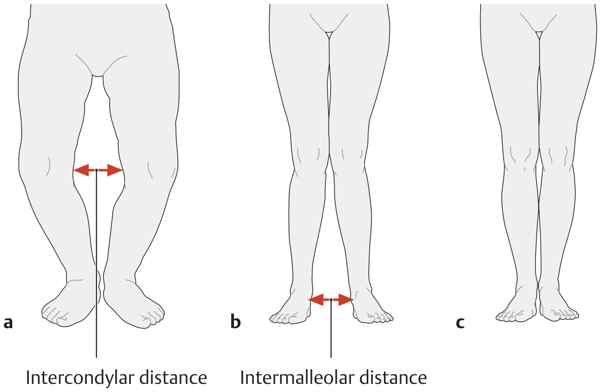
A. Intercondylar Distance - For Genu Varum
- Ask patient to stand with medial malleoli together
- Measure the gap between the medial femoral condyles at the knee
- Use a tape measure or ruler between the most prominent medial condyle points
- Normal: condyles touch (0 cm)
- Abnormal: >3 cm is considered pathological
B. Intermalleolar Distance - For Genu Valgum
- Ask patient to stand with knees together (medial condyles touching)
- Measure the gap between the medial malleoli at the ankle
- Normal: malleoli touch (0 cm)
- Abnormal: >5 cm is considered pathological
(Source: THIEME Atlas of General Anatomy and Musculoskeletal System)
Step 4 - Mechanical Axis Assessment
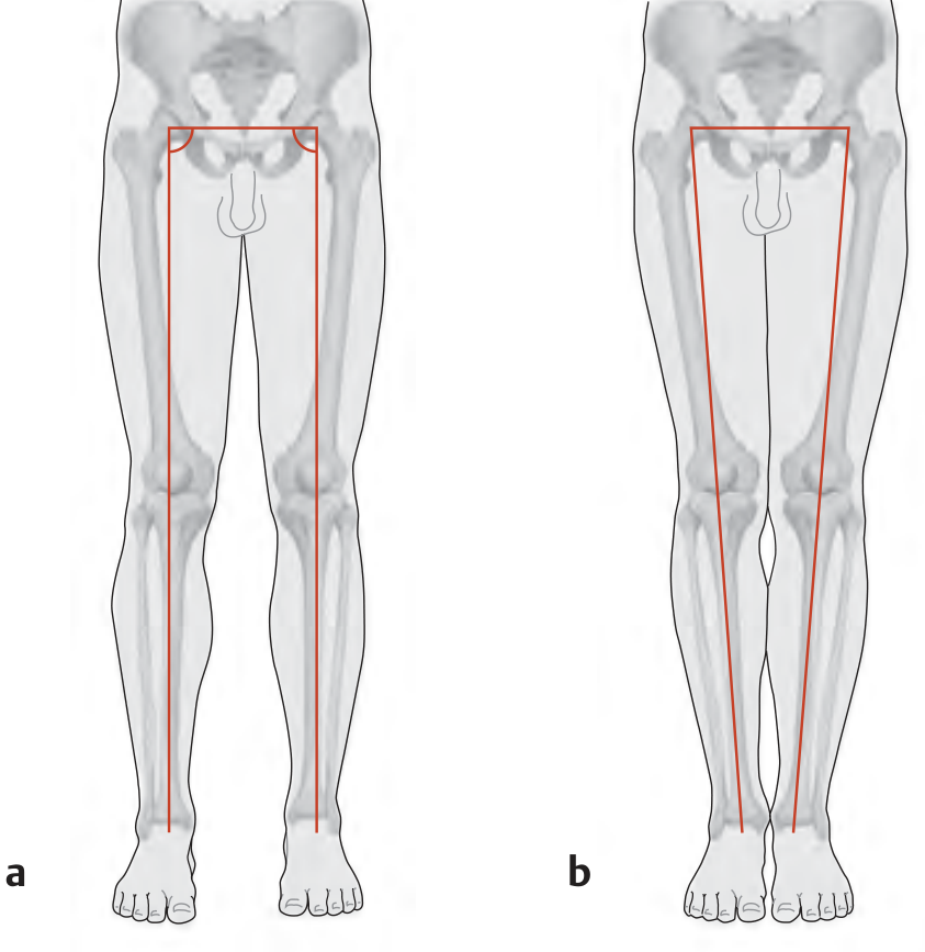
- Normal: The mechanical axis (from center of femoral head to center of ankle) passes through the center of the knee. The femorotibial angle is 174° (a slight physiological valgus).
- Genu Varum: Center of knee joint is lateral to the mechanical axis - medial compartment overloaded
- Genu Valgum: Center of knee joint is medial to the mechanical axis - lateral compartment overloaded
Step 5 - Q Angle Measurement
- Patient: Supine, quadriceps relaxed, knee in full extension
- Line 1: From ASIS (anterior superior iliac spine) to center of patella
- Line 2: From center of patella to tibial tubercle
- The angle between these two lines = Q angle
| Normal | Abnormal | |
|---|---|---|
| Male | 10-14° | >15° suggests valgus tendency |
| Female | 15-17° | >20° suggests valgus tendency |
- Increased Q angle is associated with genu valgum and patellar instability
- Decreased/negative Q angle suggests genu varum
Step 6 - Supine Examination
- Ask patient to lie supine, legs extended and relaxed
- Bring both knees together - confirm deformity persists (structural) vs disappears (postural/flexible)
- Check for fixed vs correctable deformity
- Assess whether deformity is symmetrical or asymmetrical
Step 7 - Gait Assessment
| Finding | Suggests |
|---|---|
| Waddling, wide-based gait | Genu varum |
| Scissor-like, knees crossing | Genu valgum |
| Lateral border foot wear | Genu varum |
| Medial border foot wear, pes planus | Genu valgum |
Step 8 - Associated Findings to Document
| Condition | Genu Varum | Genu Valgum |
|---|---|---|
| Compartment stress | Medial overload | Lateral overload |
| Ligament stress | Lateral collateral lax | Medial collateral lax |
| Foot | Fallen lateral arch | Pes planus |
| Pelvis | Wider-based stance | Narrow-based stance |
| Patella | Facing inward (squinting) | Laterally tilted |
Physiological (Normal) Age-Related Changes
| Age | Normal Finding |
|---|---|
| First year of life | Up to 20° genu varum is normal |
| Around age 2 | Up to 10° genu valgum is normal |
| School age (~6-7 yrs) | Legs straighten - normal adult alignment |
Any deformity persisting or worsening beyond these age norms warrants investigation for rickets, Blount's disease, or metabolic bone disease.
Summary - Quick Bedside Reference
| Test | Genu Varum | Genu Valgum |
|---|---|---|
| Feet together | Gap at knees | Normal or touching at knees |
| Knees together | Normal at ankles | Gap at ankles |
| Intercondylar distance | >3 cm = abnormal | Not applicable |
| Intermalleolar distance | Not applicable | >5 cm = abnormal |
| Q angle | Decreased/normal | Increased (>15° M, >20° F) |
| Mechanical axis deviation | Lateral to knee center | Medial to knee center |
How to measure Q angle precisely during knee examination?
The Q angle (quadriceps angle) is measured using a goniometer from the ASIS to the center of the patella, then from the center of the patella to the tibial tubercle. Normal values differ by sex.
Searching sources...
Finding Sources
Finding Sources
Finding Sources
Reading File
Reading File
Loading Image
Finding Sources
Here is a complete, precise guide to measuring the Q angle:
Q Angle - Precise Clinical Measurement
What is the Q Angle?
The Quadriceps (Q) angle is the angle formed at the patella between:
- Line 1 (Proximal): From the Anterior Superior Iliac Spine (ASIS) to the center of the patella - represents the line of pull of the quadriceps muscle
- Line 2 (Distal): From the center of the patella to the tibial tubercle - represents the line of the patellar tendon
It reflects the net lateral force vector of the quadriceps on the patella. A higher Q angle = greater lateral pull on the patella.
Equipment Needed
- Standard long-arm goniometer (preferred)
- Skin marker/pen
- Measuring tape
- Patient in appropriate position (see below)
Patient Positioning (Critical for Accuracy)
Standard Method - Supine:
- Patient supine on a flat examination table
- Knee in full extension (0°)
- Quadriceps relaxed (not contracted)
- Hip in neutral rotation - patella facing directly upward
- Feet in neutral position - not internally or externally rotated
Note: Hip rotation significantly affects the Q angle - internal rotation increases it, external rotation decreases it. Always ensure neutral hip position.
Step-by-Step Measurement Technique
Step 1 - Identify and mark 3 bony landmarks with a skin marker:
| Landmark | How to Locate |
|---|---|
| ASIS | Palpate the iliac crest anteriorly - the most prominent anterior bony point of the pelvis |
| Center of patella | Midpoint of the patella - palpate all four borders and mark the center |
| Tibial tubercle | Bony prominence on the anterior tibia just below the knee - most prominent point |
Step 2 - Draw two reference lines (optional but helps with accuracy):
- Line from ASIS to center of patella
- Line from center of patella to tibial tubercle
Step 3 - Place the goniometer:
- Fulcrum (center): Over the center of the patella
- Proximal arm: Aligned toward the ASIS
- Distal arm: Aligned toward the tibial tubercle
Step 4 - Read the angle formed between the two arms at the patella
Normal Values
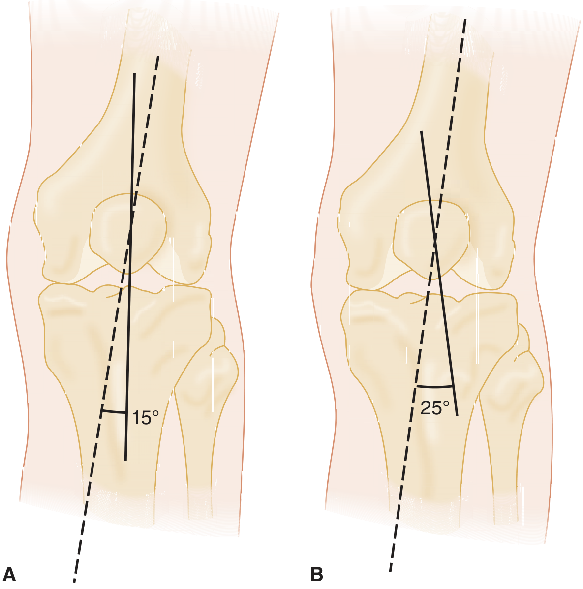
(A - Normal Q angle ~15°. B - Abnormal Q angle ~25° with increased lateral subluxation force - Tintinalli's Emergency Medicine)
| Population | Normal Range | Abnormal |
|---|---|---|
| Males | 10-14° (avg ~13°) | >15° |
| Females | 15-18° (avg ~18°) | >20° |
Females have a higher Q angle due to their broader pelvis (wider pelvis → more lateral ASIS position → greater angle).
(Source: Rheumatology 2-Volume Set, Elsevier 2022)
Why Q Angle is Higher in Females - Anatomy
| Factor | Effect |
|---|---|
| Wider pelvis | ASIS displaced more laterally → increases proximal line angle |
| Increased femoral anteversion | Adds to lateral vector |
| Genu valgum | Common in females → increases Q angle |
| Pes planus / pronated foot | Dynamic increase in Q angle during gait |
Common Errors to Avoid
| Error | Effect on Reading |
|---|---|
| Hip internally rotated | Falsely increases Q angle |
| Hip externally rotated | Falsely decreases Q angle |
| Knee not in full extension | Changes patellar position, alters reading |
| Quadriceps contracted | Patella shifts superiorly, alters angle |
| Marking tibial tubercle inaccurately | Most common source of error |
| Patient obese/difficult landmark palpation | Use fluoroscopy or CT for accuracy |
Q Angle in Standing vs Supine
| Position | Notes |
|---|---|
| Supine (standard) | Most reproducible, eliminates weight-bearing variables |
| Standing | More functional but harder to standardize; Q angle typically slightly higher due to weight-bearing and dynamic muscle activity |
| 30° knee flexion | Patella engages trochlear groove - more clinically relevant for patellar tracking assessment |
Clinical Significance of an Increased Q Angle
| Q Angle | Implication |
|---|---|
| >15° (male) / >20° (female) | Pathological lateral patellar pull |
| High Q angle | Risk of: patellofemoral syndrome, recurrent patellar dislocation, chondromalacia patellae |
| Very high Q angle (>25°) | Likely underlying structural cause - genu valgum, femoral anteversion, lateral tibial torsion, or lateralized tibial tubercle (high TT-TG distance) |
A high Q angle increases the resultant lateral subluxation force on the patella - this is the biomechanical basis for patellar instability and patellofemoral pain syndrome. (Miller's Review of Orthopaedics 9th Ed; Tintinalli's Emergency Medicine)
Associated Radiological Measurement - TT-TG Distance
When Q angle is elevated, confirm with imaging:
- TT-TG (tibial tubercle - trochlear groove) distance on CT or MRI
- Normal: <20 mm
-
20 mm: indication for tibial tubercle medialization (Fulkerson osteotomy)
- This is the radiological equivalent and more precise version of the Q angle
This is a shared conversation. Sign in to Orris to start your own chat.