
For my dermatology practical exam please give me a proper case description of “tuberous sclerosis” and also prepare me extensively for the questions that examiner can ask from medium to hard to very hard
Tuberous sclerosis complex 2021 diagnostic criteria surveillance consensus guideline
"tuberous sclerosis" AND dermatology
tuberous sclerosis facial angiofibromas shagreen patch hypomelanotic macules clinical dermatology
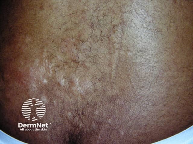
Clinical photography of a cutaneous Shagreen patch in a patient with tuberous sclerosis complex. Modality: color digital photography, noninvasive in vivo imaging of the trunk skin, most often the lumbosacral region. The patch appears as a localized, slightly raised, leathery plaque with a coarse, pebbled surface and fine reticulate skin markings. The coloration is variably hyperpigmented to brown, with subtle border irregularity and diffused beveling of surrounding skin. Histological correlation would show a dense, thickened dermal collagen network with relative paucity of elastic fibers, giving a firm, taut texture. Clinically, shagreen patches are among the major diagnostic criteria for TSC and serve as a visible surrogate marker of underlying systemic involvement. They commonly arise on the trunk, especially the lumbosacral region, and may co-develop with hypomelanotic macules, facial angiofibromas, and cortical/subependymal lesions. Differential diagnosis includes morphea, elastosis perforans serpiginosa, and other pigmented or sclerotic dermatoses. The diagnostic significance lies in reinforcing the suspicion of tuberous sclerosis and prompting targeted MRI brain, renal ultrasound, and cardiac surveillance. This image is relevant for dermatology, medical education, and radiology-pathology correlation studies, illustrating cutaneous stigmata that guide multidisciplinary evaluation and management in pediatric and adult patients with TSC. for differential diagnosis and research.
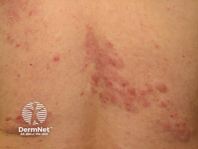
Clinical photography of a shagreen patch on the lower back illustrating a leathery, pebbled dermal plaque consistent with a connective tissue nevus. The lesion is located in the lumbosacral region of the posterior trunk. It presents as a gray‑brown, rough-surfaced plaque with irregular margins and a tactilely rugged texture, often described as resembling sharkskin or orange-peel. This cutaneous stigmata of tuberous sclerosis complex results from locally thickened dermal collagen with disorganized, dense bundles and reduced elastic fibers. Histopathology (when biopsied) shows a dense, haphazard dermal collagen matrix without cellular atypia, confirming a collagenoma phenotype characteristic of shagreen patches. Clinically, shagreen patches are harbingers of TSC and carry diagnostic significance; their presence reinforces consideration of CNS involvement (cortical tubers, subependymal nodules) and warrants comprehensive neurologic and genetic evaluation. Differential considerations include other connective tissue nevi (collagenomas) and elastin-rich nevi, but the constellation with hypomelanotic macules and facial angiofibromas strengthens the TSC diagnosis. This image serves educational and diagnostic contexts for dermatology, medical genetics, and radiology cohorts, illustrating lesion morphology, location, and relevance to systemic disease.
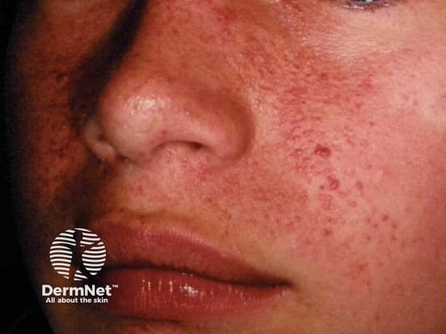
This is a high‑resolution clinical photograph of facial skin showing angiofibromas in a patient with tuberous sclerosis complex (TSC). Modality: Clinical photography, dermatologic photography; view: frontal, close‑up of the midface including the nasal bridge, cheeks, and perioral region. Anatomical localization: cutaneous integument of the face, malar and perinasal skin. Visual features: numerous small, dome‑shaped, erythematous papules (~1–3 mm) arranged in symmetrical clusters along the malar and nasolabial areas, with mild background erythema and occasional coalescence. The lesions are nonulcerative and level with the surrounding skin; vessels may appear prominent in aggregates. When biopsied histologically, angiofibromas demonstrate fibrous stroma with dilated capillaries; clinically they present as reddish papules and plaques without pigment loss. Clinical significance: a hallmark cutaneous manifestation of TSC and a major diagnostic criterion; supports diagnosis in conjunction with other TS features (hypomelanotic macules, cortical tubers, subependymal nodules, shagreen patch). Potential clinical use cases: diagnostic confirmation, monitoring of treatment response to laser or topical mTOR inhibitors, and educational reference for dermatology, pediatrics, and medical education. Differential considerations: acne vulgaris, rosacea, folliculitis, sebaceous hyperplasia. Image context notes: Ensure color fidelity for vascular intensity; include clinical history of TS; document trajectory of lesions over time to support phenotypic progression and therapeutic planning.
https://www.tsalliance.org/healthcare-professionals/diagnosi…
PMID: 39018447
PMID: 38728593
PMID: 37906297
Tuberous sclerosis complex (TSC): dermatology practical case
One-line diagnosis
Do not call it merely “tuberous sclerosis” in the exam if you can avoid it. The preferred current term is tuberous sclerosis complex (TSC).
1. Model long-case presentation
How to present the case to the examiner
Likely diagnosis
2. Complete dermatological description
A. Facial angiofibromas
- Usually begin in childhood, often becoming apparent between 2 and 5 years and increasing through adolescence.
- Multiple small, firm, smooth, dome-shaped, pink, red-brown, or yellowish-red papules.
- Symmetrical distribution over cheeks, nasolabial folds, nose, chin, and forehead.
- Histologically they are angiofibromas, not sebaceous gland tumours. Therefore, “adenoma sebaceum” is an older misnomer.
- They may bleed after trauma and may cause major cosmetic and psychosocial distress.
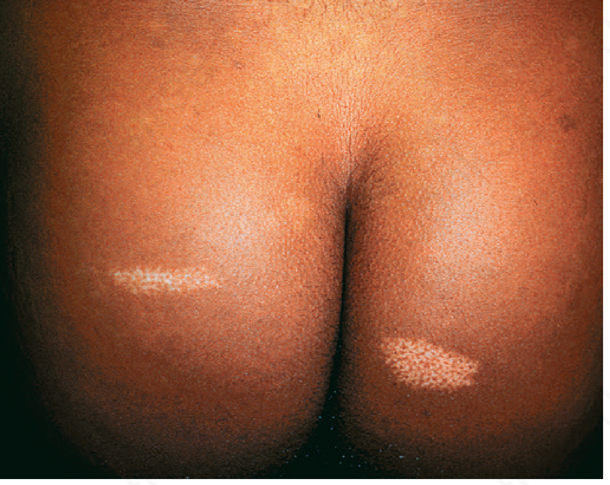
B. Hypomelanotic macules
- Earliest and commonest skin finding.
- Congenital or appearing in infancy.
- Classically oval, leaf-shaped lesions called ash-leaf macules.
- Other shapes: confetti lesions, polygonal lesions, and “thumbprint” macules.
- They become more conspicuous under Wood lamp.
- Diagnostic threshold: three or more macules, each at least 5 mm in diameter.
- A single hypomelanotic macule is not diagnostic, as isolated lesions occur in healthy individuals.
C. Shagreen patch
- A connective tissue nevus, mainly a collagenoma.
- Thickened, irregular, flesh-coloured to yellow-brown plaque with a leathery, pebbled “orange-peel” or “pigskin” surface.
- Commonly on the lower back, especially the lumbosacral region.
- Usually appears in childhood.
D. Ungual fibromas
- Also known as Koenen tumours.
- Firm, skin-coloured or pink, periungual or subungual fibrous papules.
- Usually occur around or after puberty.
- May produce nail distortion, longitudinal grooves, leukonychia, onycholysis, and pain or bleeding.
E. Fibrous cephalic plaque
- A firm, elevated, skin-coloured plaque, often on the forehead or scalp.
- “Fibrous cephalic plaque” is preferred over the older term “fibrous forehead plaque,” because it can occur elsewhere on the head.
- It is a major diagnostic feature.
F. Oral and dental findings
- Multiple dental enamel pits.
- Gingival fibromas.
- Intraoral fibromas involving gingiva, tongue, lips, or buccal mucosa.
3. Bedside examination checklist
General examination
- Developmental level, behaviour, cognition.
- Look for evidence of previous seizures or antiepileptic medication.
- Check blood pressure, especially because renal disease can cause hypertension.
- Look for pallor, abdominal mass, signs of renal impairment.
Cutaneous examination
- Examine face for angiofibromas and cephalic plaque.
- Examine under Wood lamp for hypomelanotic macules.
- Inspect lumbosacral region for shagreen patch.
- Examine all fingernails and toenails for Koenen tumours.
- Examine scalp for fibrous plaque or poliosis.
- Examine oral cavity for enamel pits and intraoral fibromas.
Systemic examination
- CNS: seizures, developmental delay, focal deficits, raised intracranial pressure.
- Eyes: retinal hamartomas.
- Cardiovascular: cardiac rhabdomyoma, especially in infants.
- Abdomen: renal angiomyolipoma, renal cysts.
- Respiratory: lymphangioleiomyomatosis, particularly in adult women.
4. Diagnostic criteria: high-yield viva answer
Genetic diagnosis
Clinical diagnosis
Major features
- Hypomelanotic macules, 3 or more, at least 5 mm.
- Angiofibromas, 3 or more, or fibrous cephalic plaque.
- Ungual fibromas, 2 or more.
- Shagreen patch.
- Multiple retinal hamartomas.
- Multiple cortical tubers and/or radial migration lines.
- Subependymal nodules, 2 or more.
- Subependymal giant cell astrocytoma, SEGA.
- Cardiac rhabdomyoma.
- Lymphangioleiomyomatosis, LAM.
- Angiomyolipomas, 2 or more.
Minor features
- Confetti skin lesions.
- Dental enamel pits, more than 3.
- Intraoral fibromas, more than 2.
- Retinal achromic patch.
- Multiple renal cysts.
- Nonrenal hamartomas.
- Sclerotic bone lesions.
Interpretation
- Definite TSC: 2 major features, OR 1 major plus 2 or more minor features.
- Possible TSC: 1 major feature, OR 2 or more minor features.
- Important exception: the combination of LAM and angiomyolipomas alone does not establish definite TSC.
5. Etiopathogenesis: medium to very hard viva
Basic answer
- TSC1 on chromosome 9q34, encoding hamartin
- TSC2 on chromosome 16p13.3, encoding tuberin
High-level answer
“Two-hit” question
A patient inherits or develops a heterozygous pathogenic variant, but a second somatic event may inactivate the remaining normal allele in particular cells. This “second hit” contributes to focal hamartoma formation.
TSC1 versus TSC2
- TSC2 mutations are generally more frequent.
- TSC2-associated disease is often clinically more severe, with more significant renal involvement and neurological manifestations, though phenotype is variable.
- A contiguous deletion involving TSC2 and PKD1 can produce severe early polycystic kidney disease with TSC.
6. Classic triad: common examiner trap
The classic Vogt triad
- Facial angiofibromas, historically called adenoma sebaceum.
- Seizures.
- Intellectual disability.
Correct modern answer
7. Systemic involvement and what it causes
| Organ system | Lesion | Clinical importance |
|---|---|---|
| Brain | Cortical tubers | Epilepsy, developmental delay, autism, intellectual disability |
| Brain | Subependymal nodules | May calcify, often along ventricular wall |
| Brain | SEGA | Can obstruct the foramen of Monro causing hydrocephalus |
| Kidney | Angiomyolipomas | Haemorrhage, pain, hypertension, renal impairment |
| Kidney | Cysts | May occur; consider TSC2-PKD1 contiguous gene syndrome if severe/early |
| Lung | LAM | Dyspnoea, recurrent pneumothorax, chylous effusion, especially adult women |
| Heart | Rhabdomyomas | Arrhythmia or obstruction in infancy; often regress later |
| Eye | Retinal hamartomas | Often asymptomatic but require ophthalmological assessment |
| Skin and mouth | Angiofibromas, shagreen patch, Koenen tumours, enamel pits | Important visible diagnostic clues |
8. Investigations
To confirm and stage disease
Dermatological
- Wood lamp examination for hypomelanotic macules.
- Clinical photography for follow-up of facial angiofibromas.
- Skin biopsy is usually not necessary if clinical findings and systemic features are characteristic.
Neurological
- MRI brain to identify cortical tubers, radial migration lines, subependymal nodules, and SEGA.
- EEG if seizures or suspicious episodes.
- Formal developmental, neuropsychological, behavioural, and autism assessment. These neuropsychiatric manifestations are collectively called TAND, or TSC-associated neuropsychiatric disorders.
Renal
- MRI abdomen is preferred for angiomyolipomas and renal cysts.
- Renal function, urinalysis, and blood pressure.
- Do not rely solely on ultrasound if detailed assessment of fat-poor angiomyolipoma is needed.
Cardiac
- Echocardiography and ECG, particularly in infants and children.
Pulmonary
- In adult females and symptomatic patients, assess for LAM. Baseline high-resolution CT chest and pulmonary-function testing are considered according to age, symptoms, and specialist guidance.
Ophthalmological
- Dilated retinal examination.
Genetics
- TSC1/TSC2 molecular testing.
- Genetic counselling and assessment of first-degree relatives.
9. Management
Core principle
A. Facial angiofibromas
- Topical sirolimus/rapamycin can reduce erythema and papule size, especially for mild to moderate lesions.
- Laser or destructive procedures for resistant or cosmetically troubling lesions:
- Pulsed dye laser or KTP laser for predominantly vascular/red lesions.
- CO2 laser or Er:YAG laser for more fibrous, raised lesions.
- Electrosurgery or shave procedures may be used selectively.
- Recurrence can occur, so repeat treatment and long-term follow-up are often needed.
B. Seizures and neurodevelopment
- Managed by paediatric neurologist or neurologist.
- Antiseizure therapy tailored to seizure type.
- Vigabatrin is commonly important for infantile spasms associated with TSC, under specialist supervision.
- Epilepsy surgery may be considered for carefully selected drug-resistant focal epilepsy.
- Everolimus may have a role in selected TSC-associated refractory focal seizures.
C. SEGA
- Surgical treatment if symptomatic, enlarging, or obstructing CSF flow.
- Everolimus can be used in appropriately selected patients, particularly when surgery is unsuitable.
D. Renal angiomyolipoma
- Regular imaging surveillance.
- mTOR inhibitor therapy may be used for enlarging or high-risk lesions.
- Embolisation is used for acute bleeding or selected high-risk lesions.
- Avoid unnecessary nephrectomy whenever feasible because preservation of renal function is important.
E. LAM
- Pulmonology referral.
- Smoking avoidance, management of pneumothorax, pulmonary function monitoring.
- Sirolimus may be used for clinically significant LAM under specialist care.
F. Counselling
- Autosomal dominant inheritance.
- Each child of an affected person has a 50% chance of inheriting the pathogenic variant.
- Many cases are de novo, so absence of family history does not exclude TSC.
- Offer genetic counselling, reproductive options discussion, and family screening.
10. Examiner questions with model answers
Medium difficulty
1. What is the diagnosis?
2. What are the facial lesions called?
3. What are ash-leaf macules?
4. How will you demonstrate ash-leaf macules?
5. What is a shagreen patch?
6. What are Koenen tumours?
7. Name the classic triad.
8. Is the classic triad required for diagnosis?
9. What inheritance pattern does TSC show?
10. Name the most important renal lesion.
Hard difficulty
11. State the current diagnostic rule for definite TSC.
12. Name the major cutaneous diagnostic features.
- Three or more hypomelanotic macules, at least 5 mm.
- Three or more facial angiofibromas or fibrous cephalic plaque.
- Two or more ungual fibromas.
- Shagreen patch.
13. Mention four minor cutaneous or oral features.
- Confetti skin lesions.
- More than three dental enamel pits.
- More than two intraoral fibromas.
- Retinal achromic patch is minor but ophthalmological rather than cutaneous.
14. Why is TSC called a phakomatosis?
15. What is the pathology of facial angiofibroma?
16. What is the pathology of a shagreen patch?
17. What is a cortical tuber?
18. What is a subependymal nodule?
19. What is SEGA, and why is it important?
20. Which pulmonary disease occurs in TSC?
21. Why do patients with TSC require blood pressure monitoring?
22. How do you differentiate facial angiofibromas from acne?
| Facial angiofibromas | Acne vulgaris |
|---|---|
| Symmetrical, fixed, smooth papules | Polymorphic lesions |
| No comedones | Comedones are typical |
| No pustules usually | Papules, pustules, nodules may occur |
| Often associated with other TSC stigmata | No systemic hamartoma syndrome |
| Childhood onset, persistent | Commonly adolescent onset |
Very hard difficulty
23. Explain the molecular mechanism in TSC.
24. Why do mTOR inhibitors work in TSC?
25. What does “TSC2/PKD1 contiguous gene syndrome” mean?
26. What is genetic mosaicism in TSC, and why does it matter?
27. If a patient has only bilateral renal angiomyolipomas and LAM, can you diagnose definite TSC?
28. Why are facial angiofibromas not “adenoma sebaceum”?
29. Outline initial surveillance after diagnosing TSC.
- MRI brain for cortical tubers, SENs, and SEGA.
- Baseline renal imaging, preferably MRI, with renal function and blood pressure.
- Detailed skin, dental, and ophthalmological examination.
- ECG and echocardiography, particularly in children.
- Assessment for seizures, TAND, development, learning, autism, and behaviour.
- Pulmonary evaluation for LAM in appropriate patients, especially adult women.
- Genetic testing and family counselling.
30. What is TAND?
31. A patient develops headache, vomiting, and drowsiness. What complication do you suspect?
32. A patient with TSC presents with sudden flank pain, hypotension, and haematuria. What is the likely diagnosis?
11. Differential diagnosis of important findings
Facial angiofibromas
- Acne vulgaris.
- Rosacea.
- Sebaceous hyperplasia.
- Fibrofolliculomas of Birt-Hogg-Dubé syndrome.
- Angiofibromas in MEN1.
- Trichoepitheliomas.
Hypomelanotic macules
- Nevus depigmentosus.
- Vitiligo.
- Pityriasis alba.
- Post-inflammatory hypopigmentation.
- Pigmentary mosaicism.
- Idiopathic guttate hypomelanosis in older patients.
Shagreen patch
- Other connective tissue nevi.
- Collagenoma in other syndromes.
- Morphea, depending on morphology.
- Focal dermal hypoplasia or scar-like lesions in unusual contexts.
12. Fast practical-exam closing statement
13. Ten facts to memorise the night before
- TSC is autosomal dominant, but many cases are de novo.
- Genes: TSC1 = hamartin, TSC2 = tuberin.
- Both normally inhibit mTORC1.
- Facial lesions are angiofibromas, not true adenoma sebaceum.
- Hypomelanotic macules: 3 or more, at least 5 mm is a major feature.
- Shagreen patch = connective tissue nevus/collagenoma, classically lumbosacral.
- Koenen tumours = periungual/subungual fibromas.
- Definite diagnosis: 2 major OR 1 major + 2 minor.
- Dangerous intracranial lesion: SEGA near foramen of Monro causing hydrocephalus.
- Dangerous renal lesion: angiomyolipoma causing haemorrhage.