
Gingival enlargement from caranza
gingival enlargement classification types clinical appearance
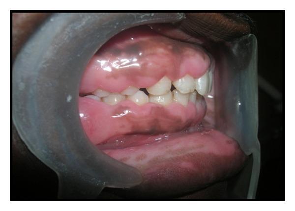
This intraoral clinical photograph, taken from a lateral perspective with a mouth retractor, demonstrates generalized gingival enlargement (gingival fibromatosis). The gingiva appears significantly thickened and bulbous, exhibiting a firm, smooth, and slightly shiny texture that lacks the normal stippled appearance. The tissue enlargement is extensive, migrating coronally and partially obscuring the clinical crowns of both the maxillary and mandibular teeth, which makes the teeth appear abnormally short. The color of the gingiva is predominantly pink, with noticeable areas of physiologic melanin hyperpigmentation presenting as brownish-black patches. The interdental papillae are severely enlarged, filling the embrasures and contributing to the overall bulky and congested appearance of the dental arches. This presentation is characteristic of hereditary gingival fibromatosis, a condition that can interfere with occlusion, speech, and oral hygiene maintenance.
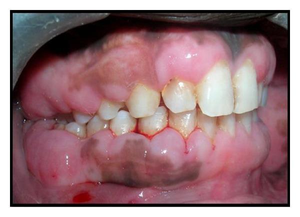
This intraoral clinical photograph displays significant generalized gingival enlargement (gingival fibromatosis) in a patient. The gingival tissue is pale pink, dense, and firm in appearance, exhibiting a bulbous, nodular, and irregular surface texture. The overgrowth is extensive, covering a substantial portion of the clinical crowns of both the maxillary and mandibular teeth, particularly in the posterior regions. Distinctive dark, pinpoint markings are visible on the gingival surface, representing bleeding points created by a pocket marker to guide surgical excision (gingivectomy). The teeth that remain visible show some crowding and plaque accumulation near the gingival margins. Physiological pigmentation (melanosis) is also present as brownish patches on the keratinized gingiva. This image serves as a clinical demonstration of hereditary or idiopathic gingival overgrowth and the preoperative marking phase of periodontal surgery.
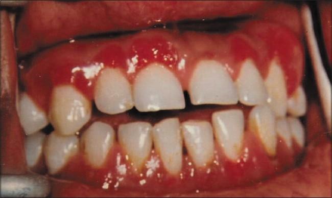
This clinical photograph displays a front-view intraoral examination showing significant gingival enlargement and inflammation. The gingival tissue across the upper and lower anterior regions, as well as the upper posterior buccal areas, exhibits a characteristic fiery red color and a distinctively irregular, granular, and pebbled surface texture. The enlargement is papillary and diffuse, significantly covering the cervical portions of the teeth. Clinical features such as the 'strawberry' or granular appearance are hallmark signs often associated with granulomatous conditions, including tuberculous gingivitis or sarcoidosis. The image serves as an educational example of atypical gingival lesions that present with minimal local deposits (plaque or calculus) but demonstrate severe morphological changes, requiring histopathological correlation to confirm systemic or infectious underlying causes such as tuberculosis.
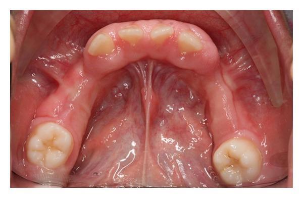
This intraoral clinical photograph provides an occlusal view of the mandibular arch in a 9-year-old pediatric patient, demonstrating generalized, severe gingival enlargement. The gingival tissue is pale pink, firm, and fibrotic in appearance, with a notable lack of inflammatory signs such as edema or bleeding. The overgrowth is most prominent in the anterior labial and lingual regions, where it significantly covers the clinical crowns of the incisors, appearing to displace them. The enlargement extends posteriorly, obscuring the alveolar ridge and surrounding the partially erupted molars. The tissue surface appears relatively smooth with diminished stippling. This presentation is consistent with hereditary gingival fibromatosis, characterized by non-hemorrhagic, slow-growing gingival hypertrophy that can interfere with tooth eruption, dental alignment, and aesthetics. The image serves as a clinical example of idiopathic or hereditary gingival overgrowth in a pediatric context.
drug induced gingival hyperplasia phenytoin cyclosporine nifedipine
inflammatory gingival enlargement pregnancy tumor epulis
Tmj Disorders from ongole ravikiran
temporomandibular joint anatomy disc condyle
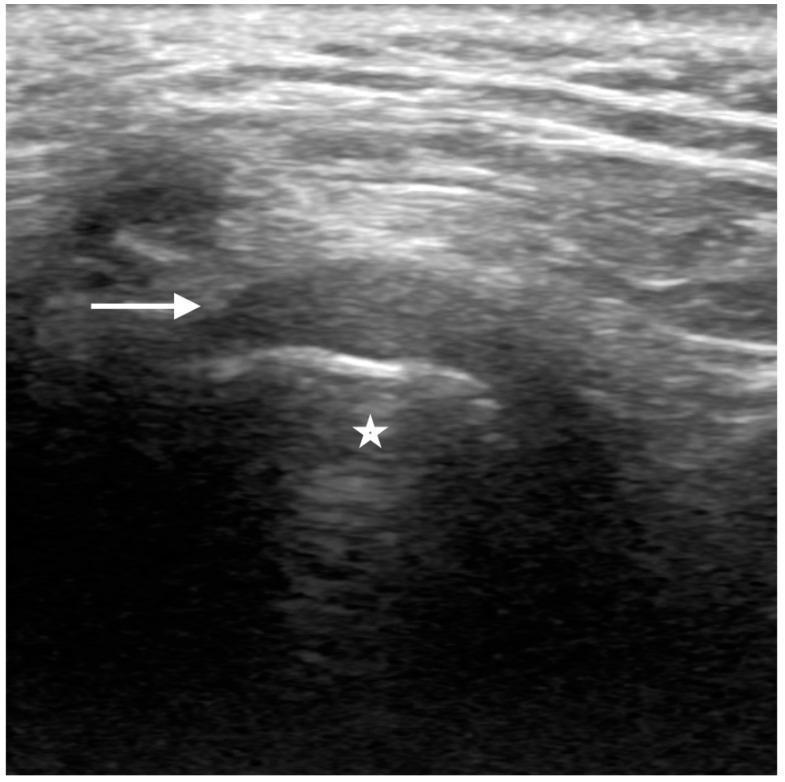
This longitudinal diagnostic ultrasound image illustrates the normal anatomy of the human temporomandibular joint (TMJ) in a closed-mouth position. The mandibular condyle is identified as a central hypoechoic region (marked with a star), bounded superiorly by a distinct, bright hyperechoic line representing the condylar cortex. Situated immediately superior to the condylar cortex is the articular disc (indicated by a white arrow), which presents as a hypoechoic, inverted C-shaped structure. This imaging modality and view are essential for evaluating the relationship between the disc and the condyle to rule out internal derangement, such as disc displacement. The image highlights the characteristic echogenicity of normal TMJ components: the hyperechoic bone cortex and the hypoechoic fibrocartilaginous disc. This content is relevant for radiology and dentistry education, specifically for teaching musculoskeletal ultrasound techniques and diagnostic criteria for normal joint function.
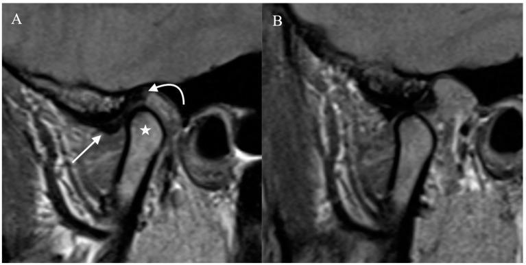
Diagnostic Image: This side-by-side sagittal proton density-weighted MRI illustrates the normal anatomy and functional dynamics of the human temporomandibular joint (TMJ). Image A shows the joint in the closed-mouth position. The mandibular condyle (marked with a star) is correctly seated within the mandibular fossa. The articular disc exhibits a classic hypointense, biconcave 'bow-tie' appearance, situated between the condyle and the temporal bone. Annotations identify the anterior band (straight arrow) and the posterior band (curved arrow). Image B demonstrates the joint in the open-mouth position, highlighting normal physiological movement. There is clear anterior translation and rotation of the mandibular condyle, which has moved forward and inferiorly toward the articular eminence. The articular disc maintains its relationship with the condyle during this transition. This comparison serves as a clinical reference for assessing disc displacement and jaw range of motion, essential in diagnosing internal derangement of the TMJ.
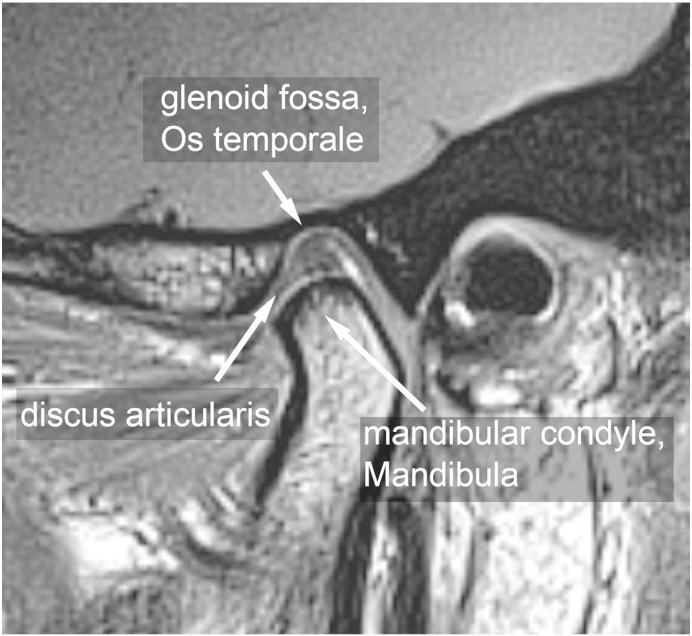
This diagnostic image is a parasagittal Magnetic Resonance Imaging (MRI) scan demonstrating the normal anatomy of the human temporomandibular joint (TMJ) in an asymptomatic individual. The image clearly labels three primary components: the glenoid fossa (Os temporale), the articular disc (discus articularis), and the mandibular condyle (Mandibula). The biconcave articular disc is shown in a physiological position, interposed between the superior glenoid fossa and the inferiorly positioned mandibular condyle. The condyle exhibits a smooth, rounded morphology sitting within the mandibular fossa, illustrating proper joint alignment. The cortical boundaries of the temporal bone and mandible appear intact. This visual serves as an educational reference for normal maxillofacial anatomy, joint articulation, and standard radiological presentation of the TMJ in the closed-mouth or neutral position. It is relevant for dental, orthodontic, and radiological training to differentiate healthy joint structures from those affected by temporomandibular disorders (TMD).
TMJ disc displacement internal derangement MRI
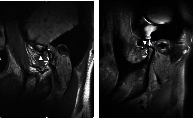
This diagnostic comparison contains two magnetic resonance imaging (MRI) views of the temporomandibular joint (TMJ) illustrating different stages of internal derangement. Image (a) demonstrates an irreducible anterior articular disc displacement. Specific pathology includes the anteriorly displaced disc (arrow), notable thickening at the attachment point of the inferior branch of the lateral pterygoid muscle (curved arrow), and secondary degenerative changes evidenced by condylar hyperosteogeny (straight arrow). Image (b) shows the contralateral side presenting with a reducible anterior articular disc displacement. While the disc is also displaced anteriorly (arrow), the lateral pterygoid muscle attachment point (curved arrow) appears normal in thickness, and there is an absence of the hyperosteogeny seen in the irreducible state. This comparison serves as an educational reference for identifying radiological signs of chronic TMJ disc displacement and associated muscle/bone remodeling, emphasizing the anatomical differences between reducible and irreducible internal derangements.
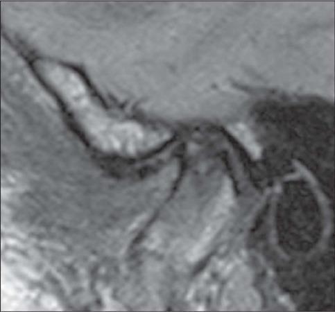
This diagnostic image is a sagittal proton density (PD)-weighted MRI of the temporomandibular joint (TMJ) in a closed-mouth position. The scan illustrates advanced internal derangement characterized by anterior disc displacement. The articular disc, which should normally reside in a biconcave configuration between the mandibular condyle and the temporal bone's articular eminence, is displaced anteriorly. Significant morphological changes indicative of degenerative disc disease are visible, including a loss of the normal biconcave shape, acquiring a more biconvex or rounded contour. The disc demonstrates thinning (tapering) of the anterior band and surface irregularities. Educational focus includes the identification of anatomical landmarks of the TMJ (mandibular condyle, articular eminence, and external auditory canal) and the assessment of disc positioning and morphology for diagnosing temporomandibular disorders (TMD). This content is relevant for intermediate to advanced medical learners in radiology, maxillofacial surgery, and dentistry.
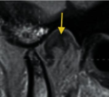
This diagnostic image is an MRI scan of the human temporomandibular joint (TMJ) in a sagittal view, demonstrating internal derangement. The primary anatomical structures visible include the mandibular condyle, the glenoid fossa of the temporal bone, and the articular disc. A yellow arrow points directly to the articular disc, which is abnormally positioned. Specifically, the image illustrates posterior disc displacement (PDD), where the posterior band of the disc is shifted behind its normal superior position relative to the condylar head, often described as being posterior to the 12 o'clock position on top of the condyle. This imaging is critical for evaluating TMJ disorders and orofacial pain, providing a clear visual contrast between the low-signal fibrocartilaginous disc and the adjacent bony and soft tissue structures. The pedagogical focus is the identification of directional disc displacement as a subset of TMJ pathologies.
TMJ osteoarthritis rheumatoid arthritis radiograph
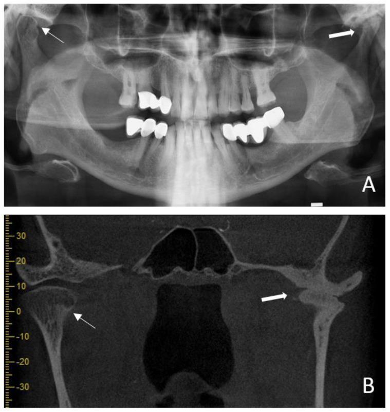
This composite diagnostic image illustrates osseous changes in the temporomandibular joint (TMJ) of a patient with long-standing rheumatoid arthritis. Panel A is a panoramic radiograph (orthopantomogram) showing bilateral condylar involvement. Key features include flattening of the condylar head, a decrease in joint space (thick arrow), and anterior condylar resorption (thin arrow) appearing as localized radiolucency. Panel B provides a coronal cone beam computed tomography (CBCT) section, which offers superior visualization of these degenerative changes. The CBCT reveals significant anterior resorption (thin arrow), multiple small subchondral radiopacities within the condylar head, and signs of bony ankylosis (thick arrow) where the condyle articulates with the temporal bone. These findings are characteristic of advanced inflammatory arthropathy of the TMJ, demonstrating the progressive bone destruction, secondary remodeling, and joint space narrowing associated with chronic systemic rheumatic disease. The imaging serves as a comparison between traditional 2D radiography and 3D cross-sectional imaging for evaluating the extent of maxillofacial pathology.
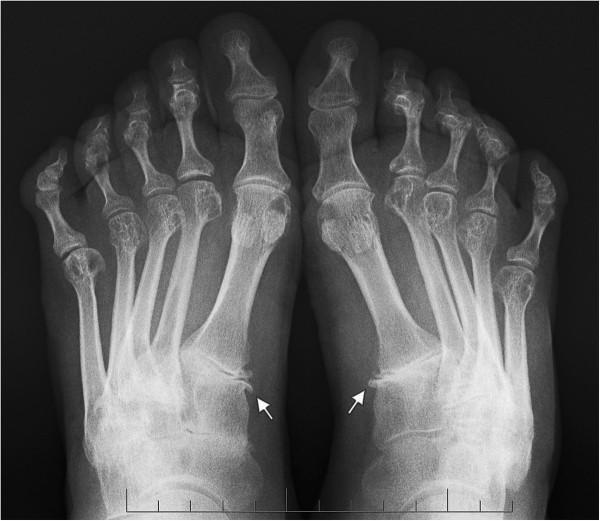
This anteroposterior (AP) radiograph of both feet demonstrates chronic inflammatory and degenerative changes characteristic of rheumatoid arthritis. The imaging shows significant forefoot involvement, including bilateral metatarsophalangeal (MTP) joint instability and subluxation of the first MTP joints. Key findings include prominent osteophyte formation at the medial aspects of the bilateral first cuneometatarsal (CM) joints, indicated by white arrows, which signifies joint hypermobility and secondary osteoarthritis. There is evidence of generalized periarticular osteopenia and joint space narrowing across the tarsal and MTP regions. The first intermetatarsal angle is increased bilateral, consistent with hallux valgus deformity development secondary to connective tissue laxity. The lesser toes exhibit deformities, including lateral deviation and clawing, typical of rheumatoid forefoot progression. This diagnostic image serves as a clinical example of midfoot and forefoot structural changes in systemic inflammatory arthritis, highlighting the relationship between joint instability and osteophyte development at the first ray.
TMJ Disorders - Ongole & Ravikiran
1. Introduction & Historical Background
- Temporomandibular Disorder (TMD) - the current preferred umbrella term (Dworkin & LeResche, 1992 - RDC/TMD criteria)
- Older terms: Myofacial Pain Dysfunction Syndrome (MPDS), Costen's Syndrome, Craniomandibular Disorder
2. Anatomy of the TMJ (Relevant to Disorders)
| Component | Details |
|---|---|
| Articular surfaces | Condylar head + articular eminence of temporal bone |
| Articular disc (meniscus) | Biconcave fibrocartilaginous structure; anterior band, intermediate zone, posterior band |
| Bilaminar zone | Posterior attachment of disc; rich in nerves and vessels |
| Joint spaces | Upper (gliding) and lower (rotational) compartments |
| Capsule & ligaments | TMJ ligament, sphenomandibular ligament, stylomandibular ligament |
| Muscles | Masseter, temporalis, medial pterygoid, lateral pterygoid |
3. Classification of TMD (Ongole & Ravikiran)
A. Muscle Disorders (Extracapsular)
- Myofascial Pain Dysfunction Syndrome (MPDS) - most common
- Myositis
- Myospasm
- Local myalgia
- Myofibrotic contracture
B. Joint (Articular) Disorders (Intracapsular)
- Internal Derangements (disc displacement)
- Inflammatory Conditions (arthritis - various types)
- Degenerative Joint Disease (osteoarthritis)
- Hypomobility (ankylosis - fibrous and bony)
- Hypermobility (subluxation and dislocation)
- Developmental disorders (condylar hyperplasia, hypoplasia, aplasia)
- Neoplasia
4. Myofascial Pain Dysfunction Syndrome (MPDS)
Etiology - Multifactorial
- Psychological factors: stress, anxiety, depression (key role - Laskin's psychophysiological theory)
- Parafunctional habits: bruxism, clenching, nail biting, pen chewing
- Occlusal factors: malocclusion, premature contacts, loss of posterior support
- Trauma: macrotrauma (blow to jaw) or microtrauma (repeated minor forces)
Laskin's Diagnostic Criteria (1969) - all 4 must be present:
- Unilateral preauricular pain / pain in muscles of mastication
- Clicking / popping of the TMJ
- Limitation / deviation of mandibular movement
- Absence of clinical, radiographic, or biochemical evidence of organic TMJ changes
Cardinal Signs and Symptoms
- Pain: dull, aching, preauricular; worse on chewing; can radiate to temple, ear, neck
- Clicking/Popping: reciprocal click; opening and closing click
- Limitation of mouth opening: normal = 35-50 mm interincisal; restricted in MPDS
- Deviation of mandible: on opening, toward the affected side
- Muscle tenderness: masseter (most common), temporalis, pterygoids
- No organic joint changes: differentiates from true arthritis
Treatment of MPDS
| Approach | Examples |
|---|---|
| Conservative (first-line) | Rest, soft diet, heat application, patient education |
| Pharmacological | NSAIDs, muscle relaxants (diazepam), tricyclic antidepressants |
| Occlusal splints | Michigan splint (stabilization splint) - most widely used |
| Physiotherapy | Jaw exercises, ultrasound, TENS |
| Psychological | Stress management, biofeedback |
| Occlusal adjustment | Only after reversible methods fail |
| Surgical | Rarely indicated for MPDS |
5. Internal Derangements (ID)
Wilkes Classification (1989) - widely used staging system:
| Stage | Description | MRI Finding | Clicking | Pain |
|---|---|---|---|---|
| I (Early) | Disc displacement with reduction | Slight forward disc displacement | Reciprocal click | Minimal |
| II (Early-Intermediate) | Disc displacement with reduction | Disc slightly folded / deformed | Click + occasional catching | Occasional pain |
| III (Intermediate) | Disc displacement without reduction (closed lock) | Disc displaced, no recapture | No click (lock) | Pain ++ |
| IV (Intermediate-Late) | Chronic DDWR with degenerative changes | Disc perforation/degeneration | Crepitus | Pain ++ |
| V (Late) | Gross anatomical derangement | Gross deformity, joint destruction | Crepitus | Variable |
Types of Disc Displacement
- Anterior disc displacement with reduction (ADDWR) - most common; reciprocal click present
- Anterior disc displacement without reduction (ADDWOR) - "closed lock"; no click, mouth opening restricted to ~25-30 mm
- Lateral, medial, and posterior displacements (less common)
MRI - Gold Standard for Disc Position
6. Arthritis of the TMJ
A. Osteoarthritis (Degenerative Joint Disease)
- Most common arthritic condition of the TMJ
- Characterized by degeneration of articular cartilage and subchondral bone changes
- Radiographic features: flattening of condylar head, osteophyte formation (anterior lipping), subchondral sclerosis, joint space narrowing, erosions, Ely's cysts (subchondral pseudocysts)
- Symptoms: crepitus (grating sound), pain, restricted movement
- Panoramic OPG - first-line imaging; CBCT / MRI for more detail
B. Rheumatoid Arthritis
- Systemic autoimmune disease; bilateral TMJ involvement in ~50% of RA cases
- Chronic synovitis → pannus formation → cartilage and bone destruction
- Clinical: bilateral pain, swelling, anterior open bite (condylar destruction causes retrognathia)
- Radiographic: condylar erosion, joint space narrowing, ankylosis in severe cases
- Lab findings: elevated RF, anti-CCP antibody, ESR, CRP
C. Psoriatic Arthritis
- Associated with psoriasis; asymmetric joint involvement
- May precede skin lesions
- Radiograph: erosions + "pencil in cup" deformity
D. Infectious Arthritis (Septic Arthritis)
- Direct spread (parotitis, otitis media, osteomyelitis) or hematogenous
- Acute: pain, swelling, trismus, fever
- Causative organisms: S. aureus most common, N. gonorrhoeae
- Treatment: systemic antibiotics + joint drainage
E. Traumatic Arthritis
- Following direct trauma; hemarthrosis, pain, restricted movement
- Self-limiting; managed with rest, NSAIDs, soft diet
7. TMJ Ankylosis
Classification:
- True ankylosis: involves the joint itself (fibrous or bony)
- False (pseudo) ankylosis / extracapsular: due to conditions outside the joint (fibrous bands from radiation, masseter hypertrophy, coronoid process elongation, depressed zygomatic arch fracture)
Etiology of True Ankylosis:
- Most common cause: Trauma (mandibular condyle fracture in children, hemarthrosis)
- Infections (suppurative arthritis)
- Rheumatoid arthritis (advanced)
- Radiation therapy
Clinical Features:
- Restricted mouth opening (trismus) - hallmark; bony ankylosis = complete inability to open
- Facial asymmetry (if unilateral and onset in childhood)
- Micrognathia / retrognathia
- Bird face deformity (bilateral bony ankylosis in children) - severe micrognathia, Class II occlusion, sleep apnea
Radiographic Features:
- OPG: loss of joint space, bone deposition, irregular condyle outline
- CT: defines the extent of bony fusion (investigation of choice)
Treatment:
- Gap arthroplasty (most common): removal of ankylosed bone + creation of a gap (>1.5 cm)
- Interpositional arthroplasty: gap filled with temporalis fascia, cartilage, dermis, alloplastic material
- Total joint replacement: in adults with severe destruction
- Aggressive post-operative physiotherapy to prevent recurrence
8. TMJ Hypermobility
Subluxation:
- Condyle moves anterior to articular eminence but self-reduces
- Click at wide opening; transient locking at full open
- Managed with physiotherapy, sclerosing agents into joint, eminectomy if severe
Dislocation:
- Condyle moves anterior to eminence and cannot self-reduce - locked in open position
- Acute dislocation: Hippocratic method - thumbs on lower molars, downward and backward pressure to reduce
- Chronic recurrent dislocation: eminectomy (Myrhaug procedure), eminoplasty, injection of sclerosing agents (sodium psylliate, sodium tetradecyl sulfate)
9. Investigations / Imaging in TMD
| Investigation | Best Used For |
|---|---|
| OPG (Panoramic) | First-line; condyle morphology, degenerative changes, fractures |
| Transcranial radiograph | Gross bony changes, but technique-sensitive |
| CBCT / CT | Bony detail - ankylosis, fractures, osteophytes, condylar morphology |
| MRI | Gold standard for disc position and morphology; soft tissue detail |
| Arthrography | Disc perforation; largely replaced by MRI |
| Arthroscopy | Diagnostic + therapeutic; direct visualization of joint compartments |
| Bone scintigraphy | Active condylar hyperplasia |
10. Research Diagnostic Criteria for TMD (RDC/TMD) - Dworkin & LeResche, 1992
- Axis I: Physical diagnosis (muscle disorders, disc displacements, arthralgia/arthritis/arthrosis)
- Axis II: Psychosocial assessment (pain-related disability, depression, somatization)
11. Treatment Summary
| Condition | Treatment |
|---|---|
| MPDS | Reversible conservative first: splint, NSAIDs, physiotherapy |
| ADDWR (click) | Physiotherapy, anterior repositioning splint |
| ADDWOR (closed lock - acute) | Manipulation under anesthesia, arthrocentesis |
| ADDWOR (chronic) | Arthroscopy, open disc repair / repositioning |
| Osteoarthritis | NSAIDs, intra-articular corticosteroids, viscosupplementation (hyaluronic acid), condyloplasty |
| RA | Disease-modifying agents (MTX, biologics) + dental splint |
| Ankylosis | Surgical - gap/interpositional arthroplasty, total joint replacement |
| Dislocation (acute) | Manual reduction (Hippocratic method) |
| Dislocation (chronic recurrent) | Eminectomy, sclerosing agents |
- MPDS = most common TMD; Laskin's criteria = 4 cardinal features; no organic changes
- Wilkes classification = 5 stages of internal derangement
- MRI = gold standard for disc position; CT = gold standard for bony ankylosis
- Reciprocal click = ADDWR; No click + restricted opening = ADDWOR (closed lock)
- Anterior open bite + bilateral TMJ pain in female = think RA
- Bird face deformity = bilateral bony ankylosis in growing child
- Hippocratic method = reduction of TMJ dislocation
- RDC/TMD (1992) and DC/TMD (2014) = research/clinical diagnostic criteria