
anatomy of upper limb
Reading File
This is a broad anatomy topic - I'll search the medical library for upper limb anatomy content in parallel.
Finding Sources
Finding Sources
Finding Sources
Finding Sources
Searching Images
upper limb anatomy bones muscles nerves diagram
Now let me read the main Upper Limb sections from Gray's Anatomy for Students:
Reading File
Reading File
Reading File
Good content found. Now let me read the key sections - bones, muscles, nerves, vessels - from Gray's Anatomy for Students (block4) which is the main Upper Limb chapter:
Reading File
Reading File
Reading File
Let me now read more sections - the arm muscles, forearm, hand, and nerves sections:
Reading File
Reading File
Reading File
Let me fetch key anatomical images to include in the answer:
Loading Image
Loading Image
Reading File
Excellent - I now have very rich content from Gray's Anatomy for Students and the THIEME Atlas. Let me fetch one more key image (brachial plexus) and read the arm muscles / nerves sections:
Loading Image
Reading File
I now have comprehensive content. Let me compile a thorough, well-illustrated answer on the anatomy of the upper limb.
Anatomy of the Upper Limb
Sources: Gray's Anatomy for Students; General Anatomy and Musculoskeletal System (THIEME Atlas of Anatomy)
Overview and Regions
The upper limb is connected to the trunk via the shoulder girdle (clavicle + scapula), which articulates with the sternum at the sternoclavicular joint. The limb itself is divided into:
- Shoulder / Pectoral region
- Arm (humerus)
- Forearm (radius + ulna)
- Hand (carpals, metacarpals, phalanges)
The gateway between the neck and the upper limb is the axillary inlet, bounded by the superior margin of the scapula (posteriorly), the clavicle (anteriorly), and the lateral margin of rib I (medially).
1. Bones
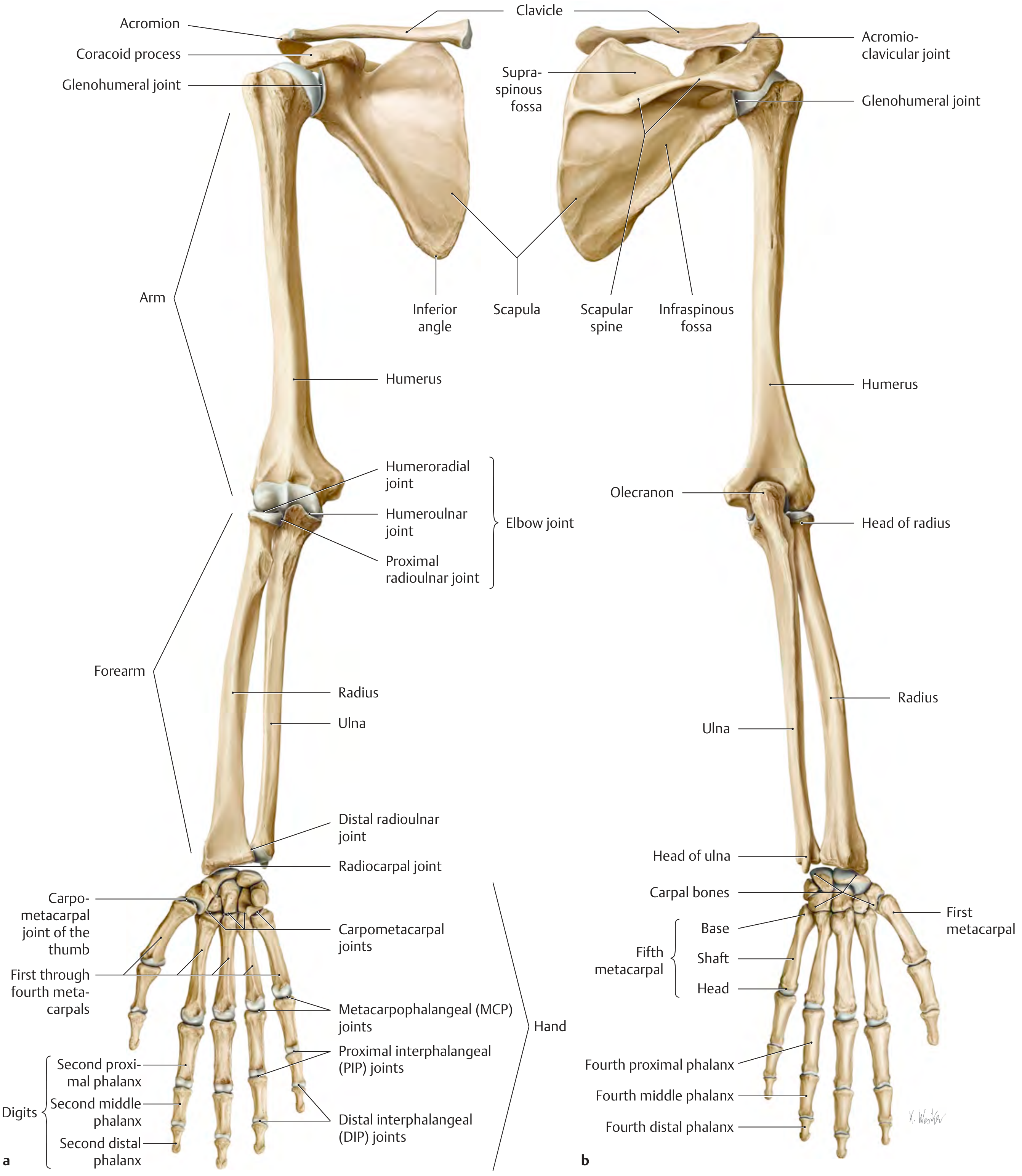
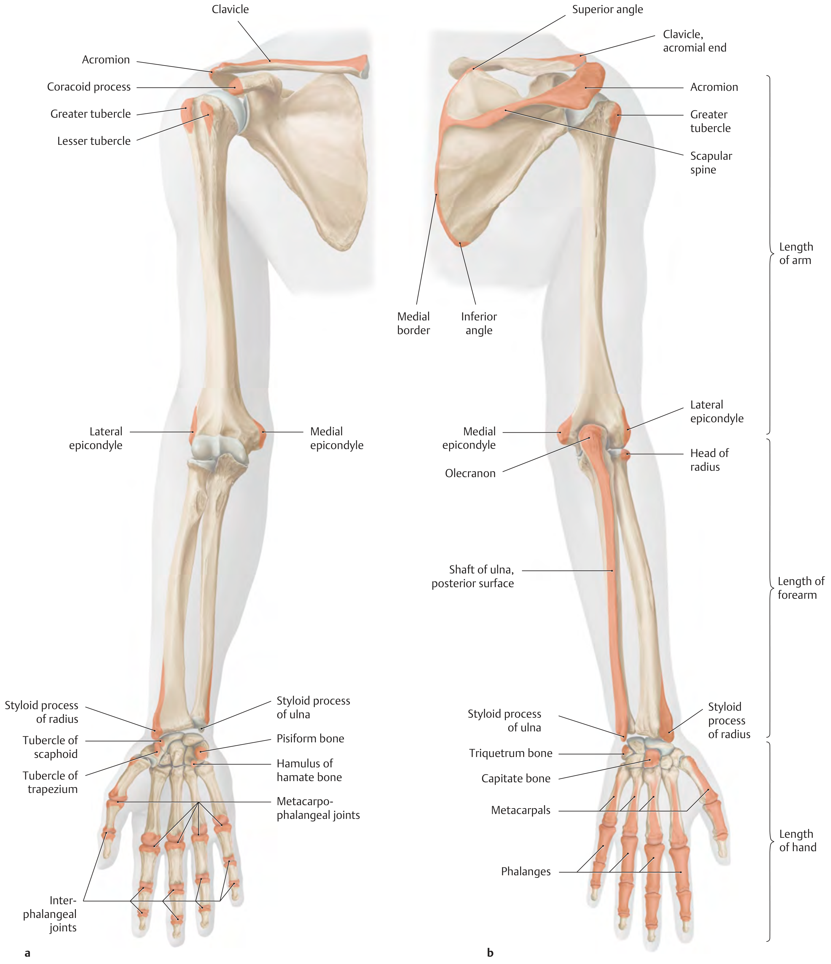
Shoulder Girdle
- Clavicle - connects the upper limb to the axial skeleton at the sternoclavicular joint
- Scapula - flat triangular bone with spine, acromion, coracoid process, supraspinous fossa, infraspinous fossa, subscapular fossa, glenoid cavity
Arm
- Humerus - proximal end has head (articulates with glenoid), anatomical neck, surgical neck, greater and lesser tubercles, intertubercular sulcus (bicipital groove); shaft has the radial (spiral) groove for the radial nerve; distal end has capitulum, trochlea, medial and lateral epicondyles, coronoid fossa, radial fossa (anteriorly), olecranon fossa (posteriorly)
Forearm
- Radius (lateral) - has radial head, radial tuberosity (biceps insertion), styloid process, dorsal tubercle (Lister's tubercle)
- Ulna (medial) - has olecranon, trochlear notch, coronoid process, styloid process
Segment lengths (standard reference points):
- Length of arm = acromion to lateral epicondyle
- Length of forearm = lateral epicondyle to styloid process of radius
- Length of hand = styloid process of radius to tip of third finger
Wrist and Hand
- 8 Carpal bones (proximal row lateral to medial): Scaphoid, Lunate, Triquetrum, Pisiform; (distal row): Trapezium, Trapezoid, Capitate, Hamate
- Mnemonic: "Some Lovers Try Positions That They Can't Handle"
- 5 Metacarpals (I-V)
- 14 Phalanges - thumb has 2 (proximal + distal); fingers 2-5 each have 3 (proximal, middle, distal)
2. Joints
| Joint | Bones | Type | Key Movements |
|---|---|---|---|
| Sternoclavicular | Clavicle + manubrium | Synovial, saddle | Elevation, depression, protraction, retraction |
| Acromioclavicular | Acromion + clavicle | Synovial, plane | Gliding |
| Glenohumeral (shoulder) | Glenoid + humeral head | Synovial, ball-and-socket | Flexion, extension, abduction, adduction, rotation, circumduction |
| Elbow (humeroulnar + humeroradial) | Humerus + ulna + radius | Synovial, hinge | Flexion, extension |
| Proximal radioulnar | Radial head + radial notch of ulna | Synovial, pivot | Pronation / supination |
| Distal radioulnar | Ulna head + ulnar notch of radius | Synovial, pivot | Pronation / supination |
| Radiocarpal (wrist) | Radius + scaphoid, lunate, triquetrum | Synovial, condyloid | Flexion, extension, abduction (radial deviation), adduction (ulnar deviation) |
| Carpometacarpal (CMC) of thumb | Trapezium + 1st metacarpal | Synovial, saddle | Wide range: opposition |
| MCP joints | Metacarpal heads + proximal phalanges | Synovial, condyloid | Flexion, extension, abduction, adduction |
| PIP / DIP joints | Phalanges | Synovial, hinge | Flexion, extension |
The elbow joint capsule contains fat pads overlying the coronoid, radial, and olecranon fossae. The anular ligament of the radius encircles the radial head, allowing pronation/supination. The ulnar and radial collateral ligaments stabilize the elbow.
3. Muscles
Shoulder / Rotator Cuff Muscles (SITS)
| Muscle | Origin | Insertion | Nerve | Action |
|---|---|---|---|---|
| Supraspinatus | Supraspinous fossa | Greater tubercle | Suprascapular | Initiates abduction (first 15°) |
| Infraspinatus | Infraspinous fossa | Greater tubercle | Suprascapular | Lateral rotation |
| Teres minor | Lateral border of scapula | Greater tubercle | Axillary | Lateral rotation |
| Subscapularis | Subscapular fossa | Lesser tubercle | Upper + lower subscapular | Medial rotation |
Other Shoulder Muscles
- Deltoid (anterior, middle, posterior parts) - axillary nerve (C5, C6); abducts arm (>15°), flexion (anterior), extension (posterior)
- Teres major - lower subscapular nerve; medial rotation, adduction, extension
- Serratus anterior - long thoracic nerve (C5, C6, C7); protracts and rotates scapula; loss causes winging of scapula
- Pectoralis major / minor - medial + lateral pectoral nerves; adduction, medial rotation, flexion
- Latissimus dorsi - thoracodorsal nerve (C6-C8); extension, adduction, medial rotation
Arm Muscles
Anterior compartment (flexors, musculocutaneous nerve):
- Biceps brachii - flexes elbow and supinates forearm; long head from supraglenoid tubercle, short head from coracoid process; inserts on radial tuberosity
- Brachialis - primary elbow flexor; deep to biceps; inserts on ulnar tuberosity
- Coracobrachialis - flexes and adducts arm at shoulder; musculocutaneous nerve pierces it
Posterior compartment (extensor, radial nerve):
- Triceps brachii (3 heads - long, medial, lateral) - extends elbow; radial nerve (C6-C8); inserts on olecranon
Forearm Muscles
Anterior compartment (mostly median nerve; ulnar nerve for 1.5 muscles):
- Superficial: Pronator teres, Flexor carpi radialis, Palmaris longus, Flexor carpi ulnaris (ulnar nerve), Flexor digitorum superficialis
- Deep: Flexor digitorum profundus (lateral half = median nerve/AIN; medial half = ulnar nerve), Flexor pollicis longus, Pronator quadratus
Posterior compartment (all radial nerve / deep branch = posterior interosseous nerve):
- Superficial: Brachioradialis, Extensor carpi radialis longus, Extensor carpi radialis brevis, Extensor digitorum, Extensor digiti minimi, Extensor carpi ulnaris
- Deep: Supinator, Abductor pollicis longus, Extensor pollicis brevis, Extensor pollicis longus, Extensor indicis
Hand Muscles
Thenar eminence (median nerve, unless noted):
- Abductor pollicis brevis, Opponens pollicis, Flexor pollicis brevis (superficial head median; deep head ulnar), Adductor pollicis (ulnar nerve)
Hypothenar eminence (all ulnar nerve):
- Abductor digiti minimi, Flexor digiti minimi brevis, Opponens digiti minimi
Intrinsic muscles:
- 4 Dorsal interossei (DAB - abduct fingers) - ulnar nerve
- 3 Palmar interossei (PAD - adduct fingers) - ulnar nerve
- 4 Lumbricals (flex MCP, extend IP joints) - lateral 2 by median nerve, medial 2 by ulnar nerve
4. The Axilla
The axilla is the pyramidal space acting as gateway to the upper limb. Its walls are:
- Anterior wall: Pectoralis major, pectoralis minor, subclavius
- Posterior wall: Subscapularis, teres major, latissimus dorsi
- Medial wall: Serratus anterior over ribs 1-4
- Lateral wall: Intertubercular sulcus of humerus
- Apex (inlet): Bounded by clavicle, superior scapular border, rib I
- Floor (base): Dome of axillary skin between anterior and posterior folds
Key apertures through the posterior wall:
- Quadrangular space: axillary nerve + posterior circumflex humeral artery
- Triangular space: circumflex scapular artery
- Triangular interval: radial nerve + profunda brachii artery
5. Nerves - Brachial Plexus
The brachial plexus (C5-T1) is the nerve supply to the entire upper limb.
Structure: Roots → Trunks (Upper C5-C6, Middle C7, Lower C8-T1) → Divisions → Cords (Lateral, Medial, Posterior) → Branches
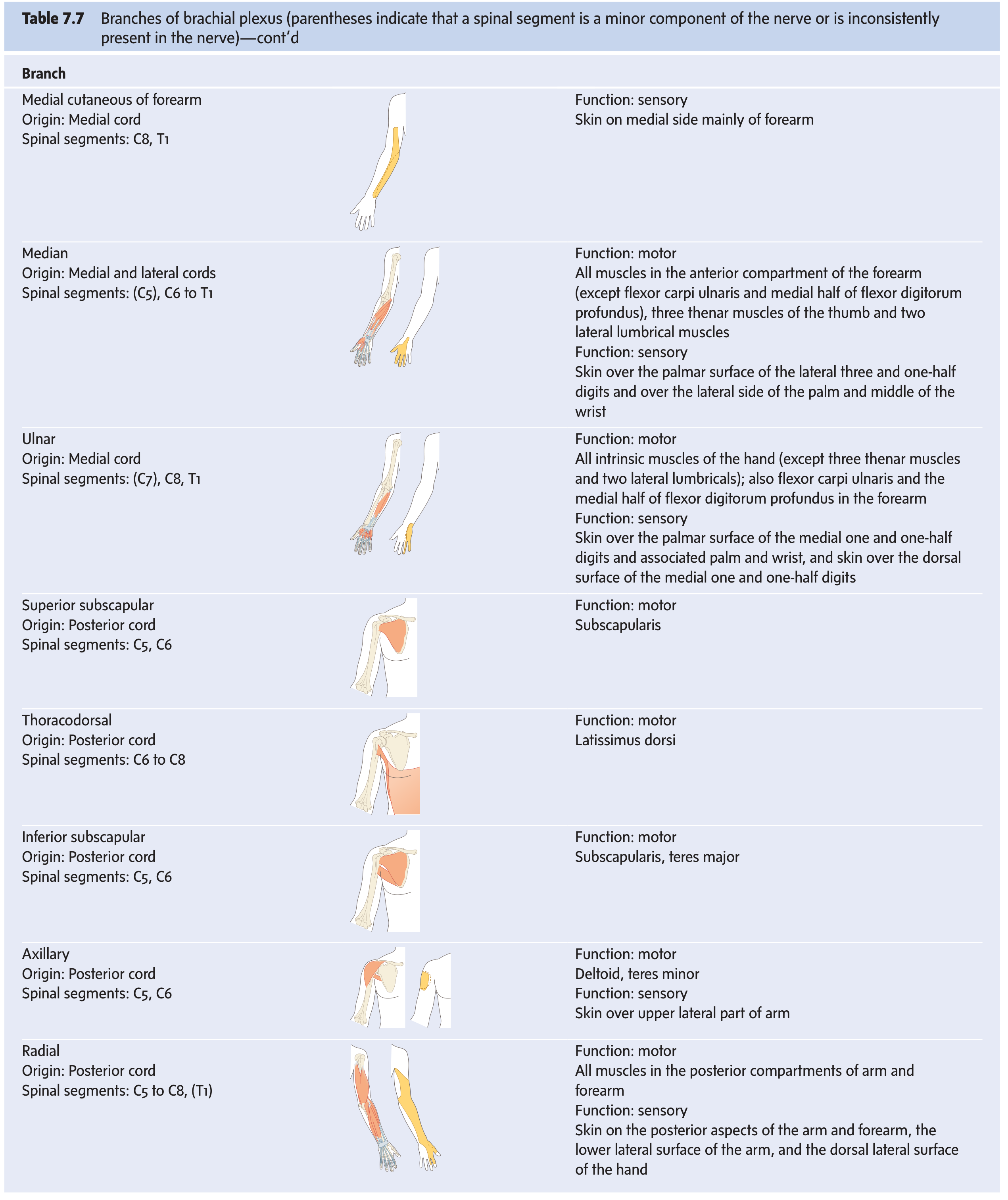
Terminal Branches
| Nerve | Cord of Origin | Spinal Segments | Motor Supply | Sensory Supply |
|---|---|---|---|---|
| Musculocutaneous | Lateral | C5-C7 | Biceps, brachialis, coracobrachialis | Lateral forearm (lateral cutaneous nerve of forearm) |
| Median | Lateral + Medial | C6-T1 | Anterior forearm (except FCU + medial FDP), thenar muscles (LOAF: lateral 2 lumbricals, opponens pollicis, abductor pollicis brevis, flexor pollicis brevis) | Lateral 3½ digits (palmar), lateral palm |
| Ulnar | Medial | C8-T1 | FCU, medial FDP, most intrinsic hand muscles | Medial 1½ digits (palmar + dorsal), medial palm |
| Radial | Posterior | C5-T1 | All posterior compartments of arm + forearm (triceps, brachioradialis, extensors) | Posterior arm + forearm, dorsolateral hand |
| Axillary | Posterior | C5-C6 | Deltoid, teres minor | Upper lateral arm |
Clinically Important Nerve Injuries
- Long thoracic nerve (C5-C7) damage → winging of scapula (serratus anterior paralysis)
- Axillary nerve damage at surgical neck of humerus → loss of deltoid (can't abduct), loss of sensation over lateral arm
- Radial nerve in radial groove → wrist drop (extensors paralyzed); sensation preserved over posterior forearm
- Median nerve at wrist (carpal tunnel) → ape hand (thenar wasting), loss of LOAF muscles
- Ulnar nerve at medial epicondyle → claw hand (ring and little fingers), loss of intrinsics
- Intercostobrachial nerve (T2 lateral cutaneous) passes through the medial axillary wall to supply posteromedial arm skin
6. Blood Supply
Axillary Artery (continuation of subclavian)
Three parts, relative to pectoralis minor:
- Part 1 (medial to PM): Superior thoracic artery
- Part 2 (behind PM): Thoracoacromial artery, Lateral thoracic artery
- Part 3 (lateral to PM): Subscapular artery (→ thoracodorsal + circumflex scapular), Anterior circumflex humeral artery, Posterior circumflex humeral artery (exits with axillary nerve through quadrangular space)
Brachial Artery
Runs in the medial bicipital groove accompanied by the median nerve. Divides at the cubital fossa into:
- Radial artery (lateral) → crosses anatomical snuffbox → deep palmar arch (mainly)
- Ulnar artery (medial) → superficial palmar arch (mainly)
Palmar Arches
| Arch | Dominant supply | Branches |
|---|---|---|
| Superficial | Ulnar artery + superficial branch of radial | 3 common palmar digital arteries → proper digital arteries (fingers 2-5) |
| Deep | Radial artery + deep branch of ulnar | 3 palmar metacarpal arteries; princeps pollicis (to thumb); radialis indicis |
Allen's test assesses adequacy of communication between the two arches.
Major Veins
- Cephalic vein - lateral, runs in deltopectoral groove, drains into axillary vein
- Basilic vein - medial, pierces deep fascia mid-arm, becomes axillary vein
- Median cubital vein - connects cephalic and basilic in cubital fossa (common IV access site)
- Deep veins follow the arteries as venae comitantes
7. Lymphatics
Lymph from the upper limb drains primarily to the axillary lymph nodes (5 groups: anterior/pectoral, posterior/subscapular, lateral, central, apical). The apical nodes drain into the subclavian lymphatic trunk.
8. Key Surface Anatomy Landmarks
- Cubital fossa: Triangle anterior to elbow; base = epicondyle line; medial border = pronator teres; lateral border = brachioradialis; contains biceps tendon (lateral), brachial artery (medial), median nerve (most medial) - mnemonic TAN (Tendon, Artery, Nerve)
- Anatomical snuffbox: Bordered laterally by abductor pollicis longus + extensor pollicis brevis tendons, medially by extensor pollicis longus tendon; floor = scaphoid + trapezium; radial artery crosses its floor; scaphoid fracture causes tenderness here
- Carpal tunnel: Bounded by flexor retinaculum (transverse carpal ligament) anteriorly and carpal bones posteriorly; transmits 4 tendons of FDS, 4 of FDP, flexor pollicis longus, and the median nerve (but NOT the ulnar nerve)
- Guyon's canal: Transmits the ulnar nerve and ulnar artery at the wrist (medial to carpal tunnel)
Summary Diagram
| Region | Bones | Key Muscles | Key Nerve | Key Artery |
|---|---|---|---|---|
| Shoulder girdle | Clavicle, Scapula | Rotator cuff, Deltoid | Axillary, Suprascapular | Axillary |
| Arm | Humerus | Biceps, Brachialis, Triceps | Musculocutaneous (ant.), Radial (post.) | Brachial |
| Forearm | Radius, Ulna | Flexors (ant.), Extensors (post.) | Median + Ulnar (ant.), Radial / PIN (post.) | Radial, Ulnar |
| Hand | 8 Carpals, 5 MC, 14 Phalanges | Thenar, Hypothenar, Interossei, Lumbricals | Median (lateral), Ulnar (medial) | Superficial + Deep palmar arches |
Gray's Anatomy for Students; THIEME Atlas of Anatomy - General Anatomy and Musculoskeletal System
This is a shared conversation. Sign in to Orris to start your own chat.