
Levatir ani muscles
levator ani muscle anatomy pelvic floor
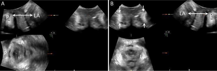
This diagnostic ultrasound image utilizes multiplanar mode (MP mode) to display the normal anatomy of the female pelvic floor across three orthogonal planes: midsagittal, coronal, and axial. The image is divided into panels A and B, illustrating the methodology for assessing the levator ani (LA) muscle and its insertion point on the pubic symphysis (P). In the midsagittal plane (top left of panel A; top right of panel B), a double-sided arrow indicates the minimal anteroposterior (AP) diameter, which serves as a reference for the minimal axial plane. The coronal plane (top left of panel B) highlights the levator ani muscles as symmetrical, crescent-shaped hyperechogenic structures (indicated by white arrows). In the axial plane (bottom left), the relationship between the levator ani and the pubic symphysis is visualized, showing the intact U-shaped morphology of the pelvic floor muscles. This imaging technique is critical in urogynecology for diagnosing levator ani muscle injuries (LAMI) or avulsions, which are key risk factors for pelvic organ prolapse (POP).
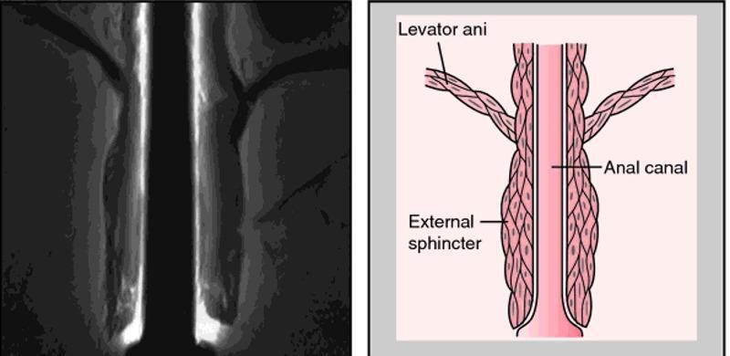
A composite image for medical education comprising a diagnostic radiological image (MRI) on the left and a corresponding anatomical diagram on the right, illustrating the pelvic floor and anal canal. The MRI shows a coronal view of the anorectal region, highlighting the muscular layers of the pelvic outlet. The anatomical diagram provides a schematic representation of the same structures for clarity. It labels the levator ani as a broad, striated muscle group superiorly that forms the pelvic diaphragm and descends to blend with the longitudinal muscle layer of the anal canal. The external sphincter is depicted as a distinct, thicker muscular cuff surrounding the distal portion of the anal canal, located inferior to the levator ani's main attachment points. This visual comparison is designed to teach the spatial relationships between the pelvic floor muscles and the terminal digestive tract, emphasizing the functional anatomy required for fecal continence. The educational focus is on identifying the levator ani and external sphincter during pelvic imaging and understanding their relative morphology.
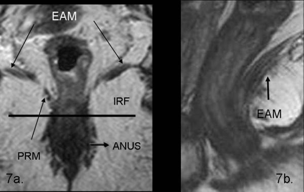
This diagnostic imaging set consists of two T2-weighted turbo spin-echo (TSE) MRI scans of the pelvic floor, demonstrating normal rectal and perianal anatomy. Image 7a is a coronal view illustrating the spatial relationship between the levator ani muscle (EAM), visible as a low-signal intensity band extending laterally and superiorly, and the puborectalis muscle (PRM) forming a muscular sling around the anal canal. The ischio-rectal fossa (IRF) is identified as a hyperintense region of fat lateral to the sphincter complex. The anal canal (ANUS) is shown in the midline. Image 7b is a sagittal view providing a longitudinal perspective of the levator ani muscle (EAM), appearing as a hypointense linear structure supporting the pelvic viscera. These images serve as an educational reference for identifying normal pelvic floor musculature, including the internal and external anal sphincters, and are critical for the staging and preoperative planning of rectal carcinomas and pelvic floor disorders.
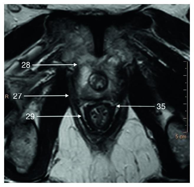
This T2-weighted axial magnetic resonance imaging (MRI) scan displays the cross-sectional anatomy of the male pelvic floor at the level of the prostate apex. The image highlights the musculoskeletal and fascial relationships critical for pelvic surgery. The central feature is the prostate gland, which exhibits a hyperintense (bright) signal relative to the surrounding pelvic musculature. Key anatomical landmarks are labeled: 28 indicates the puboprostatic (levator prostate) muscle located anterior to the gland; 27 denotes the pubococcygeus muscle situated laterally; 29 marks the puborectalis muscle located inferiorly, forming part of the levator ani complex; and 35 identifies the pararectal fascia positioned posteriorly, separating the rectum from the levator ani. The pelvic muscles generally appear hypointense (dark), providing high contrast against the glandular tissue and periprostatic fat. This diagnostic imaging serves as an educational tool for understanding the zonal anatomy of the prostate and the complex fascial planes of the lesser pelvis.
Levator Ani Muscles
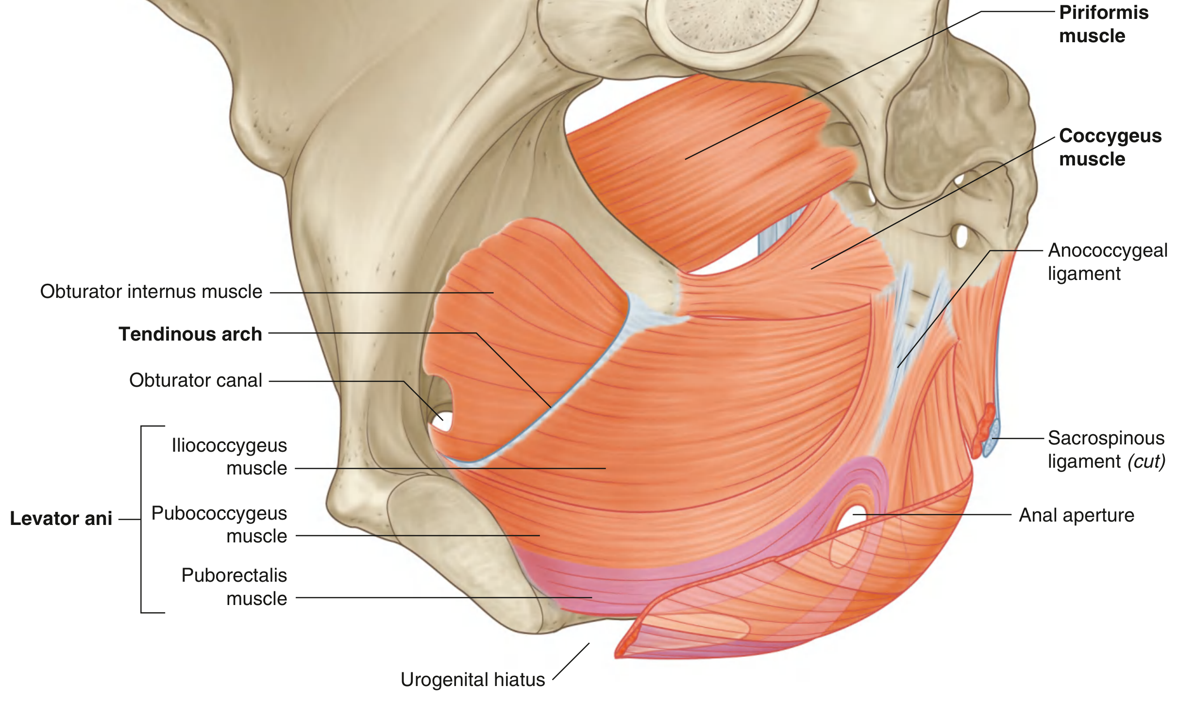
Origin
- Posterior aspect of the body of the pubic bone
- Tendinous arch - a linear thickening in the fascia covering the obturator internus muscle
- Spine of the ischium
Three Parts of Levator Ani
1. Pubococcygeus
- Origin: Body of pubis (lateral to origin of puborectalis)
- Insertion: Anococcygeal ligament and coccyx (courses posteriorly attaching along the midline)
- Subdivisions (based on midline association):
- Puboprostaticus (levator prostatae) - in males
- Pubovaginalis - in females
- Puboanalis
2. Puborectalis
- Origin: Superior pubic ramus on both sides of the pubic symphysis
- Insertion: Forms a loop/sling around the anorectal junction, interwoven with the deep part of the external anal sphincter
- Key function: Maintains the anorectal angle (perineal flexure) - approximately 90° at rest - which acts as a "pinch valve" to maintain fecal continence
- During defecation, relaxation of puborectalis widens the anorectal angle to ~130-140°
3. Iliococcygeus
- Origin: Tendinous arch of the obturator internus fascia
- Insertion: Levator (iliococcygeal) raphe, anococcygeal ligament, and coccyx
- Joins its partner from the opposite side to form the anococcygeal ligament (anococcygeal body)
Key Relationships
| Feature | Detail |
|---|---|
| Urogenital hiatus | Anterior U-shaped gap between the two muscles; transmits the urethra (both sexes) and vagina (females) |
| Anococcygeal ligament | Posterior midline raphe where muscles meet, attaches to coccyx |
| Perineal body | Midline junction posterior to vagina (females) |
Innervation
- Direct branches from anterior ramus of S4
- Inferior rectal branch of the pudendal nerve (S2-S4)
Functions
- Supports pelvic viscera - forms a muscular floor holding bladder, rectum, uterus/prostate in place
- Maintains anorectal angle - puborectalis sling keeps rectum-anal canal angle ~90° for continence
- Reinforces external anal sphincter
- Acts as vaginal sphincter in females (pubovaginalis fibers)
- Participates in defecation - relaxes and straightens the anorectal angle; then contracts to return anal canal to position post-defecation
Summary Table
| Part | Origin | Insertion | Innervation |
|---|---|---|---|
| Pubococcygeus | Pubis (lateral) | Anococcygeal ligament, coccyx | Pudendal nerve (S2-S4) |
| Puborectalis | Superior pubic ramus (both sides) | Sling around anorectal junction | Pudendal nerve + sacral plexus (S2-S4) |
| Iliococcygeus | Tendinous arch of obturator internus fascia | Iliococcygeal raphe, anococcygeal ligament, coccyx | Pudendal nerve (S2-S4) |
Clinical Notes
- Levator ani syndrome - chronic anorectal pain from spasm of the levator ani, presenting as a dull ache/pressure in the rectum
- Pelvic organ prolapse - levator ani injury (commonly from obstetric trauma) weakens the pelvic floor support, leading to bladder, uterine, or rectal prolapse. MRI is used to assess levator ani muscle avulsions as key predictors of prolapse risk
- Defecation disorders - failure of puborectalis to relax (anismus) causes obstructed defecation