
Management of patient in sickle cell crisis
"sickle cell" AND "vaso-occlusive crisis" AND management
sickle cell disease pathophysiology management
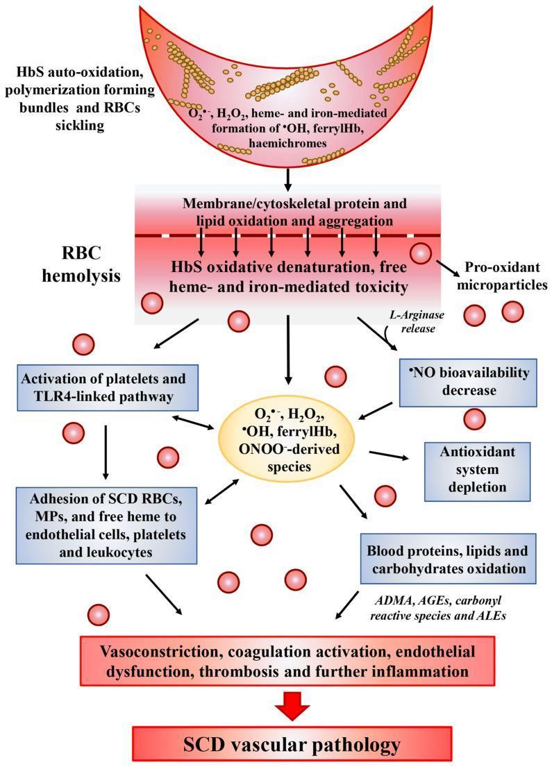
A medical pathophysiology diagram illustrating the oxidative stress cascade in Sickle Cell Disease (SCD) and its progression to vascular pathology. The diagram follows a vertical flow starting with a sickled red blood cell (RBC) undergoing HbS auto-oxidation and polymerization, leading to the generation of reactive oxygen species (ROS) such as H2O2, ferrylHb, and hemichromes. This triggers a central event of RBC hemolysis, characterized by membrane lipid oxidation, protein aggregation, and the release of pro-oxidant microparticles and L-arginase. Downstream pathways depict the systemic consequences, including the activation of platelets and the TLR4-linked inflammatory pathway, adhesion of SCD RBCs to endothelial cells and leukocytes, and a significant decrease in nitric oxide (NO) bioavailability. The lower section highlights the depletion of the antioxidant system and the oxidative modification of blood components into ADMA, AGEs, and ALEs. The cascade culminates in clinical manifestations such as vasoconstriction, endothelial dysfunction, and thrombosis, collectively summarized as SCD vascular pathology. This illustration is designed for advanced medical education regarding the molecular mechanisms of hematologic disorders.
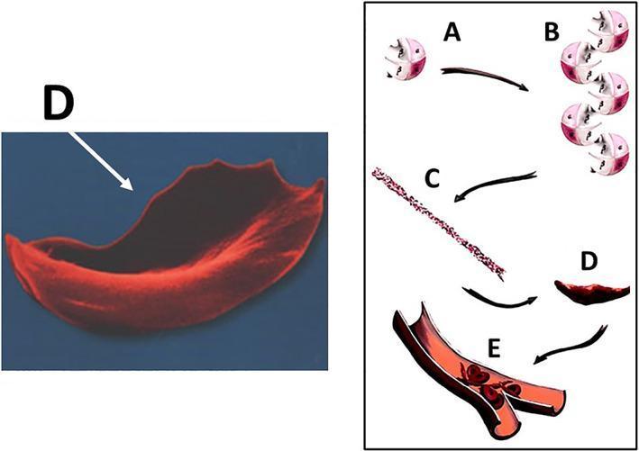
This educational graphic illustrates the molecular and cellular pathophysiology of sickle cell disease (SCD) leading to vaso-occlusion. The diagram is divided into a microscopic anatomical illustration and a high-magnification scanning electron micrograph. Steps A through D depict the hemoglobin S (HbS) polymerization process: (A) individual deoxygenated hemoglobin tetramers, (B) aggregation of hemoglobin molecules, (C) formation of long, rigid hemoglobin polymers or strands, and (D) the resulting transformation of a flexible biconcave red blood cell into a rigid, crescent or sickle-shaped erythrocyte. A corresponding scanning electron micrograph (labeled D) highlights the characteristic elongated, spicular morphology of a sickled cell. Section E shows a cross-section of a small bifurcating blood vessel where these rigid, abnormally shaped cells are aggregating, causing mechanical obstruction (vaso-occlusion) of the lumen. This process demonstrates the mechanism behind tissue ischemia and subsequent complications such as osteonecrosis in SCD patients. The content is suitable for medical students and clinicians studying hematology and musculoskeletal pathology.
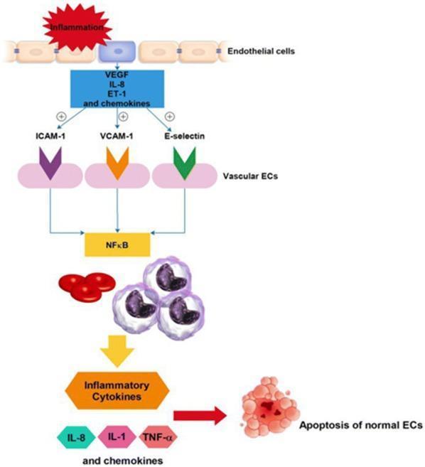
This pathophysiology diagram illustrates the signaling pathway of endothelial cell (EC) activation and apoptosis, particularly in the context of sickle cell disease (SCD) interactions with the vascular wall. The process begins with 'Inflammation' triggering the release of mediators including VEGF, IL-8, ET-1, and chemokines. These factors upregulate adhesion molecules on vascular ECs, specifically ICAM-1, VCAM-1, and E-selectin (represented by colored chevron icons). Activation of these molecules converges on the NF-̄Κβ signaling hub. This pathway facilitates the recruitment and interaction of homozygotic SS red blood cells (RBCs) and leukocytes (white blood cells). The subsequent cascade leads to the secretion of inflammatory cytokines, notably IL-8, IL-1, and TNF-α. The final stage of this inflammatory loop is the induction of apoptosis in normal endothelial cells, shown as a cluster of fragmented cells. The diagram serves as a model for understanding vaso-occlusive mechanisms and potential therapeutic targets like NF-̄Κβ inhibition in hematological disorders.
Management of Sickle Cell Crisis
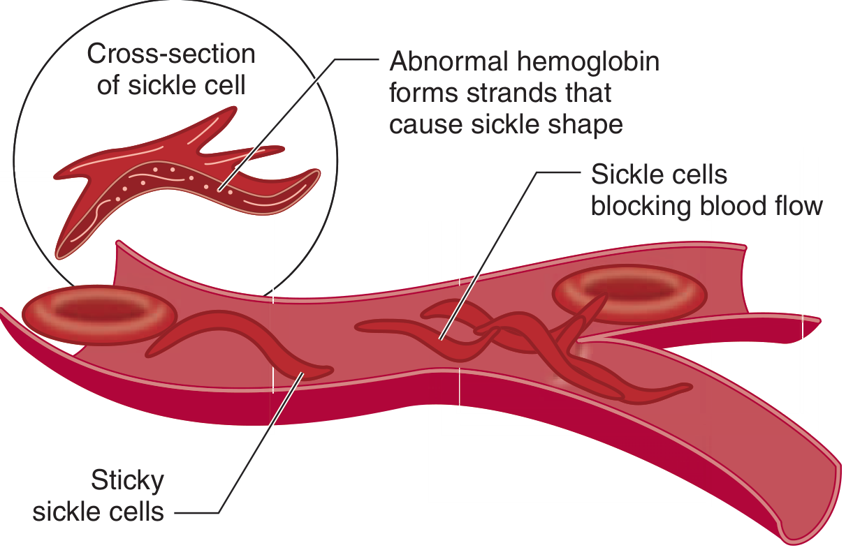
Background & Pathophysiology
- Increased blood viscosity
- Microvascular occlusion
- Tissue ischemia and infarction
- Chronic hemolysis
Types of Sickle Cell Crisis
| Crisis Type | Key Features |
|---|---|
| Vaso-occlusive (VOC) / Pain crisis | Most common ED presentation; bone/muscle/joint pain |
| Acute Chest Syndrome (ACS) | New lung infiltrate + fever, hypoxia, chest pain |
| Splenic Sequestration | Rapidly enlarging spleen, Hb ≥2 g/dL below baseline |
| Aplastic Crisis | Parvovirus B19; low retic count, severe anemia |
| Stroke | Focal neurologic signs; TCD detects increased velocities |
| Priapism | Painful erection >4 hours |
1. Vaso-Occlusive Pain Crisis - Management
Initial Assessment (within 30 minutes)
- Duration, location, and character of pain
- Precipitating factors; fever, cough, respiratory symptoms
- Medications already taken
- How this episode compares to prior episodes
- Vital signs: temperature, SpO2, pulse
- Respiratory exam (rule out ACS)
- Abdominal exam (spleen size, hepatomegaly)
- Assess hydration status
- CBC + reticulocyte count
- Metabolic panel (renal function, electrolytes)
- LFTs, LDH, bilirubin (hemolysis markers)
- Urinalysis
- Blood cultures + CXR if febrile
- CXR if any respiratory symptoms
A mildly elevated WBC (up to ~15,000) and low-grade fever are common during uncomplicated VOC. A WBC >20,000 with bandemia suggests concurrent infection and needs further workup.
A. Pain Management
- NSAIDs (e.g., ketorolac 15-30 mg IV or ibuprofen orally): reduce inflammation, opioid-sparing effect; well tolerated by patients; avoid with renal impairment
- Acetaminophen 500-1000 mg orally/IV every 6 hours (can combine with NSAIDs)
- Oral opioids (e.g., codeine, oxycodone) if home NSAIDs have failed
- Morphine 0.1-0.15 mg/kg IV/SC every 2-4 hours (most widely used)
- Hydromorphone 0.015-0.02 mg/kg IV every 3-4 hours (preferred in opioid-tolerant or chronic pain patients; useful in children)
- Patient-controlled analgesia (PCA): preferred for consistent dosing when hospitalized
- Reassess pain every 30-60 minutes and titrate; do not underdose
- Avoid meperidine (pethidine) - seizure risk from normeperidine accumulation
- Avoid mixed agonist-antagonists (e.g., pentazocine) in opioid-tolerant patients
- NSAIDs added to opioids reduce opioid requirements and are well-accepted by patients
- Non-pharmacologic: warmth, rest, distraction (TV, music), calm environment
- Patients on chronic hydroxyurea: continue it
Recent meta-analysis (PMID 42190636, 2026) found that early opioid administration in pediatric VOC is associated with faster pain relief, supporting timely escalation to opioids in children.
B. Hydration
- Oral fluids: ~3 L/day if tolerated
- IV fluids (0.9% saline or 0.45% saline) if:
- Unable to maintain oral intake
- Clinically dehydrated
- Moderate-severe crisis
- Avoid overhydration, especially in ACS (risk of pulmonary edema)
- Target urine output ~1 mL/kg/hour
- D5 0.45% NaCl is commonly used in pediatric patients
C. Oxygen
- Supplemental O2 only if SpO2 <95% or patient is hypoxic
- Do NOT routinely administer oxygen to all patients - it does not abort sickling in non-hypoxic patients and may suppress erythropoiesis with prolonged use
D. Transfusion
- Do NOT transfuse for asymptomatic anemia - these patients have chronic anemia and are compensated
- Simple transfusion is indicated for:
- Aplastic crisis with severe symptomatic anemia
- Splenic sequestration (use 5-10 mL/kg aliquots; avoid over-transfusion as autotransfusion from spleen can cause rebound rise in Hb)
- Symptomatic severe anemia (Hb significantly below baseline)
- Exchange transfusion (red cell exchange): preferred over simple transfusion for:
- Stroke (maintains HbS <30%)
- Severe ACS not responding to simple transfusion
- Multi-organ failure
E. Antibiotics / Infection
- Sickle cell patients are functionally asplenic - high risk for encapsulated organisms (S. pneumoniae, H. influenzae, N. meningitidis)
- Fever ≥38.5°C → treat as medical emergency
- Blood cultures before antibiotics
- Empiric broad-spectrum antibiotics (e.g., ceftriaxone 50-75 mg/kg in children; ceftriaxone or amoxicillin-clavulanate in adults) should be started promptly
- Children: prophylactic penicillin V (125 mg BID up to age 3; 250 mg BID age 3-5) should be confirmed as ongoing
2. Acute Chest Syndrome (ACS)
- Hospital admission
- IV fluids (cautiously - avoid overhydration)
- Supplemental oxygen to maintain SpO2 ≥95%
- Incentive spirometry (prevents atelectasis)
- Broad-spectrum antibiotics covering atypical organisms (e.g., ceftriaxone + azithromycin/erythromycin)
- Simple transfusion (or exchange transfusion if severe) to raise Hb to ~10 g/dL and reduce HbS
- Bronchodilators if wheezing
- Exchange transfusion if rapidly deteriorating, SpO2 <90% despite O2, or multi-lobar involvement
- Avoid NSAIDs (can worsen pulmonary function in ACS)
3. Splenic Sequestration
- Rapidly enlarging spleen with Hb ≥2 g/dL below baseline
- Management: type and screen, serial abdominal exams, IV fluid resuscitation, transfusion in 5-10 mL/kg aliquots (beware rebound hyperviscosity as spleen re-releases blood)
- Splenectomy considered after recurrent episodes
4. Aplastic Crisis
- Usually triggered by Parvovirus B19, which halts RBC production
- CBC shows: Hb well below baseline + low or zero reticulocyte count
- Management: type and screen, parvovirus serology/PCR, simple transfusion if anemia is severe/symptomatic, isolate patient (parvovirus is infectious), IV fluids
5. Stroke
- Ischemic stroke is 300x more common in SCD children than the general population
- Management:
- Urgent MRI brain + MRA
- Emergency exchange transfusion (preferred) OR simple transfusion (reduces HbS to <30%)
- Chronic transfusion program to maintain HbS <30% post-stroke
- Transcranial Doppler (TCD) screening annually from age 2: if velocities >200 cm/s, start prophylactic transfusion
6. Priapism
- Sustained painful erection >4 hours in SCD males
- Management:
- Analgesia (IV opioids as per VOC protocol)
- Oral/IV hydration
- Supplemental oxygen
- Urology consult for corporal aspiration and phenylephrine irrigation if does not resolve within a few hours
- Avoid exchange transfusion (risk of worsening with rapid Hb rise)
7. Discharge vs Admission Criteria
- Pain not controlled after 4-6 hours of IV analgesia
- Fever ≥38.5°C (especially in children)
- New respiratory symptoms or drop in SpO2
- Aplastic crisis, ACS, stroke, or sequestration
- Inability to tolerate oral fluids
- Management needs observation setting until stable
8. Long-term / Chronic Management
| Agent | Indication |
|---|---|
| Hydroxyurea | Reduces VOC frequency by increasing HbF; indicated in moderate-severe SCD |
| Prophylactic penicillin | Children up to age 5 (functionally asplenic) |
| Folic acid | Daily supplementation for all SCD patients (high RBC turnover) |
| Pneumococcal, meningococcal, Hib vaccines | Essential due to functional asplenia |
| Transcranial Doppler screening | Annual from age 2; high-risk children get chronic transfusion |
| Chronic transfusion program | Post-stroke; high TCD velocities; recurrent ACS |
| Bone marrow transplant | Only curative therapy (allogeneic HSCT) |
| Voxelotor, crizanlizumab, L-glutamine | Newer FDA-approved agents for SCD management |
Key Points Summary
- Pain crisis: analgesia within 30-60 min; use NSAIDs + opioids (avoid meperidine); PCA for severe/admitted patients
- Hydration: ~3 L/day orally or IV; avoid over-hydration
- Oxygen: only if SpO2 <95%
- Fever is a medical emergency in SCD - early cultures + empiric antibiotics
- ACS = new infiltrate + respiratory symptoms: antibiotics + exchange transfusion if severe
- Stroke = emergency exchange transfusion
- Do not transfuse for chronic baseline anemia alone
- Hydroxyurea remains the cornerstone of long-term disease modification