
Difference between classical and non classical migraine..refer to Harrison textbook of medicine .
migraine with aura visual scotoma scintillating
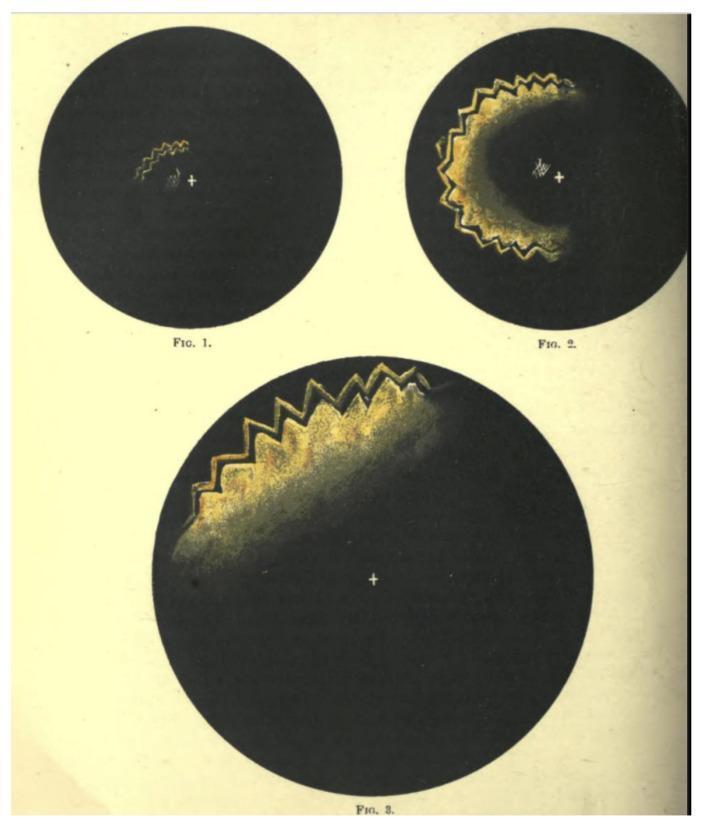
This sequence of three historical illustrations, labeled Figure 1 through Figure 3, depicts the temporal and spatial progression of a visual migraine aura (fortification spectra). Each figure presents a circular visual field with a central white fixation crosshair against a black background. The images demonstrate the characteristic 'march' of the aura from a central point toward the periphery. In Figure 1, a small, luminous, yellow-gold zig-zagged arc appears near the fixation point. In Figure 2, the disturbance has expanded into a larger crescent shape with a distinct crenellated or 'fortress-wall' border, accompanied by a trailing area of relative scotoma or blurred vision. Figure 3 illustrates the advanced stage, where the jagged, iridescent arc has migrated to the superior periphery of the visual field, significantly increasing in scale. These illustrations by James Mackenzie serve as a classic clinical representation of the neurological phenomenon of cortical spreading depression as perceived by a patient, highlighting the hallmark characteristics of teichopsia: jagged morphology, peripheral expansion, and associated visual field deficit.
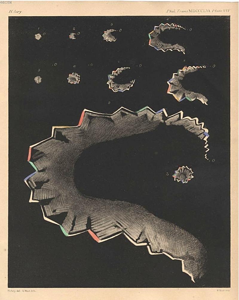
This historical medical illustration, titled 'Stages of Teichopsia' (1870), is a landmark clinical diagram representing the progression of a migraine aura across the visual field. The image depicts nine chronological stages (numbered 1-9) of a visual disturbance known as a scintillation scotoma or 'fortification spectrum'. The sequence begins with a small, central point of light and evolves into an expanding, crescentic arc. The figures are characterized by their jagged, crenellated edges, resembling the bastions of a fortified town, which Hubert Airy termed 'teichopsia'. These edges feature a prismatic or polychromatic effect, with alternating segments of red, green, blue, and yellow against a dark background, illustrating the 'shimmering' or 'boiling' nature of the aura. The diagram is an important record in the history of neurology and ophthalmology, providing a subjective yet scientifically detailed mapping of transient hemiopsia and the physiological manifestation of cortical spreading depression as perceived by a patient. This content is relevant for studying the clinical presentation of migraine with aura and the history of diagnostic visualization in neurology.
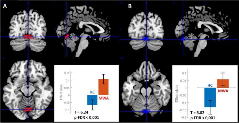
This diagnostic image displays resting-state functional MRI (rs-fMRI) seed-to-voxel connectivity analysis results for patients with migraine with aura (MWA). The visual is divided into two sections, A and B, each showing three orthogonal brain views (coronal, sagittal, and axial) and an accompanying bar graph. Section A illustrates increased functional connectivity between the right antero-dorsal insula (ROI 1 R) and a posterior cluster colored in red/orange. Section B illustrates increased functional connectivity between the left antero-dorsal insula (ROI 1 L) and a similar cluster colored in blue. Blue crosshairs in both sections localize the primary cluster to the cerebellar vermis (Vermis VI/VII) and the lingual gyrus. The bar graphs compare 'Effect Sizes' between Healthy Controls (HC) and Migraine with Aura (MWA) patients, indicating significantly higher connectivity in the MWA group (p-FDR < 0.001). Anatomical landmarks visible include the cerebral cortex, corpus callosum, brainstem, and cerebellar hemispheres. This image is used to demonstrate neuro-pathophysiological differences in functional brain networks associated with migraine disorders.
Classical vs. Non-Classical Migraine - Harrison's Principles of Internal Medicine, 22nd Edition (2025)
Terminology note: The older terms "classical migraine" and "non-classical (common) migraine" have been replaced in modern classification. The International Classification of Headache Disorders-III (ICHD-III), as reproduced in Harrison's Table 441-1, now uses:
- Classical migraine = Migraine with aura (ICHD 1.2)
- Non-classical (common) migraine = Migraine without aura (ICHD 1.1)
Overview
ICHD Classification (Harrison's Table 441-1)
| Code | Type |
|---|---|
| 1.1 | Migraine without aura (non-classical) |
| 1.2 | Migraine with aura (classical) |
| 1.2.1 | Migraine with typical aura |
| 1.2.1.1 | Typical aura with headache |
| 1.2.1.2 | Typical aura without headache (acephalgic migraine) |
| 1.2.2 | Migraine with brainstem aura |
| 1.2.3 | Hemiplegic migraine |
| 1.2.4 | Retinal migraine |
| 1.3 | Chronic migraine |
Key Differences at a Glance
| Feature | Classical Migraine (with aura) | Non-Classical Migraine (without aura) |
|---|---|---|
| Old term | Classical migraine | Common migraine |
| Prevalence | ~20-25% of migraineurs | ~75-80% of migraineurs (majority) |
| Aura | Present - focal neurologic symptoms preceding headache | Absent |
| Aura type | Visual (most common: scintillating scotoma, zigzag/fortification spectra), sensory, speech | None |
| Aura duration | Typically 20-30 min, by definition reversible and <60 min | N/A |
| Onset of headache | Follows or overlaps with aura | No aura precedes it |
| Headache character | Throbbing/pulsating, often unilateral | Throbbing/pulsating, often unilateral |
| Associated features | Nausea, vomiting, photophobia, phonophobia, allodynia | Same |
| Stroke risk | Elevated (especially in women using combined OCP) | Lower |
| OCP contraindication | Yes - estrogen-containing contraceptives are contraindicated | No absolute contraindication |
| Triptan timing | Triptans NOT effective during aura; must be given after aura ends and headache begins | Triptans effective at headache onset |
| Genetics | Ion channel mutations implicated (FHM genes: CACNA1A, ATP1A2, SCN1A) | Polygenic susceptibility |
The Aura - Detailed (Harrison's)
- Sensory aura - paresthesias (pins and needles) spreading in a characteristic march
- Motor aura - weakness (hemiplegic migraine - a special subtype)
- Speech/language aura - dysphasia
- Brainstem aura - dysarthria, vertigo, tinnitus, diplopia (replaced older term "basilar artery migraine")
Phases of a Migraine Attack (both types share these, except aura)
-
Premonitory/Prodromal phase (hours to days before headache):
- Neck discomfort, cognitive impairment ("brain fog"), mood change, fatigue, yawning/sleepiness, polyuria/polydipsia, food cravings
-
Aura (classical migraine only):
- Neurologic disturbance, typically scintillating scotoma, lasting 20-30 min
-
Headache phase:
- Pain (typically unilateral, throbbing), nausea/vomiting, photophobia, phonophobia, osmophobia, allodynia, vertigo
-
Postdrome:
- Tiredness, weariness, concentration impairment - lasting hours to up to a day
Pathophysiology (both types share the same core mechanism)
- FHM1: CACNA1A (Ca2.1 P/Q-type calcium channel)
- FHM2: ATP1A2 (Na+/K+ ATPase)
- FHM3: SCN1A (neuronal voltage-gated sodium channel)
Clinical Implications
-
Contraception: Harrison's explicitly states that migraine with aura is a contraindication to estrogen-containing contraceptives (combined OCP, patch, ring), due to increased stroke risk. Progestin-only methods are safe.
-
Triptan use: "Triptans are generally not effective in migraine with aura unless given after the aura is completed and the headache initiated." - Harrison's 22E. In migraine without aura, triptans can be given at headache onset.
-
Acephalgic migraine (aura without headache, ICHD 1.2.1.2): A patient can have the full aura of classical migraine with little or no head pain. This is particularly relevant in older patients and must be differentiated from TIA.
-
Stroke association (migrainous infarction): A complication listed under ICHD 1.4.3 - occurs almost exclusively in migraine with aura.