
Portal vein
Reading File
The portal vein is a major venous structure carrying nutrient-rich blood from the GI tract and spleen to the liver. Let me pull comprehensive content from the medical library.
Searching sources...
Finding Sources
Finding Sources
Good hits. Now let me read the most authoritative anatomy and surgical sections in parallel.
Reading File
Reading File
Reading File
Excellent content. Now let me fetch the anatomy diagram and also get portal hypertension / portosystemic anastomoses data.
Loading Image
Finding Sources
Finding Sources
Now let me get the portosystemic anastomoses details and the portal vein thrombosis section.
Reading File
Reading File
I now have comprehensive content. Let me also fetch the portosystemic anastomoses diagram.
Loading Image
I now have everything needed for a thorough, well-illustrated answer. Here is the complete coverage of the portal vein:
Portal Vein
Formation
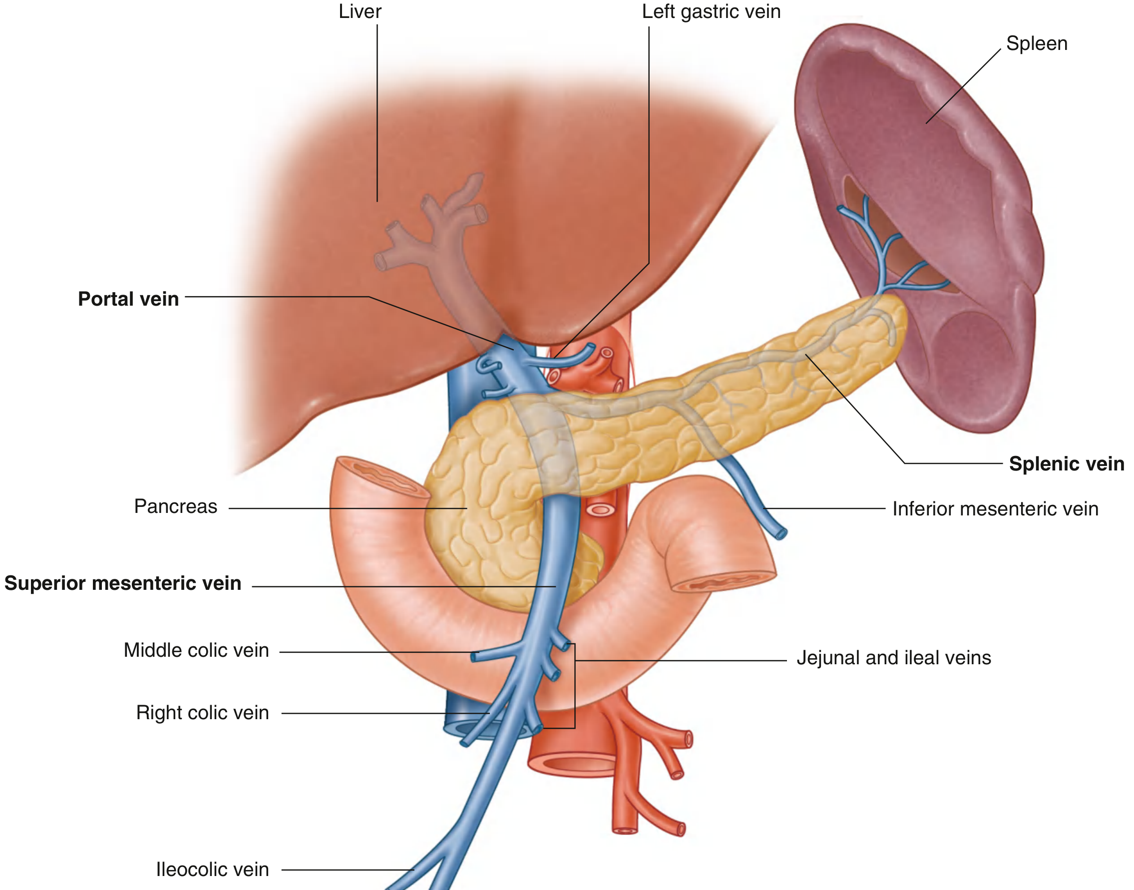
The portal vein is formed by the union of the superior mesenteric vein (SMV) and the splenic vein, posterior to the neck of the pancreas at the level of vertebra L2. The inferior mesenteric vein (IMV) usually drains into the splenic vein just upstream from this confluence. The main portal vein ranges from 5.5 to 8 cm in length and approximately 1 cm in diameter.
- Gray's Anatomy for Students, p. 417
- Sabiston Textbook of Surgery, p. 1864
Course
After formation, the portal vein:
- Passes posterior to the superior part of the duodenum (first part of duodenum)
- Enters the right margin of the lesser omentum (hepatoduodenal ligament)
- Within the hepatoduodenal ligament it occupies the posterior position - with the bile duct to its right and the hepatic artery proper to its left; the vein is anterior to the omental (epiploic) foramen of Winslow
- Ascends to the porta hepatis where it divides into right and left branches that enter the liver parenchyma
Portal Vein Anatomy Diagram (Gray's Anatomy for Students)
Tributaries
| Tributary | Drains |
|---|---|
| Splenic vein | Spleen, pancreas, fundus/greater curvature of stomach (via short gastric + left gastro-omental veins) |
| Superior mesenteric vein | Small intestine, right/transverse colon (via jejunal, ileal, ileocolic, right colic, middle colic veins) |
| Inferior mesenteric vein | Left colon, sigmoid, upper rectum - usually joins splenic vein |
| Right and left gastric (coronary) veins | Lesser curvature of stomach and abdominal esophagus |
| Cystic veins | Gallbladder |
| Para-umbilical veins | Connect to anterior abdominal wall veins via ligamentum teres |
The left gastric (coronary) vein inserts variably: at the SMV-splenic junction (~60%), in the portal vein proper (~25%), or in the splenic vein (~15%).
Intrahepatic Distribution
The portal vein bifurcates at the liver hilum into:
- Left portal vein: runs transversely along the base of segment IV, then bends 90° into the umbilical fissure, giving off branches to segments II, III, and IV; also supplies the left caudate lobe
- Right portal vein: shorter extrahepatic course, divides into right anterior and right posterior sectoral branches supplying the right lobe
Standard anatomy (main portal vein → left + right, then right → anterior + posterior sectoral) is present in ~70% of individuals. The most common variant is portal vein trifurcation (main portal vein → left + right anterior + right posterior), and the second most common is the right posterior portal vein arising as the first branch of the main portal vein.
The portal vein is unique in having both tributaries (from splanchnic organs) and branches (into the liver).
- Sabiston Textbook of Surgery, p. 1864
- Schwartz's Principles of Surgery, p. 1376
Physiology
| Parameter | Value |
|---|---|
| Normal portal pressure | 3-5 mmHg |
| Contribution to hepatic blood flow | ~75% |
| Contribution to hepatic oxygen supply | 50-70% |
| Valves | None (valveless throughout) |
Despite being postcapillary and largely de-oxygenated blood, the high flow rate means the portal vein delivers 50-70% of the liver's oxygen. The absence of valves allows pressure measurement at any point along the system and also allows reversal of flow in portal hypertension.
Portosystemic Anastomoses
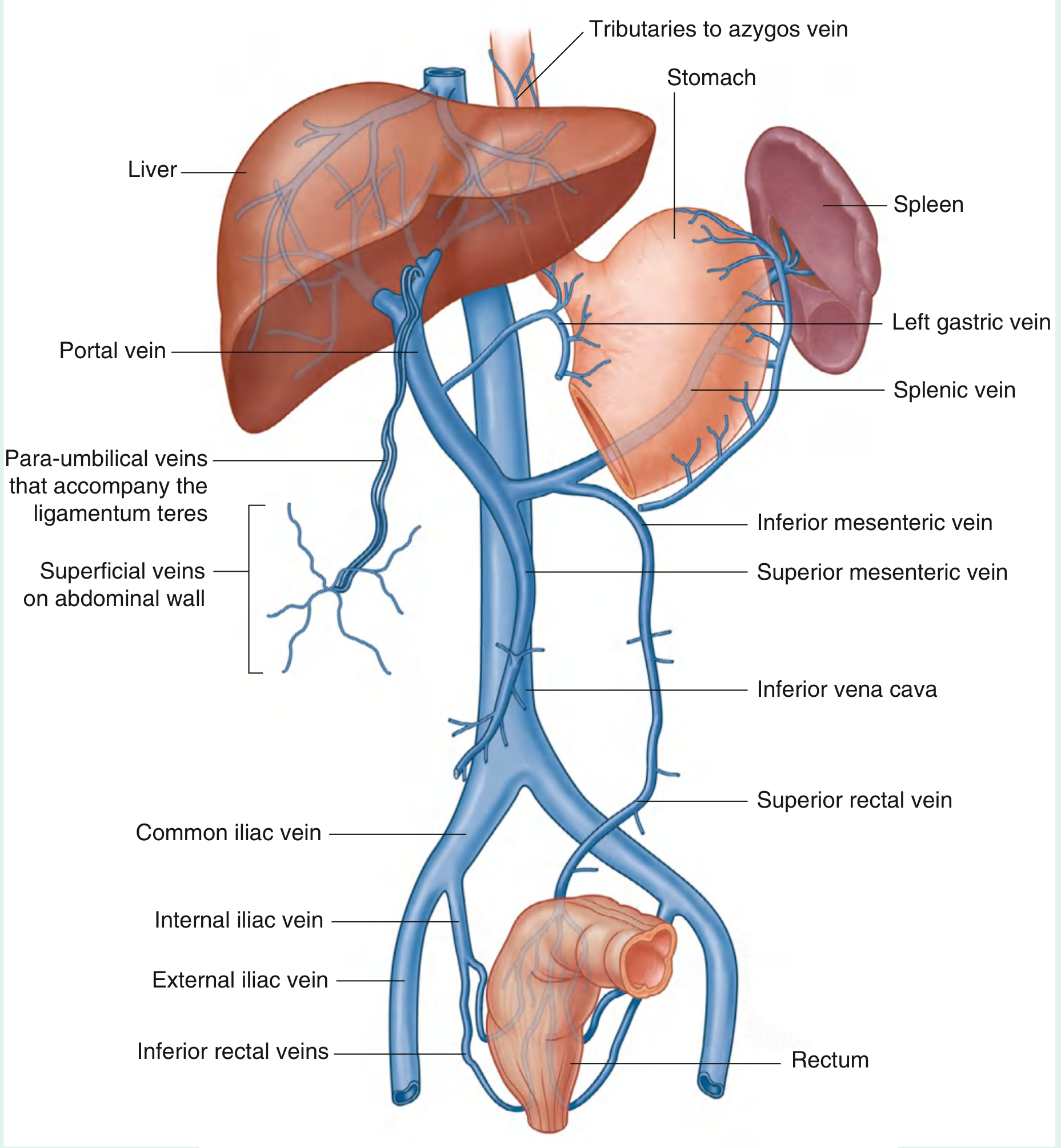
Because the portal system is valveless, elevated portal pressure forces blood to decompress into the systemic venous system at sites where the two systems communicate:
| Site | Portal side | Systemic side | Clinical consequence |
|---|---|---|---|
| Gastroesophageal junction | Left gastric (coronary) + short gastric veins | Azygos system via esophageal veins | Esophageal/gastric varices - most dangerous; can cause exsanguinating hemorrhage |
| Umbilicus | Para-umbilical veins (via ligamentum teres) | Superficial epigastric/thoracoepigastric veins | Caput medusae |
| Anorectal junction | Superior rectal vein (IMV tributary) | Middle/inferior rectal veins (internal iliac) | Anorectal varices (distinct from hemorrhoids) |
| Retroperitoneal | Veins of ascending/descending colon, bare area of liver | Retroperitoneal systemic veins | Retroperitoneal collaterals; hazardous during surgery |
- Gray's Anatomy for Students, p. 419-420
- Fischer's Mastery of Surgery, p. 5294
Portal Vein Thrombosis (PVT)
- Most common cause of prehepatic portal hypertension (~50% of cases in children)
- Chronic occlusion leads to cavernous transformation - formation of collateral vessels in/around the occluded vein
- Portal pressure in portal hypertension rises to 20-30 mmHg
- Ultrasound findings: hepatofugal flow (flow away from liver), splenomegaly, portosystemic collaterals
Portal Hypertension - Key Features
When portal pressure exceeds ~10 mmHg (hepatic venous pressure gradient >5 mmHg), clinically significant consequences emerge:
- Esophageal/gastric varices with risk of massive hemorrhage
- Splenomegaly with hypersplenism
- Ascites (from sinusoidal hypertension + hypoalbuminemia)
- Caput medusae
- Hepatic encephalopathy (portosystemic shunting of ammonia)
- Hepatorenal syndrome in advanced disease
Common causes: prehepatic (PVT, splenic vein thrombosis), intrahepatic (cirrhosis - most common overall, schistosomiasis), posthepatic (Budd-Chiari, right heart failure).
Clinical Correlates
- Hepatoduodenal ligament content ("Portal Triad" in the free edge of lesser omentum): Portal vein (posterior), Hepatic artery proper (left/anterior), Common bile duct (right/anterior) - "Love Affair Gone Wrong" mnemonic: Left Artery, right biLe duct, posteriorly vein
- Pringle maneuver: manual compression of the hepatoduodenal ligament to control hepatic inflow during liver surgery
- TIPS (Transjugular Intrahepatic Portosystemic Shunt): creates an artificial shunt between the hepatic vein and portal vein within the liver to decompress portal hypertension
- Portal vein gas (pneumatosis portalis): seen on CT as air in the portal venous system - indicates bowel ischemia/infarction and is an ominous sign
Extra hepatic billary appretus
The extrahepatic biliary apparatus consists of the bile ducts outside the liver - the hepatic ducts, common bile duct, gallbladder, and cystic duct. Let me pull comprehensive textbook content.
Searching sources...
Finding Sources
Finding Sources
Reading File
Finding Sources
Reading File
Reading File
Finding Sources
Finding Sources
Finding Sources
Finding Sources
Reading File
Reading File
Reading File
Reading File
Loading Image
Loading Image
Loading Image
I now have all the content and diagrams needed for a comprehensive answer.
Extrahepatic Biliary Apparatus
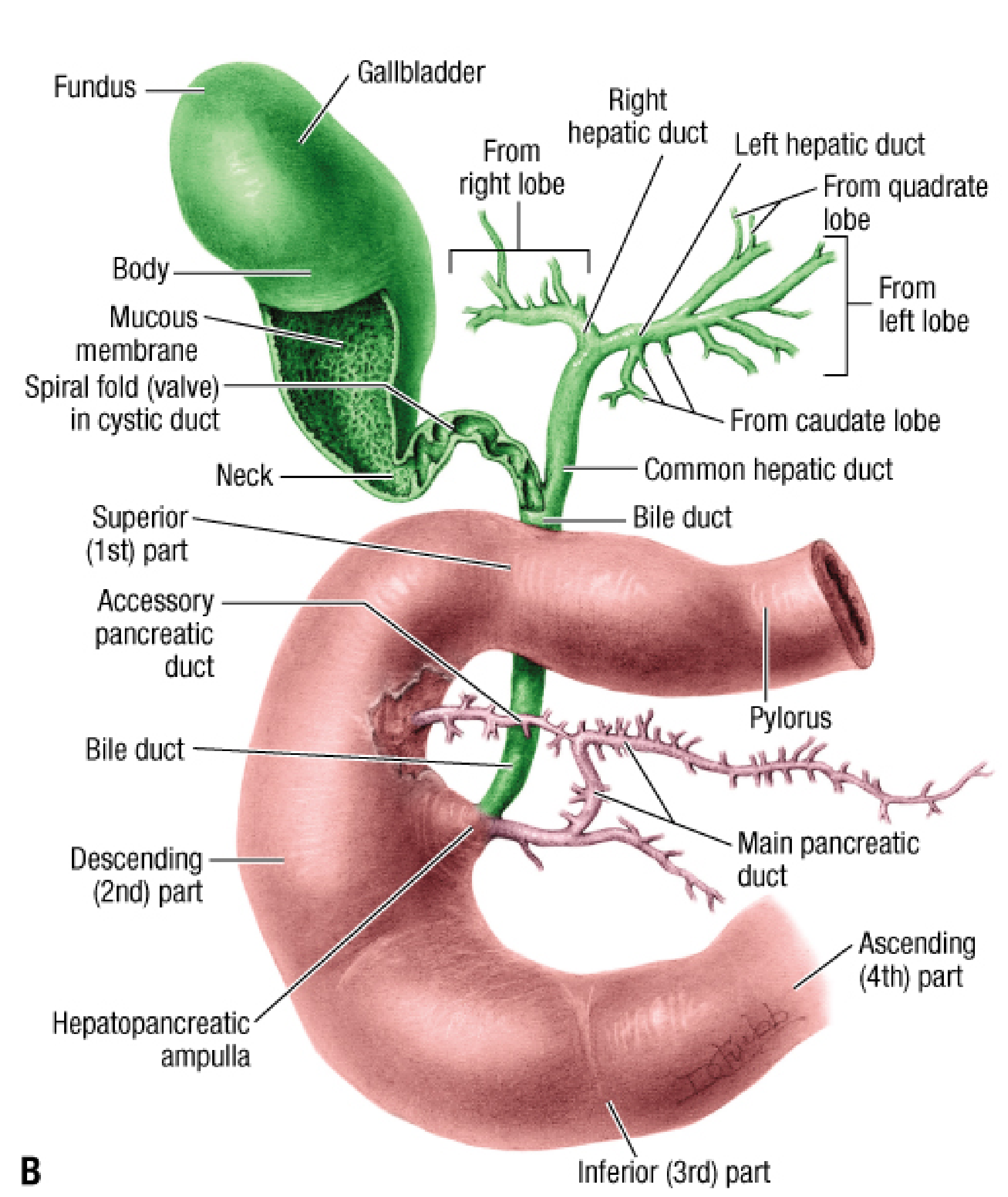
The extrahepatic biliary apparatus is the system of ducts and storage organs that collect bile from the liver, store it in the gallbladder, and deliver it to the duodenum. It consists of: the right and left hepatic ducts, the common hepatic duct (CHD), the cystic duct, the gallbladder, and the common bile duct (CBD).
1. Right and Left Hepatic Ducts
- Formed within the liver parenchyma by union of segmental ducts
- They emerge from the liver and unite 0.25 to 2.5 cm from the liver surface to form the common hepatic duct
- Accessory hepatic ducts (normal segmental ducts that join the biliary tract extrahepatically rather than intrahepatically) may be present - usually with about half the diameter of the main duct; they are surgically important as they can be mistaken for the cystic duct
2. Common Hepatic Duct (CHD)
- Formed by union of right and left hepatic ducts
- Usually less than 2.5 cm long
- Runs in the free edge of the lesser omentum (hepatoduodenal ligament)
- Becomes the common bile duct when joined by the cystic duct
3. Gallbladder
A pear-shaped saccular organ attached to the inferior (visceral) surface of the liver in the gallbladder fossa (between segments IV and V).
Parts
| Part | Description |
|---|---|
| Fundus | Wide, rounded end; projects below the inferior liver border; contacts the abdominal wall at the right 9th costal cartilage in the midclavicular line; related to hepatic flexure and 1st part of duodenum |
| Body | Main tapering portion; contacts visceral surface of liver, transverse colon, and superior duodenum |
| Neck | Makes an S-shaped bend with spirally arranged mucosal folds forming the valve of Heister; lies immediately superior to the duodenum |
| Infundibulum (Hartmann's pouch) | Asymmetric bulge on the right side of the neck; may adhere to the first part of duodenum via the cholecystoduodenal ligament; a stone impacted here can cause Mirizzi syndrome |
Relations
- Posteriorly: rests on Gerota's fascia of the right kidney
- Inferiorly: related to the duodenum and hepatic flexure of colon
Blood Supply
- Cystic artery - most commonly a branch of the right hepatic artery, arising posterior to the common hepatic duct; bifurcates into anterior (superficial) and posterior (deep) branches at the neck
- In 15% of cases the right hepatic artery and/or cystic artery cross in front of the common hepatic duct - a surgically important variant
Venous Drainage
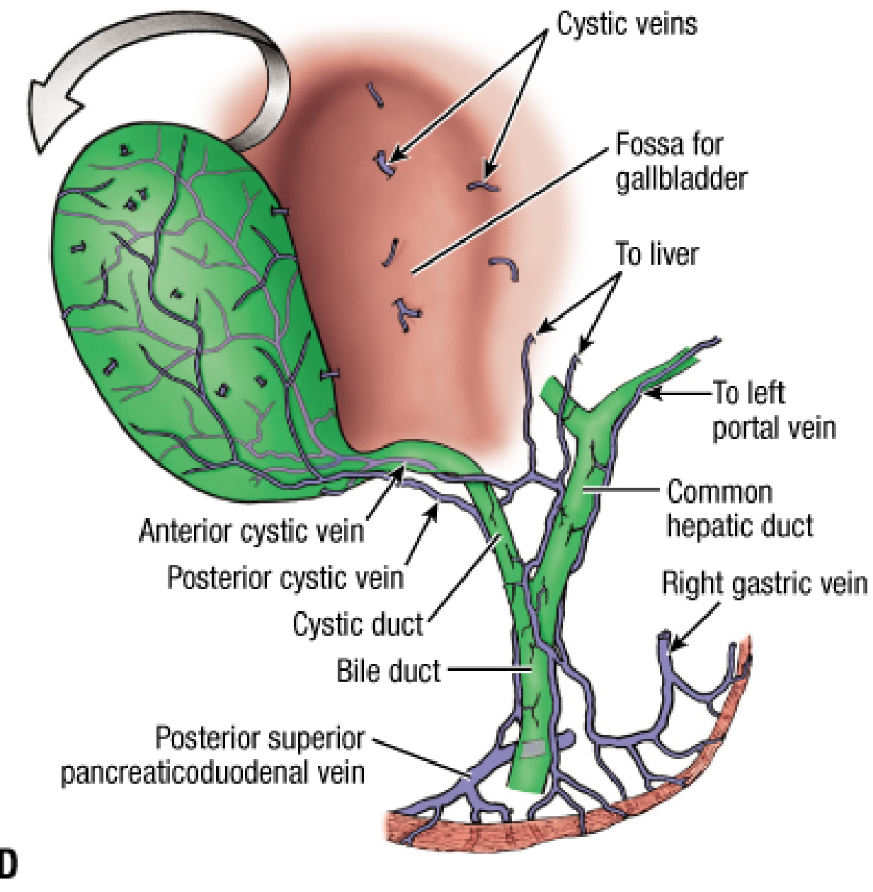
- Anterior and posterior cystic veins drain the gallbladder; they follow the cystic artery and then drain directly into the cystic plate, connecting to the middle hepatic vein within the intrahepatic parenchyma
Nerve Supply
- Sympathetic: from the 9th thoracic segment and celiac plexus; the right phrenic nerve also contributes (explains referred pain to the right shoulder tip in cholecystitis)
- Parasympathetic: hepatic branch of the anterior vagus nerve
Function
- Stores and concentrates bile during fasting (sphincter of Oddi closed → bile fills gallbladder)
- Cholecystokinin (CCK) released with enteral feeding causes gallbladder contraction + sphincter of Oddi relaxation → bile bolus delivered to duodenum
- Main function of bile: emulsification of ingested fats to allow pancreatic lipase action
Lymphatic Drainage
The cholecystic-retropancreatic pathway is the major route: cystic duct → node of Calot → pericholedochal nodes → portal vein nodes → superior retropancreatic nodes → periaortic nodes.
4. Cystic Duct
- Connects gallbladder neck to the common hepatic duct
- Contains the spiral valve of Heister (spirally arranged mucosal folds that prevent sudden emptying or overfilling)
- Joins the CHD to form the CBD in the supraduodenal segment of CHD in 80% of cases
- Variable junction: may join as low as the retroduodenal or retropancreatic part; occasionally joins the right hepatic duct or a right sectoral duct
Cystic Duct Variations
| Variant | Description |
|---|---|
| Direct junction (most common) | Joins CHD directly |
| Low junction | Joins far down near pancreas |
| Anterior spiral | Spirals anteriorly before joining |
| Posterior spiral | Spirals posteriorly before joining |
| Joins right hepatic duct | Risk of inadvertent clipping |
| Bifurcated or double cystic duct | Rare |
| Absent cystic duct | Very rare |
5. Common Bile Duct (CBD)
- Formed by union of the cystic duct + common hepatic duct
- Length: approximately 7.5 cm; mean diameter: ~6 mm (may dilate significantly with distal obstruction)
- Divided into four parts:
| Part | Length | Relations |
|---|---|---|
| 1. Supraduodenal | ~2.5 cm | In the free right border of the lesser omentum; anterior to portal vein, to the right of hepatic artery |
| 2. Retroduodenal | ~2.5-4 cm | Posterior to the 1st part of duodenum; just to the right of the gastroduodenal artery; loosely attached by areolar tissue |
| 3. Pancreatic (infraduodenal) | Variable | In a groove or tunnel on the posterior surface of the head of pancreas; may be entirely retropancreatic or within pancreatic substance; obstructed in carcinoma of the pancreatic head → painless jaundice |
| 4. Intraduodenal | ~2 cm | Turns 90° right; passes obliquely through the wall of the 2nd part of duodenum; narrows from ~10 mm to ~5 mm; surrounded by the sphincter of Oddi |
The CBD enters the posteromedial wall of the 2nd part of the duodenum, approximately 7-10 cm distal to the pylorus, opening at the summit of the major duodenal papilla (ampulla of Vater).
Blood Supply to the Bile Duct
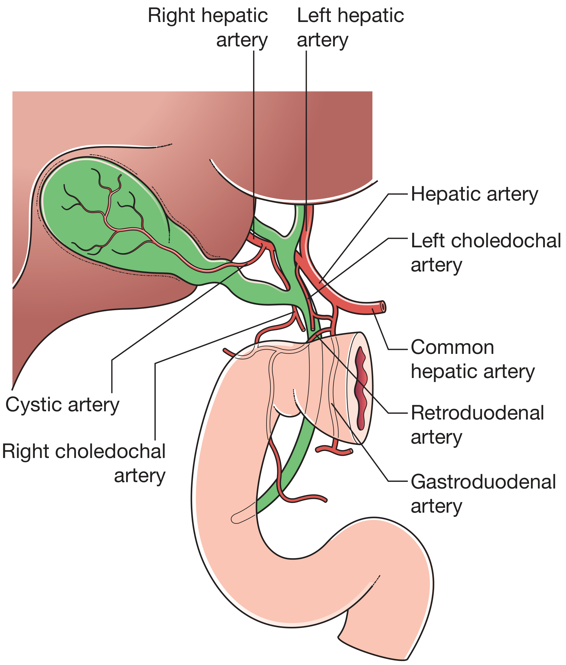
- Supraduodenal CBD: small branches from the cystic artery (from above) and gastroduodenal/retroduodenal artery (from below) - forming an axial blood supply running at 3 o'clock and 9 o'clock positions along the duct
- Retroduodenal CBD: branches from the gastroduodenal and retroduodenal arteries
- This axial supply is vulnerable during high bile duct injury, leading to ischemic strictures
6. Hepatopancreatic Ampulla (Ampulla of Vater) and Sphincter of Oddi
The CBD joins the main pancreatic duct (of Wirsung) at the ampulla of Vater. Their union can occur:
- Outside the duodenal wall (most common) - they then travel together through the wall
- At the ampulla itself
- Independently, opening separately on the papilla (when a complete septum is present)
The intramural common channel can be as narrow as 2 mm. This narrowing is why gallstones tend to impact just proximal to the ampulla.
The sphincter of Oddi has two functional components:
- Choledochal sphincter: surrounds the terminal CBD - the main regulator of bile flow into the duodenum
- Sphincter of the hepatopancreatic ampulla: surrounds the ampulla itself
7. Triangle of Calot (Hepatocystic Triangle)
A key surgical landmark during cholecystectomy.
Original description (Calot): bounded by:
- Inferiorly - cystic duct
- Medially - common hepatic duct
- Superiorly - superior border of the cystic artery
Modern/contemporary definition (hepatocystic triangle):
- Superiorly - inferior surface of the liver
- Laterally - cystic duct and medial border of gallbladder
- Medially - common hepatic duct
Contents: cystic artery (usually found within its boundaries), right hepatic artery, right hepatic duct, and the node of Calot (a cystic lymph node that acts as a landmark for finding the cystic artery in difficult cases).
The goal of laparoscopic cholecystectomy is to achieve the Critical View of Safety - complete dissection of the hepatocystic triangle with only two structures (cystic duct and cystic artery) entering the gallbladder, to avoid bile duct injury.
Summary Diagram
| Structure | Length | Key Relation |
|---|---|---|
| Right + Left hepatic ducts | Variable | Emerge from liver |
| Common hepatic duct | <2.5 cm | Free edge of lesser omentum |
| Cystic duct | Variable | Contains valve of Heister |
| Common bile duct | ~7.5 cm | 4 parts: supra-, retro-, pancreatic, intraduodenal |
| Gallbladder | 7-10 cm long | Segments IV-V fossa |
Sources: Fischer's Mastery of Surgery, p. 4552-4559; Bailey and Love's Short Practice of Surgery 28th ed., p. 1254; Yamada's Textbook of Gastroenterology, p. 228-229; Current Surgical Therapy 14e, p. 560
Ischiorectal fossa
The ischiorectal (ischioanal) fossa is a fat-filled wedge-shaped space lateral to the anal canal, important for its contents and as a site of abscess formation. Let me pull comprehensive textbook content.
Searching sources...
Finding Sources
Finding Sources
Reading File
Reading File
Finding Sources
Reading File
Finding Sources
Finding Sources
Reading File
Reading File
Reading File
Loading Image
Loading Image
Finding Sources
Excellent - I now have comprehensive content from multiple authoritative sources and two excellent diagrams. Let me compile the full answer.
Ischiorectal (Ischioanal) Fossa
Nomenclature note: The term ischioanal fossa is now preferred over ischiorectal fossa in modern anatomy, as the space is related to the anal canal - not directly to the rectum. Both terms remain in clinical use.
Overview
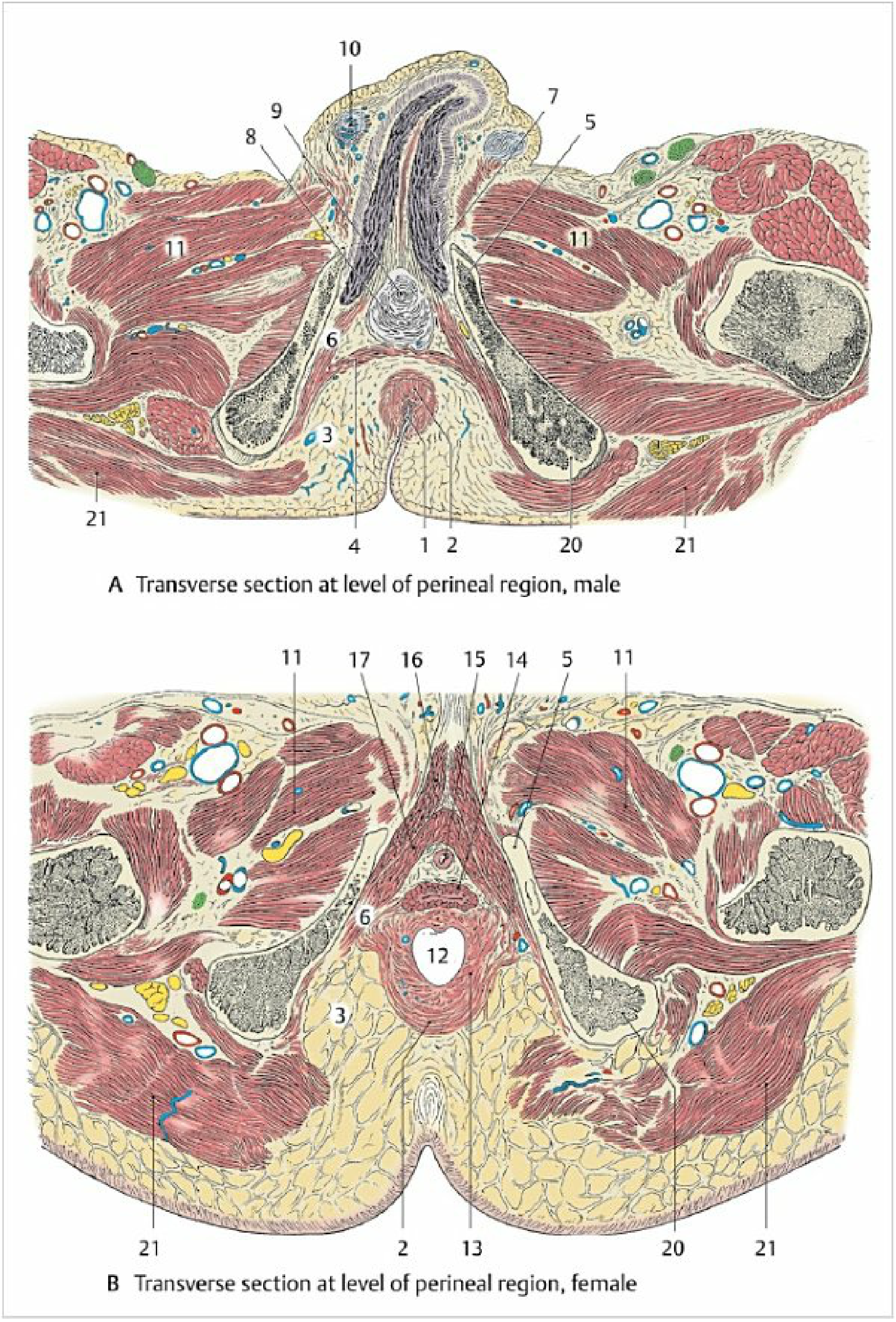
The ischioanal fossa is a pyramid-shaped (wedge-shaped) fat-filled space located on each side of the anal canal, outside the pelvic floor. The two fossae are separated in the midline anteriorly by the anal canal, but communicate posteriorly via the deep postanal space (behind the anal canal), which is important for horseshoe abscess formation.
Shape and Orientation
The fossa is described as a three-sided pyramid:
- The tip (apex) points superiorly, near the junction of the levator ani and obturator internus
- The base faces inferiorly, covered by perineal skin
- The tip of the pyramid points toward the pubic symphysis (anteriorly)
Boundaries
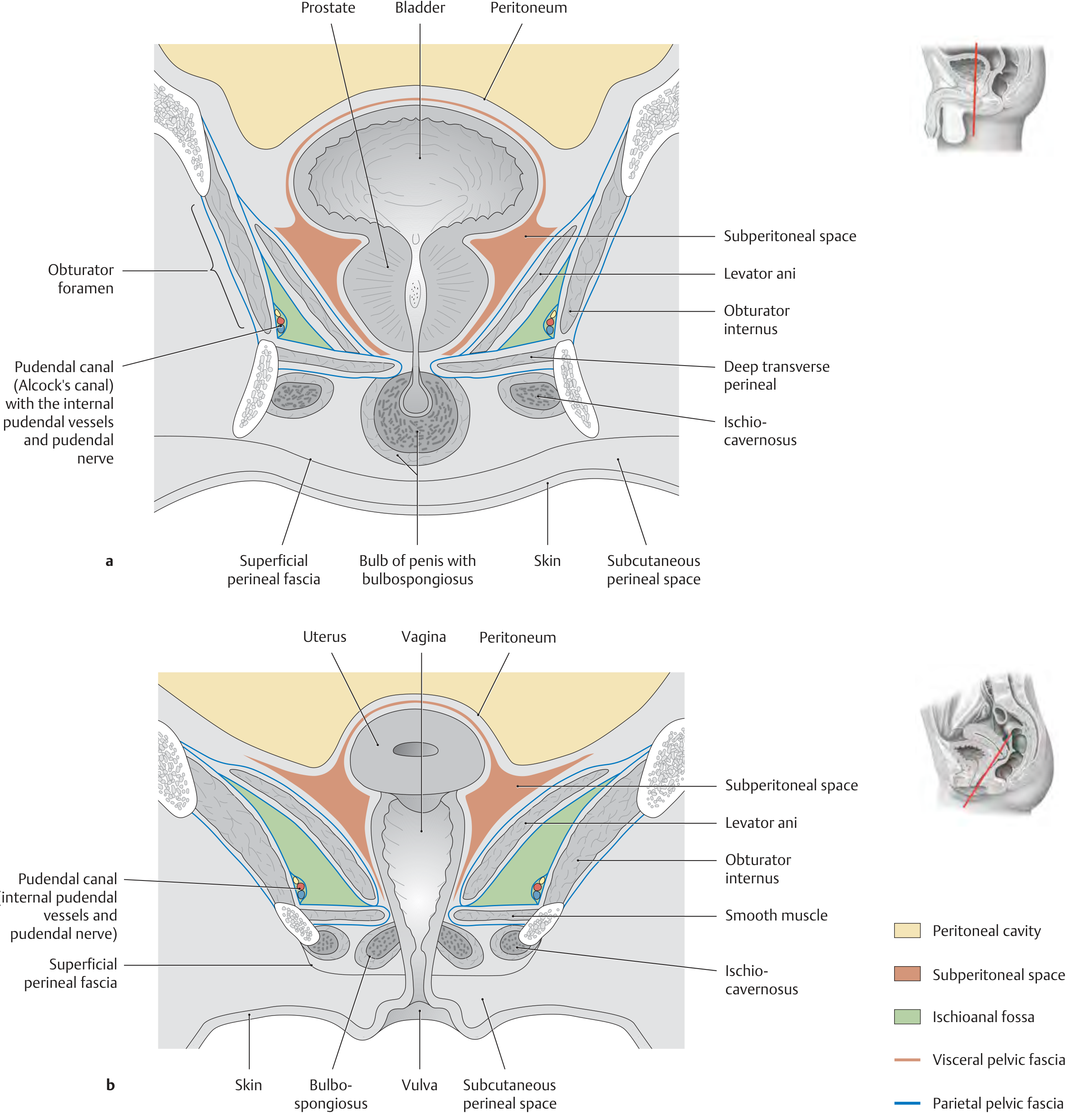
| Wall | Boundary |
|---|---|
| Medial (superomedial) | External anal sphincter + levator ani (inferior fascia of pelvic diaphragm) |
| Lateral | Obturator internus muscle and its fascia + ischial tuberosity |
| Inferior (base) | Perineal skin |
| Superior (apex) | Junction of levator ani and obturator internus (tendinous arch) |
| Posterior | Gluteus maximus muscle and sacrotuberous ligament |
| Anterior | Posterior border of the urogenital diaphragm (deep transverse perineal muscle) |
The anterior recess of the ischioanal fossa extends forward above the urogenital diaphragm between the levator ani and the superior surface of the perineal membrane.
Contents
1. Fat Body of the Ischioanal Fossa
- The fossa is mainly filled with fat (the fat pad / fat body)
- This fat acts as a mobile pad that slides downward and backward during bowel evacuation and during labor - allowing the anal canal to distend
- The fat is loose and areolar, making it easily displaced and able to accommodate large abscesses
2. Pudendal Canal (Alcock's Canal) - Key Structure
Running in the lateral wall of the ischioanal fossa, within a fascial sheath of the obturator internus fascia, is the pudendal canal (Alcock's canal).
- Formation: formed by a duplication/fold of the obturator internus fascia
- Course: begins just below the ischial spine → courses along the lateral wall of the ischioanal fossa below the tendinous arch of the levator ani → passes toward the pubic symphysis
- Entry: neurovascular structures exit the lesser pelvis through the greater sciatic foramen, loop around the ischial spine/sacrospinous ligament, and re-enter the perineum through the lesser sciatic foramen into the pudendal canal
Contents of the pudendal canal:
- Internal pudendal artery
- Internal pudendal vein(s)
- Pudendal nerve (S2-S4)
3. Inferior Rectal (Inferior Anal) Vessels and Nerve
- The inferior rectal vessels and inferior rectal nerve (branch of pudendal nerve) cross the fossa transversely from the pudendal canal to reach the external anal sphincter and perianal skin
- They traverse the fat of the ischioanal fossa
4. Perineal Branch of S4
- A small nerve crossing the fossa contributing to innervation of the external sphincter
Transverse Section Diagram
Communication Between the Two Fossae
The left and right ischioanal fossae are anatomically separate in their main portions but communicate via the:
- Deep postanal space (also called the retroanal space) - posterior to the anal canal, between the external sphincter and the anococcygeal ligament
- This communication is why abscesses can spread from one side to the other forming a horseshoe abscess
Clinical Significance
1. Ischiorectal (Ischioanal) Abscess
- Most common type of anorectal abscess (after perianal abscess)
- Origin: usually from cryptoglandular infection - an intersphincteric abscess that penetrates the external sphincter below the puborectalis level and expands into the fat of the ischioanal fossa
- Why it can be large and insidious: the levator ani (upper border of the fossa) slopes upward, so the abscess expands upward rather than pointing through the skin; it may be palpable as a bulge above the puborectalis but actually lies below the levator ani
- Presentation: pain, redness, and fluctuance in the perirectal/buttock region; fever; inability to sit
2. Horseshoe Abscess
- A horseshoe-shaped infection that starts in the posterior midline intersphincteric space, enters the deep postanal space (incising the anococcygeal ligament), and spreads bilaterally to involve both ischioanal fossae
- Treatment: drainage of the deep postanal space + counter-drainage incisions for each limb of the ischiorectal space
3. Anal Fistula
- An ischiorectal (trans-sphincteric) fistula has its external opening typically over the ischioanal fossa skin
- The fistula tract crosses the external sphincter
4. Pudendal Nerve Block
- Performed by anesthetizing the pudendal nerve at the level of the ischial spine - before it enters the pudendal canal - using a transvaginal or transperineal approach
- Used in obstetrics for perineal analgesia; anesthetizes the inferior rectal, perineal, dorsal clitoral/penile, and posterior labial/scrotal nerves
5. Surgical Caution
- The internal pudendal vessels and pudendal nerve in the lateral wall of the fossa are at risk during ischiorectal abscess drainage; incisions should be kept medial (closer to the anal canal) to avoid injury
Summary Table
| Feature | Detail |
|---|---|
| Shape | Pyramid / wedge |
| Medial wall | External anal sphincter + levator ani fascia |
| Lateral wall | Obturator internus + ischial tuberosity |
| Key structure in lateral wall | Pudendal canal (Alcock's canal) |
| Contents of pudendal canal | Internal pudendal a/v + pudendal nerve (S2-S4) |
| Fills with | Fat (fat body - mobile pad) |
| Posterior communication | Deep postanal space (connects both fossae) |
| Clinical importance | Ischiorectal abscess, horseshoe abscess, anal fistula, pudendal block |
Sources: General Anatomy and Musculoskeletal System - THIEME Atlas, p. 581-583; Color Atlas of Human Anatomy Vol. 2, p. 476-479; Campbell-Walsh Urology, p. 3938; Maingot's Abdominal Operations, p. 1939-1944; Yamada's Textbook of Gastroenterology, p. 180
Kidney
The kidney is a paired retroperitoneal organ responsible for filtration, excretion, and hormonal functions. Let me pull comprehensive textbook content covering anatomy, relations, blood supply, and clinical correlates.
Searching sources...
Finding Sources
Finding Sources
Finding Sources
Finding Sources
Reading File
Reading File
Reading File
Reading File
Finding Sources
Finding Sources
Reading File
Reading File
Reading File
Excellent content gathered. Now let me fetch the key diagrams - the kidney blood supply, internal structure, and posterior relations.
Loading Image
Loading Image
Loading Image
I now have thorough content and excellent diagrams. Let me compile the complete answer.
Kidney
Overview
The kidneys are paired retroperitoneal organs responsible for blood filtration, urine production, fluid/electrolyte balance, acid-base regulation, and hormone production (erythropoietin, renin, active vitamin D).
Gross Anatomy
Size, Weight, and Position
| Feature | Value |
|---|---|
| Length | 11-12 cm (MRI mean: 12.4 cm men, 11.6 cm women) |
| Width | 5.0-7.5 cm |
| Thickness | 2.5-3.0 cm |
| Weight (male) | 125-170 g |
| Weight (female) | 115-155 g |
| Vertebral level | T12 to L3 |
| Right vs Left | Right is slightly more caudal (displaced by liver) |
The long axis of each kidney is oblique - the upper poles are closer to the midline and lie more posteriorly than the lower poles. The renal pelvis faces anteromedially (~30° anterior rotation from the coronal plane).
Internal Structure
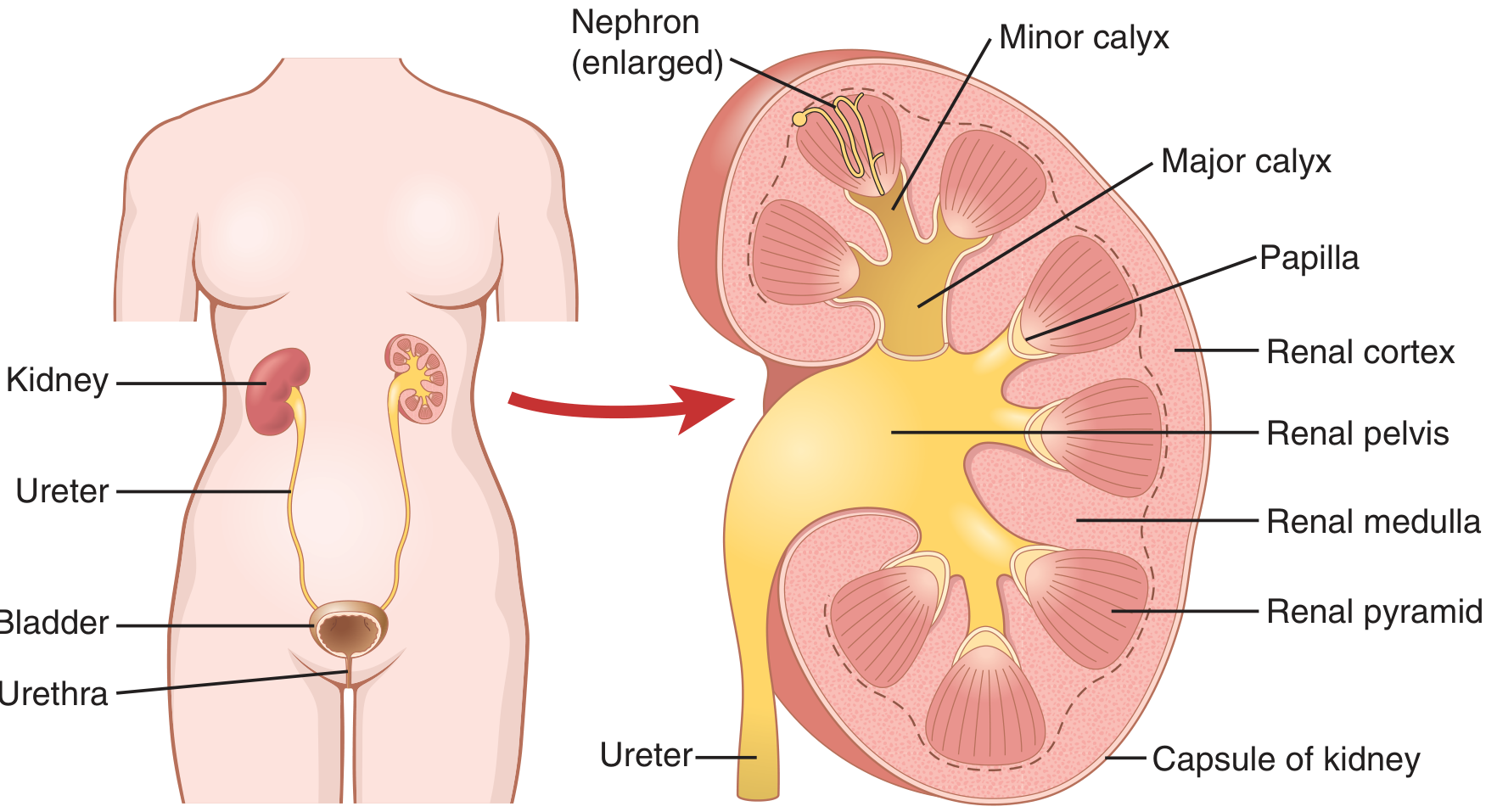
On a bisected kidney, two zones are visible:
Cortex (Outer)
- Granular-appearing outer zone
- Contains the glomeruli, proximal convoluted tubules, distal convoluted tubules, and columns of Bertin (cortical tissue projecting between pyramids)
Medulla (Inner)
- Striated inner zone; divided into 8-18 renal pyramids
- Each pyramid is a cone with:
- Base at the corticomedullary junction
- Apex (papilla) pointing toward the renal pelvis
- Contains loops of Henle and collecting ducts
- Papillary ducts (~20 per papilla) open at the area cribrosa on the papillary tip
- Columns of Bertin are cortical invaginations that separate adjacent pyramids
Collecting System
| Structure | Detail |
|---|---|
| Minor calyces | 7-9 per kidney (range 5-14); cup-shaped; each receives 1-3 papillae |
| Major calyces | 2-3; formed by union of minor calyces |
| Renal pelvis | Expanded proximal ureter; exits at the ureteropelvic junction (UPJ) |
| Ureter | 28-34 cm long; mean diameter 1.8 mm (max 3 mm) |
Coverings (from inside out)
- Fibrous capsule - thin, tough; strips easily from a healthy kidney
- Perirenal fat (perinephric fat) - fine, light yellow; fills the perirenal space
- Gerota's fascia (renal fascia) - surrounds the kidney + adrenal gland + perirenal fat
- Anterior lamina = fascia of Toldt (prerenal fascia)
- Posterior lamina = fascia of Zückerkandl (retrorenal fascia)
- The two laminae merge laterally to form the lateroconal fascia
- Medially: continuous with adventitia of aorta, IVC, and renal vessels
- Superiorly: closed (fused above the adrenal)
- Inferiorly: open (cone-shaped opening into extraperitoneal pelvis) - explains how perirenal collections can track inferiorly
- Paranephric (pararenal) fat - coarser, yellow-orange; external to Gerota's fascia; most prominent posteriorly
Clinical note: Gerota's fascia contains perinephric fluid collections (urinoma, hematoma, abscess) and is used as the plane of dissection in radical nephrectomy.
Relations
Posterior Relations
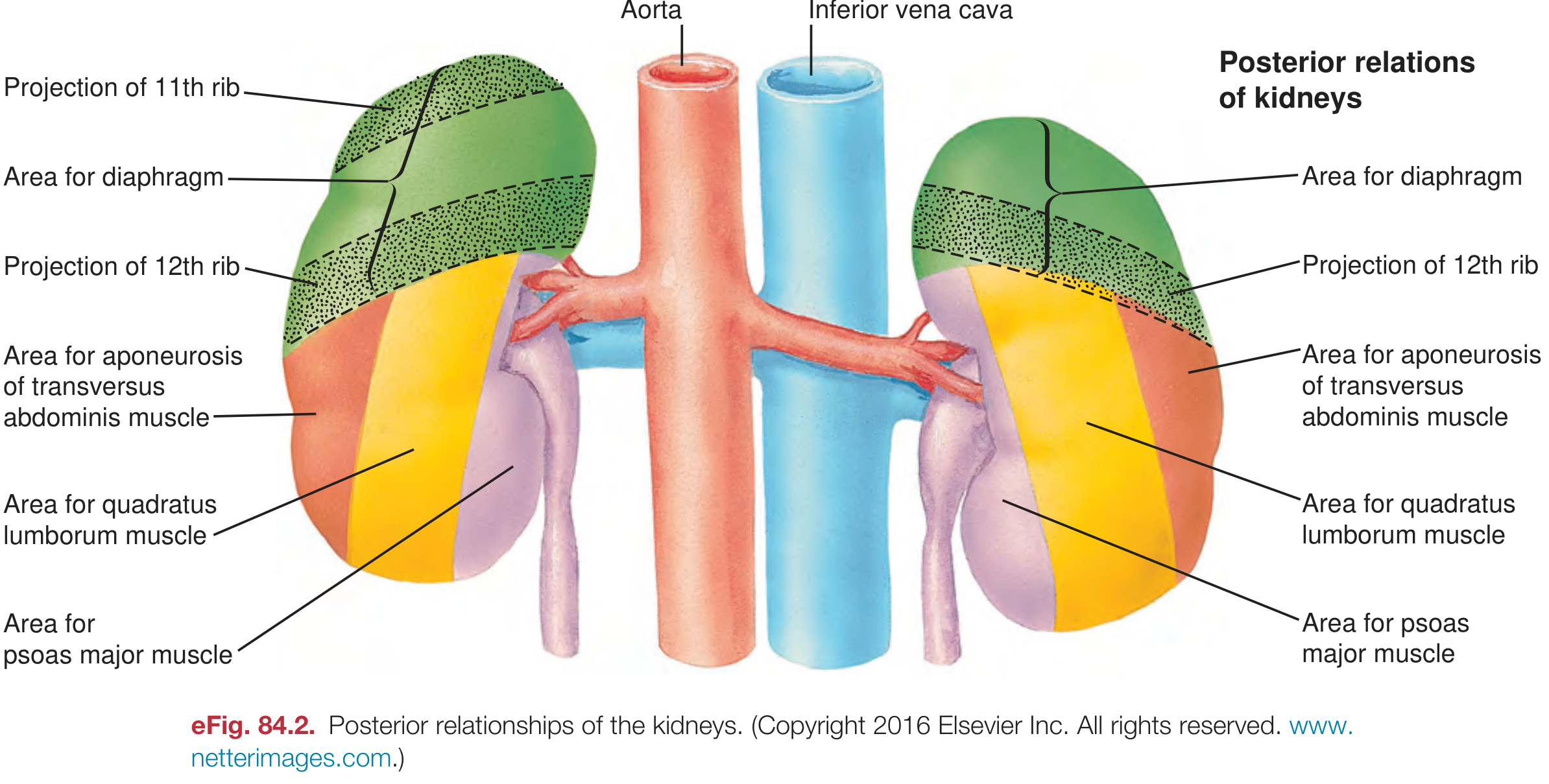
| Region | Structure |
|---|---|
| Upper pole (both) | Diaphragm |
| Upper portion (lateral) | 11th and 12th ribs |
| Medial | Psoas major |
| Lateral | Quadratus lumborum |
| Lateral to quadratus | Aponeurosis of transversus abdominis |
Anterior Relations
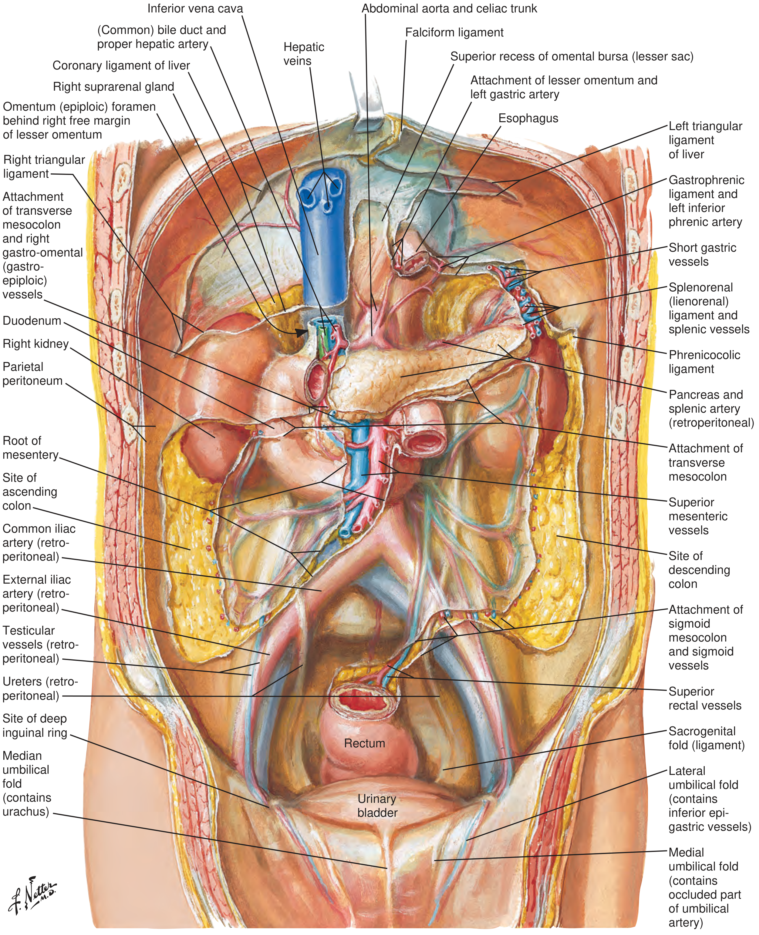
| Kidney | Anterior Relations |
|---|---|
| Right | Right lobe of liver (upper 2/3), hepatic flexure of colon (lower pole), 2nd part of duodenum (medial), gallbladder (anterior/medial) |
| Left | Spleen (upper lateral), body/tail of pancreas + splenic vessels (anterior/medial), stomach (upper), jejunum (lower), splenic flexure of colon (lateral) |
Medial Relations (both sides)
- Right: Right adrenal gland (superomedial), IVC, duodenum
- Left: Left adrenal gland (superomedial), aorta, tail of pancreas
Blood Supply
Renal Artery
- Arises from the abdominal aorta at L1-L2
- Right renal artery is longer and passes posterior to the IVC, right renal vein, head of pancreas, and descending duodenum
- Left renal artery is shorter; passes posterior to the left renal vein and body of pancreas
- Each renal artery divides at the hilum into:
- Anterior division (larger): gives 3-4 segmental arteries supplying upper, middle, lower + apical segments
- Posterior division (smaller): supplies posterior segment (>50% of posterior surface)
Segmental arteries are end arteries - no collateral circulation between segments. Occlusion → segmental infarction.
Accessory renal arteries occur in up to 25% of kidneys; they typically enter at the poles (usually lower pole) directly from the aorta.
Intrarenal Arterial Hierarchy
Renal artery
→ Segmental (lobar) arteries
→ Interlobar arteries (in columns of Bertin)
→ Arcuate arteries (at corticomedullary junction)
→ Interlobular (radial) arteries (through cortex)
→ Afferent arterioles → Glomerular capillaries
→ Efferent arterioles → Peritubular capillaries (cortex)
→ Vasa recta (medulla)
The renal circulation has two capillary beds in series (glomerular + peritubular), separated by the efferent arteriole:
- Glomerular capillary pressure: ~60 mmHg (drives filtration)
- Peritubular capillary pressure: ~13 mmHg (drives reabsorption)
Renal Vein
- The left renal vein is longer (~7.5 cm vs ~2.5 cm right); crosses anterior to the aorta
- Left renal vein receives: left gonadal vein (inferiorly), left suprarenal vein (superiorly), and left inferior phrenic vein
- Right gonadal and right suprarenal veins drain directly into the IVC
Renal Blood Flow
- ~22% of cardiac output (~1100 mL/min in adults)
- Cortex receives ~90% of total renal blood flow
The Nephron - Functional Unit
Each kidney contains ~800,000-1,000,000 nephrons (range: 200,000 to >2.5 million). Nephrons cannot regenerate. After age 40, ~10% are lost every decade.
Each nephron comprises:
| Component | Location | Function |
|---|---|---|
| Glomerulus (Bowman's capsule) | Cortex | Filtration |
| Proximal convoluted tubule | Cortex | Reabsorption (Na, glucose, amino acids, HCO₃, water) |
| Loop of Henle (thin + thick limbs) | Medulla | Countercurrent multiplication; concentration |
| Distal convoluted tubule | Cortex | Fine-tuning Na/K/Ca; aldosterone-sensitive |
| Collecting duct | Medulla → papilla | Water reabsorption (ADH-sensitive); acid-base |
Nephron Types
- Cortical nephrons (~85%): short loops of Henle, peritubular capillaries
- Juxtamedullary nephrons (~15%): long loops extending deep into medulla; associated with vasa recta - critical for urinary concentration
Lymphatic Drainage
- Renal lymphatics follow the renal vessels
- Drain to lateral aortic (para-aortic) lymph nodes at the level of L1-L2
- Important in staging of renal cell carcinoma
Nerve Supply
- Sympathetic: from T10-L1 via the celiac and renal plexuses; vasomotor (vasoconstriction of afferent arterioles); pain fibers
- Parasympathetic: vagus nerve (minor functional role)
- Referred pain from the kidney: loin to groin (T10-L1 dermatomes); upper ureteric colic → anterior abdominal wall; lower ureteric colic → scrotum/labia
Juxtaglomerular Apparatus (JGA)
Located at the vascular pole of each glomerulus; consists of:
- Juxtaglomerular (granular) cells - modified smooth muscle cells of the afferent arteriole; secrete renin
- Macula densa - specialised cells of the thick ascending limb / early distal tubule; detect tubular NaCl concentration
- Extraglomerular mesangial (Lacis) cells
Renin release is triggered by: decreased renal perfusion pressure, decreased NaCl at macula densa, sympathetic stimulation (β1). This activates the RAAS (renin → angiotensin I → ACE → angiotensin II → aldosterone).
Key Clinical Correlates
| Condition | Anatomy Involved |
|---|---|
| Renal cell carcinoma | Spreads within Gerota's fascia; classic triad: hematuria, loin pain, palpable mass |
| Renal colic | Ureteric stones cause severe loin-to-groin pain; T10-L1 referral |
| Percutaneous nephrolithotomy | Posterior calyces preferred; risk of pleural injury with upper pole (supracostal) access |
| Perinephric abscess | Contained within Gerota's fascia; can track inferiorly into pelvis |
| Horseshoe kidney | Lower poles fused across midline; lie lower (L4-L5); incomplete ascent due to mesenteric artery; predisposed to pelviureteric junction obstruction and infection |
| Renovascular hypertension | Renal artery stenosis → reduced renal perfusion → renin-angiotensin activation |
| Left renal vein compression | "Nutcracker syndrome" - left renal vein compressed between aorta and SMA → left-sided varicocele, hematuria |
| Retrorenal colon | Colon posterior to kidney in ~16% (left) at lower pole - at risk during percutaneous access |
Sources: Brenner and Rector's The Kidney, p. 38-82; Guyton and Hall Medical Physiology, p. 326-328; Campbell-Walsh-Wein Urology, p. 2152-2159, 2222-2223; Grainger & Allison's Diagnostic Radiology, p. 2162
This is a shared conversation. Sign in to Orris to start your own chat.