
Bulky uterus adenomyosis
Reading File
Finding Sources
Searching PubMed
adenomyosis
Searching the Web
adenomyosis NHS symptoms diagnosis treatment
Bulky uterus with adenomyosis usually means the uterine muscle has become thickened and enlarged because tissue similar to the uterine lining has grown within the muscle wall. It is a benign condition, not cancer.
It can cause:
- Heavy or prolonged periods, sometimes with clots
- Severe period cramps
- Pelvic heaviness, bloating, or lower abdominal pain
- Pain during sex
- Anemia from heavy bleeding
- Sometimes difficulty conceiving, though many people can still become pregnant
A pelvic ultrasound, preferably transvaginal, often suggests adenomyosis. MRI can clarify uncertain cases or distinguish it from fibroids. Fibroids can coexist and also make the uterus bulky. Berek & Novak's Gynecology notes that diagnosis is increasingly made using ultrasound or MRI rather than only after hysterectomy.
Treatment depends on symptoms, anemia, age, and whether pregnancy is desired:
- NSAID pain medicines during periods if safe for you
- Tranexamic acid for heavy menstrual bleeding, if prescribed
- Hormonal treatment, especially a levonorgestrel intrauterine system such as Mirena, or other progestin/combined hormonal options
- Procedures or surgery for persistent severe symptoms. Hysterectomy is definitive only when childbearing is complete and conservative treatment has not helped. The NHS adenomyosis guidance describes these options.
Please see a gynecologist promptly if bleeding is very heavy, pain is worsening, you feel faint or breathless, or you may be pregnant. A complete blood count and iron studies are often appropriate if periods are heavy, to check for anemia.
Adenomyosis pg review
Reading File
For a postgraduate review, adenomyosis is best covered as definition and pathogenesis, clinical features, imaging diagnosis, fertility impact, medical treatment, uterine-sparing procedures, and definitive surgery.
Asking for Preferences
Which PG-review format do you want?
Bulky uterus with adenomyosis usually means the uterine muscle has become thickened and enlarged because tissue similar to the uterine lining has grown within the muscle wall. It is a benign condition, not cancer.
It can cause:
- Heavy or prolonged periods, sometimes with clots
- Severe period cramps
- Pelvic heaviness, bloating, or lower abdominal pain
- Pain during sex
- Anemia from heavy bleeding
- Sometimes difficulty conceiving, though many people can still become pregnant
A pelvic ultrasound, preferably transvaginal, often suggests adenomyosis. MRI can clarify uncertain cases or distinguish it from fibroids. Fibroids can coexist and also make the uterus bulky. Berek & Novak's Gynecology notes that diagnosis is increasingly made using ultrasound or MRI rather than only after hysterectomy.
Treatment depends on symptoms, anemia, age, and whether pregnancy is desired:
- NSAID pain medicines during periods if safe for you
- Tranexamic acid for heavy menstrual bleeding, if prescribed
- Hormonal treatment, especially a levonorgestrel intrauterine system such as Mirena, or other progestin/combined hormonal options
- Procedures or surgery for persistent severe symptoms. Hysterectomy is definitive only when childbearing is complete and conservative treatment has not helped. The NHS adenomyosis guidance describes these options.
Please see a gynecologist promptly if bleeding is very heavy, pain is worsening, you feel faint or breathless, or you may be pregnant. A complete blood count and iron studies are often appropriate if periods are heavy, to check for anemia.For a postgraduate review, adenomyosis is best covered as definition and pathogenesis, clinical features, imaging diagnosis, fertility impact, medical treatment, uterine-sparing procedures, and definitive surgery.
Finding Sources
Searching PubMed
adenomyosis diagnosis treatment
Searching the Web
adenomyosis diagnosis management guideline ultrasound MRI
Finding Sources
Finding Sources
Searching Images
MRI pelvis adenomyosis thickened junctional zone T2 image
Reading Web Page
https://www.aafp.org/afp/2022/0100/p33
Adenomyosis: Postgraduate Review
Definition
Adenomyosis is a benign uterine disorder in which endometrial glands and stroma are present within the myometrium, accompanied by surrounding myometrial smooth-muscle hypertrophy and hyperplasia. This produces a diffusely enlarged, globular, often tender uterus.
It is classified under the FIGO PALM-COEIN system of abnormal uterine bleeding as AUB-A. It frequently coexists with fibroids and endometriosis.
Pathogenesis
The exact cause is uncertain. Proposed mechanisms:
- Invagination theory: basalis endometrium extends downward through a disrupted endometrial-myometrial junction.
- Tissue-injury-and-repair mechanism: repeated uterine peristalsis, menstruation, pregnancy, curettage, or uterine surgery causes junctional-zone injury, inflammation, and local estrogen-mediated repair.
- Metaplasia theory: embryonic Müllerian remnants or myometrial stem cells undergo metaplasia into endometrial tissue.
- Local estrogen excess, progesterone resistance, inflammation, neuroangiogenesis, and abnormal uterine contractility contribute to symptoms.
Epidemiology and risk factors
- Increasingly diagnosed in reproductive-age women due to improved imaging.
- Commonly associated with increasing age, multiparity, previous uterine surgery or instrumentation, and coexisting endometriosis/fibroids.
- It may occur in younger women, including those presenting with infertility.
Types
- Diffuse adenomyosis: widespread involvement of the myometrium, causing a bulky globular uterus.
- Focal adenomyosis / adenomyoma: localized mass-like lesion, which may mimic a leiomyoma.
- Cystic adenomyosis: rare, may be seen in younger patients and can cause severe cyclical pain.
Clinical features
Many patients are asymptomatic. Typical symptoms are:
- Heavy menstrual bleeding, often with anemia
- Progressive secondary dysmenorrhea
- Chronic pelvic pain
- Dyspareunia
- Irregular uterine bleeding
- Pelvic pressure or heaviness due to a bulky uterus
- Subfertility, recurrent implantation failure, or adverse reproductive outcomes in some patients
Examination
- Uniformly enlarged, globular, soft or “boggy” uterus
- Uterine tenderness, particularly premenstrually
- Unlike fibroids, the uterus is usually not irregularly enlarged, unless both conditions coexist.
Differential diagnosis
- Uterine leiomyoma
- Endometriosis
- Endometrial polyp
- Endometrial hyperplasia or malignancy
- Pregnancy-related bleeding
- Pelvic inflammatory disease
- Dysfunctional or non-structural abnormal uterine bleeding
Diagnosis
Initial assessment
- Menstrual and pain history, fertility wishes, prior uterine surgery
- Pregnancy test when relevant
- CBC and ferritin to assess anemia from heavy menstrual bleeding
- Evaluate for other PALM-COEIN causes of AUB.
- Endometrial sampling should be considered in abnormal bleeding in women aged 45 years or older, or younger women with persistent bleeding or risk factors for endometrial hyperplasia/cancer.
Transvaginal ultrasound
First-line investigation. Typical MUSA features include:
Direct features
- Myometrial cysts
- Hyperechogenic islands
- Echogenic subendometrial lines and buds
Indirect features
- Globular uterus
- Asymmetrical myometrial thickening
- Fan-shaped shadowing
- Translesional vascularity
- Irregular or interrupted junctional zone
MRI pelvis
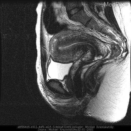
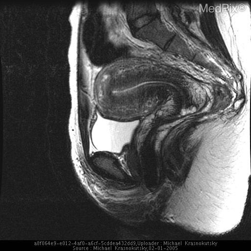
MRI is useful when ultrasound is equivocal, there are coexisting fibroids, or for preoperative mapping.
Findings:
- Thickened or irregular low-signal-intensity junctional zone on T2-weighted imaging
- Myometrial heterogeneity
- Tiny high-signal foci representing ectopic endometrium or hemorrhage
- Focal adenomyoma may appear as an ill-defined lesion without the pseudocapsule typical of a fibroid
A junctional-zone thickness greater than 12 mm is a traditional MRI sign, but diagnosis should use the overall imaging appearance rather than one cutoff alone.
Histopathology
Histology after hysterectomy remains the definitive diagnostic standard, showing endometrial glands and stroma embedded in myometrium with surrounding smooth-muscle hypertrophy.
Adenomyosis versus fibroid
| Feature | Adenomyosis | Leiomyoma |
|---|---|---|
| Enlargement | Diffuse, globular | Localized, irregular |
| Consistency | Boggy, tender | Firm |
| Lesion margin | Poorly defined | Well circumscribed, pseudocapsule |
| Ultrasound | Heterogeneous myometrium, cysts, shadowing | Discrete whorled mass |
| MRI | Thickened junctional zone | Well-defined T2-low lesion |
| Fertility-sparing surgery | Difficult in diffuse disease | Myomectomy often feasible |
Management
Management is individualized according to symptom severity, anemia, uterine size, coexisting disease, fertility wishes, and desire for uterine preservation.
1. Expectant treatment
Appropriate for asymptomatic patients or those with mild symptoms approaching menopause.
2. Medical treatment
For pain
- NSAIDs during menstruation
- They improve dysmenorrhea but do not eliminate adenomyotic tissue.
For heavy menstrual bleeding
- Tranexamic acid during menses, if suitable
- Hormonal menstrual suppression is usually more effective for combined pain and bleeding symptoms.
Hormonal options
-
Levonorgestrel-releasing intrauterine system, LNG-IUS
- Often the preferred uterus-preserving treatment for heavy bleeding and dysmenorrhea.
- May reduce uterine volume in some patients.
- Expulsion or insertion difficulty may occur with a markedly enlarged or distorted uterine cavity.
-
Combined oral hormonal contraception
- Useful for menstrual suppression and pain control.
-
Progestogens
- Dienogest, oral progestins, or depot medroxyprogesterone can reduce bleeding and pain.
- Recent meta-analyses support symptom benefit from dienogest, though choice must consider adverse effects and individual tolerance. A 2024 systematic review/meta-analysis is indexed as PMID 38729430.
-
GnRH agonists
- Can reduce uterine volume and symptoms.
- Usually short-term because of hypoestrogenic adverse effects.
- Add-back therapy is generally needed for longer courses.
- May be used as a bridge to surgery or before fertility treatment in selected patients.
-
GnRH antagonists
- May be considered in selected symptomatic patients where locally approved, but longer-term adenomyosis-specific evidence and cost considerations remain relevant.
3. Uterus-sparing interventions
These require careful counselling, especially if future pregnancy is desired.
- Uterine artery embolization: may improve pain and bleeding, but its role in women seeking future fertility is uncertain.
- High-intensity focused ultrasound, HIFU/MRgFUS: non-incisional option in selected focal or diffuse disease; availability and long-term fertility data vary.
- Adenomyomectomy or cytoreductive surgery: can be considered for focal adenomyoma or selected severe disease. Diffuse disease is technically difficult to excise completely.
- Subsequent pregnancy requires specialist surveillance because uterine rupture and placental complications have been reported after extensive myometrial surgery.
4. Definitive treatment
Hysterectomy is the definitive treatment for severe symptoms refractory to medical treatment in women who have completed childbearing. Removal of the ovaries is not routinely required for adenomyosis alone.
The AAFP clinical review supports TVUS or MRI for noninvasive diagnosis, LNG-IUS for bleeding and pain control in those not seeking pregnancy, and hysterectomy when conservative treatment fails.
Adenomyosis and infertility
Adenomyosis may impair fertility through altered uterine peristalsis, inflammation, abnormal endometrial receptivity, and distortion of the junctional zone. It may coexist with endometriosis.
For patients planning pregnancy:
- Refer early to a gynecologist or reproductive-medicine specialist.
- Avoid assuming that symptom suppression equals improved fertility.
- IVF planning should be individualized. Current evidence for prolonged medical suppression before IVF is mixed and depends on disease extent and treatment protocol. A recent systematic review/meta-analysis on this topic is PMID 39666211.
Exam-oriented conclusion
Adenomyosis is a benign estrogen-dependent disorder characterized by ectopic endometrial glands and stroma within the myometrium. It causes a bulky globular uterus, heavy menstrual bleeding, progressive dysmenorrhea, chronic pelvic pain, and sometimes infertility. TVUS is the usual first-line diagnostic test, while MRI is useful for confirmation and mapping. LNG-IUS and progestogen-based therapy are main conservative treatments; hysterectomy remains definitive management after childbearing is complete.
Create a one-page adenomyosis exam revision sheet
Reading File
Finding Sources
Searching PubMed
adenomyosis treatment
Searching the Web
adenomyosis diagnosis management transvaginal ultrasound LNG IUS clinical review
ADENOMYOSIS: ONE-PAGE EXAM REVISION SHEET
Definition
Endometrial glands and stroma within the myometrium, causing surrounding smooth-muscle hypertrophy, inflammation, and a bulky, globular uterus.
- FIGO PALM-COEIN category: AUB-A
- May coexist with endometriosis and leiomyomas
- Histology is definitive, but diagnosis is now commonly made by imaging.
Risk factors / associations
- Increasing reproductive age
- Multiparity
- Previous uterine surgery or instrumentation
- Endometriosis and fibroids
- Often improves after menopause
Pathogenesis
- Down-growth/invagination of basal endometrium into myometrium
- Tissue injury, repair, inflammation, local hyperestrogenism, and progesterone resistance
- Myometrial hypertrophy results in uterine enlargement
Clinical presentation
Classic triad
- Heavy menstrual bleeding
- Progressive secondary dysmenorrhea
- Bulky, tender, globular uterus
Also:
- Chronic pelvic pain
- Dyspareunia
- Irregular bleeding
- Anemia
- Subfertility / adverse reproductive outcomes
- May be asymptomatic
Examination
- Diffusely enlarged, globular, soft/boggy uterus
- May be tender, particularly premenstrually
- Contrast with fibroid: fibroids more often produce a firm, irregularly enlarged uterus.
Diagnosis
Baseline work-up
- Pregnancy test where relevant
- CBC ± ferritin for anemia
- Evaluate other causes of AUB
- Endometrial sampling if indicated by age, risk factors, or persistent abnormal bleeding
First-line: transvaginal ultrasound
Features:
- Globular uterus
- Asymmetric myometrial thickening
- Heterogeneous myometrium
- Myometrial cysts
- Fan-shaped shadowing
- Echogenic subendometrial lines/buds
- Irregular or interrupted junctional zone
- Translesional vascularity
MRI pelvis
Use if ultrasound is equivocal, fibroids coexist, or mapping is needed.
Features:
- Thickened/irregular junctional zone
- T2-low-signal myometrial thickening
- Small T1/T2 high-signal hemorrhagic foci
- Ill-defined adenomyoma, unlike the circumscribed pseudocapsule of a fibroid
Differential diagnosis
- Leiomyoma
- Endometriosis
- Endometrial polyp
- Endometrial hyperplasia/malignancy
- Pregnancy-related bleeding
- Other PALM-COEIN causes of AUB
Management: tailor to symptoms and fertility wishes
Mild symptoms / no fertility plans
- Reassurance and expectant management
- NSAIDs for dysmenorrhea
- Tranexamic acid during menses for heavy bleeding, if appropriate
Medical hormonal treatment
- LNG-IUS (Mirena): preferred conservative option for heavy bleeding and dysmenorrhea
- Combined hormonal contraception
- Oral or injectable progestogens, including dienogest
- GnRH agonist with add-back therapy for short-term use, severe symptoms, or preoperative treatment
Uterus-preserving procedures
Selected patients only:
- Adenomyomectomy, mainly for focal adenomyoma
- High-intensity focused ultrasound
- Uterine artery embolization
Counsel carefully regarding uncertain fertility outcomes and, after extensive uterine surgery, risks in a subsequent pregnancy.
Definitive treatment
- Hysterectomy for refractory symptoms when childbearing is complete.
- Ovarian removal is not routinely necessary for adenomyosis alone.
Adenomyosis vs Leiomyoma
| Feature | Adenomyosis | Leiomyoma |
|---|---|---|
| Uterus | Globular, diffuse enlargement | Irregular enlargement |
| Consistency | Boggy, tender | Firm |
| Lesion | Diffuse or ill-defined focal lesion | Well-circumscribed mass |
| Imaging | Myometrial cysts, disrupted JZ | Whorled, discrete fibroid |
| Surgery | Difficult to excise if diffuse | Myomectomy often feasible |
Exam conclusion
Adenomyosis is a benign structural cause of AUB characterized by ectopic endometrium within myometrium. Suspect it in a patient with heavy painful periods and a bulky tender globular uterus. Confirm noninvasively with TVUS, use MRI selectively, treat conservatively with LNG-IUS or hormonal suppression, and offer hysterectomy as definitive treatment after completion of childbearing.
The key imaging and treatment approach aligns with the AAFP review. Recent meta-analyses support symptom improvement with dienogest, including PMID 38729430.
Palm coein classification
Reading File
Finding Sources
Searching PubMed
abnormal uterine bleeding PALM COEIN
Searching the Web
FIGO PALM COEIN classification abnormal uterine bleeding
PALM-COEIN Classification of Abnormal Uterine Bleeding
FIGO PALM-COEIN classifies causes of abnormal uterine bleeding in non-pregnant women of reproductive age.
Structural causes: PALM
Usually identified by imaging or histopathology.
| Code | Cause | Key point |
|---|---|---|
| P | Polyp | Endometrial or cervical polyp |
| A | Adenomyosis | Endometrial glands/stroma within myometrium |
| L | Leiomyoma | Fibroids: submucosal, intramural, subserosal |
| M | Malignancy and hyperplasia | Endometrial hyperplasia or genital tract malignancy |
Non-structural causes: COEIN
| Code | Cause | Examples / key point |
|---|---|---|
| C | Coagulopathy | von Willebrand disease, platelet disorders, anticoagulants |
| O | Ovulatory dysfunction | PCOS, adolescence, perimenopause, thyroid disease, hyperprolactinemia |
| E | Endometrial | Primary endometrial haemostatic/inflammatory dysfunction, with regular ovulatory cycles and no structural cause |
| I | Iatrogenic | Hormonal contraception, IUCD, anticoagulants, drugs affecting dopamine metabolism |
| N | Not yet classified | AV malformation, chronic endometritis, cesarean scar defect, other emerging entities |
Easy mnemonic
“PALM” = things you can usually see or feel.
“COEIN” = functional/systemic causes.
“COEIN” = functional/systemic causes.
Example
A patient with heavy menstrual bleeding, dysmenorrhea, and a bulky globular uterus due to adenomyosis is classified as AUB-A.
This classification is listed in Berek & Novak’s Gynecology under the AUB differential diagnosis and is widely recognized by FIGO.
This is a shared conversation. Sign in to Orris to start your own chat.