
what is the use of magnesium oxide in super refectory status epilepticus
"magnesium" AND "super-refractory status epilepticus"
magnesium oxide super refractory status epilepticus mechanism use
PMID: 39216051
PMID: 36979664
https://pmc.ncbi.nlm.nih.gov/articles/PMC8219503
Magnesium in Super-Refractory Status Epilepticus (SRSE)
What is Super-Refractory Status Epilepticus?
Mechanism of Action
- In prolonged seizures, GABA-A receptors are downregulated and internalized from the synapse
- Simultaneously, NMDA and AMPA (glutamate) receptors are upregulated
- This shift explains why benzodiazepines (which act on GABA-A) lose efficacy, while NMDA antagonists become particularly relevant
- Magnesium blocks the NMDA receptor channel pore in a voltage-dependent manner, reducing excitatory glutamatergic transmission
- This complements the mechanism of ketamine (another NMDA antagonist also used in SRSE)
Clinical Use and Dosing Protocol
| Parameter | Detail |
|---|---|
| Loading dose | 4 g IV bolus |
| Maintenance infusion | 2-6 g/hour |
| Target plasma level | 3.5 mmol/L (some case reports titrated up to 3.8-6.5 mg/dL) |
| Route | Intravenous (MgSO4) |
| Monitoring | Continuous cardiovascular monitoring, deep tendon reflexes, serum levels |
Why it is Used in SRSE Specifically
- Complementary mechanism - targets glutamate excitotoxicity, which is dominant in prolonged SE when GABAergic drugs have lost efficacy
- Relative safety - compared to deep anesthetic agents (propofol, pentobarbital), magnesium carries lower risk of hemodynamic compromise at standard doses
- Adjuvant to anesthetics - can be combined with ongoing anesthetic infusions to allow dose reduction
- Potential neuroprotection - NMDA blockade may reduce excitotoxic neuronal injury in prolonged seizures
- Specific utility in autoimmune SRSE - case series document benefit in NORSE (New Onset Refractory SE) and FIRES (Febrile Infection-Related Epilepsy Syndrome)
Adverse Effects / Contraindications
| Adverse Effect | Note |
|---|---|
| Hypotension | Especially at high infusion rates |
| Cardiac arrhythmia | Monitor continuously |
| Neuromuscular blockade / respiratory depression | Check deep tendon reflexes as a clinical proxy |
| Hypermagnesemia | Requires serum level monitoring |
| Contraindicated in severe renal failure (CrCl <30 mL/min) | Magnesium is renally excreted |
Place in the Therapeutic Algorithm
- Repeat burst suppression
- Additional non-anesthetic AEDs (e.g., topiramate, carbamazepine)
- Ketamine (another NMDA antagonist, often preferred as first NMDA agent)
- Pyridoxine (for pyridoxine-dependent SE)
- Immunomodulation (steroids, IVIG, plasma exchange) for autoimmune causes
- Ketogenic diet
- Hypothermia, neurosurgery, or deep brain stimulation in extreme cases
Treatment for the hsv encephalitis
"herpes simplex encephalitis" AND treatment
HSV encephalitis treatment guidelines 2024 acyclovir duration management
Treatment of HSV Encephalitis
The Golden Rule: Start Empirically - Don't Wait
- Harrison's Principles of Internal Medicine 22E, p.1506
Diagnostic Approach (Flowchart)
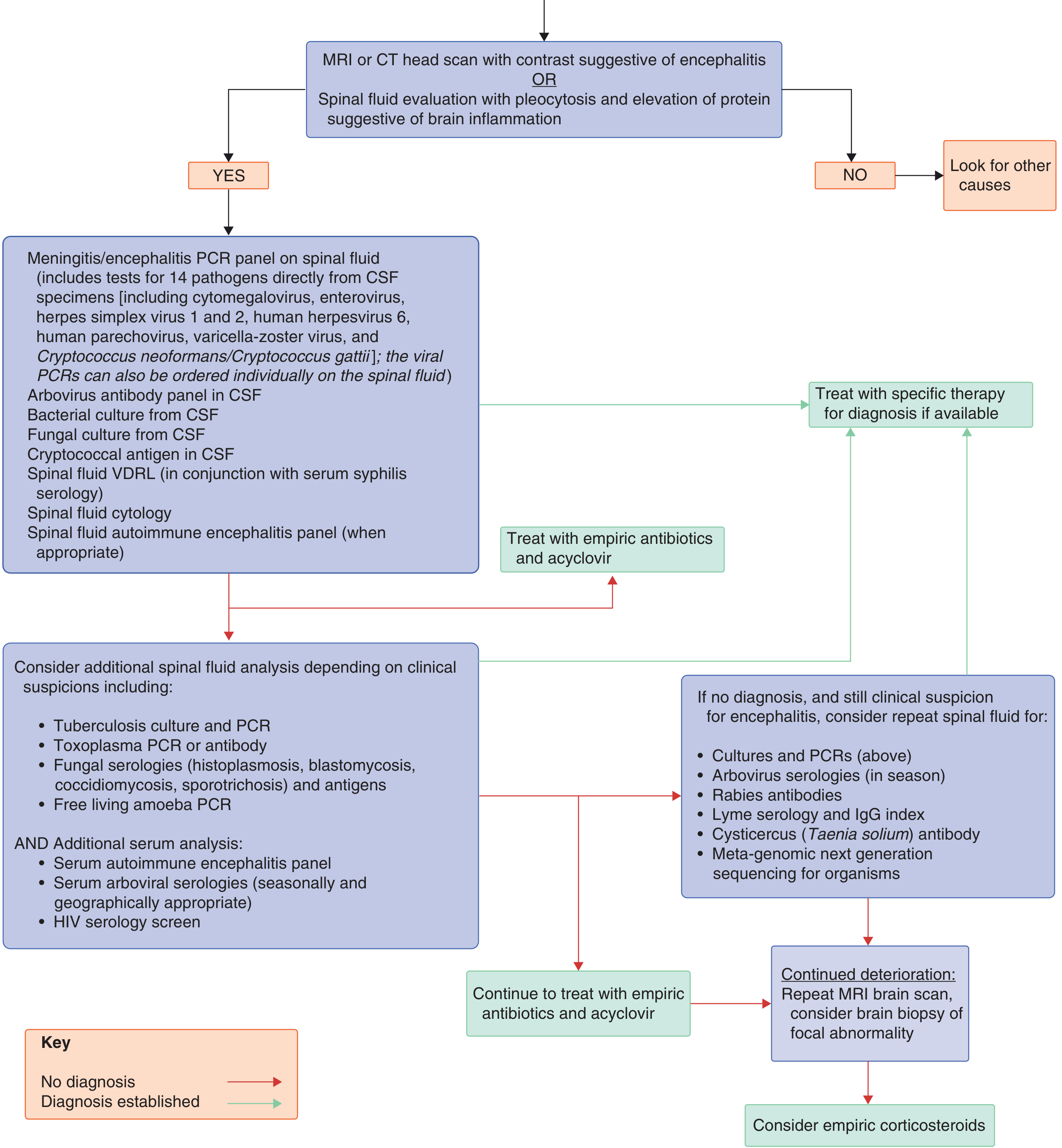
1. Specific Antiviral Therapy
| Parameter | Detail |
|---|---|
| Drug of choice | IV Acyclovir |
| Dose | 10 mg/kg every 8 hours (= 30 mg/kg/day) |
| Duration | 14-21 days |
| Route | Intravenous only (inadequate CNS penetration with oral formulations at standard doses) |
| Renal impairment | Dose must be reduced; monitor serum creatinine |
| Obese patients | Dose based on ideal body weight |
- Goldman-Cecil Medicine, p.3299
- Adams and Victor's Principles of Neurology 12e, p.451
- Katzung's Basic and Clinical Pharmacology 16e
2. When to Stop or Continue Acyclovir
- If CSF HSV PCR is positive: complete full 14-21 day IV course, then perform repeat LP to confirm PCR negativity before stopping
- If CSF is still PCR-positive at end of treatment: continue IV acyclovir with weekly PCR until negative
- If PCR is negative but clinical suspicion remains high (e.g., normal early CSF in first 24 hours, classic MRI pattern): continue acyclovir and repeat LP after 24-48 hours
- If PCR is negative and alternative diagnosis is established: acyclovir can be discontinued
- Immunocompromised patients: treat for at least 21 days; consider long-term oral suppression until CD4 >200
3. Mechanism of Acyclovir
- Harrison's Principles of Internal Medicine 22E, p.1506
4. Supportive Management
| Issue | Management |
|---|---|
| ICP elevation | Fluid restriction, avoid hypotonic IV solutions, careful ICP monitoring; all standard measures for cerebral edema apply |
| Seizures | Treat with standard anticonvulsant regimens; prophylactic antiepileptics are reasonable given high seizure frequency in severe encephalitis |
| Fever | Active suppression |
| Respiratory/BP | Continuous monitoring; ICU care in severe/early stages |
| Complications | Aspiration pneumonia prevention, DVT prophylaxis, pressure ulcer care, catheter infection vigilance |
| Mass effect / hemorrhagic necrosis | Large-volume temporal lobe involvement can behave like an expanding lesion; coma + pupillary changes may indicate brainstem compression or direct spread - assess with imaging |
5. Corticosteroids - Controversial
- Significant cerebral edema with mass effect
- Post-HSE autoimmune encephalitis (see below): methylprednisolone 1 g IV daily for 5 days
6. Post-HSE Autoimmune Encephalitis - An Important Complication
- Relapse of symptoms (behavioral changes, movement disorders, seizures in children; delirium in adults)
- Worsening MRI findings
- Positive CSF/serum NMDA-receptor antibodies
- Goldman-Cecil Medicine, p.3306
- Adams and Victor's Principles of Neurology 12e, p.457
7. Relapses After Acyclovir
8. Acyclovir Side Effects to Monitor
| Side Effect | Notes |
|---|---|
| Renal dysfunction (transient) | Most important - ensure adequate hydration during infusion |
| Local vein irritation / phlebitis | Common with peripheral IV |
| Elevated hepatic enzymes (mild) | Usually transient |
| Nausea, vomiting, tremor | Uncommon |
| Encephalopathy | Rare; difficult to distinguish from the encephalitis itself |
- Adams and Victor's Principles of Neurology 12e, p.451
Prognosis
-
Less than 20% return to work
-
~50% are classified as moderately to severely disabled
-
Early treatment (before coma) is the strongest predictor of a good outcome - the level of consciousness at initiation of treatment is critical
-
Goldman-Cecil Medicine, p.3304
How do we manage the vancomycin and ceftrixone resistance in the bacterial meningities and what about the treatment
"bacterial meningitis" AND "antibiotic resistance" AND treatment
vancomycin ceftriaxone resistant pneumococcal meningitis management treatment options 2024
Bacterial Meningitis: Management of Vancomycin + Ceftriaxone Resistance
Understanding the Problem
- Penicillin/cephalosporin resistance: alteration of penicillin-binding proteins (PBPs) by transformation and horizontal DNA transfer from related streptococci. Altered PBPs have lower affinity for beta-lactams, causing resistance ranging from intermediate to high level.
- Vancomycin penetration problem: even when S. pneumoniae is "susceptible" to vancomycin in vitro, dexamethasone (used adjunctively) reduces blood-brain barrier inflammation and thereby reduces CSF vancomycin penetration, potentially creating a pharmacokinetic failure even with a susceptible organism.
-
Penicillin susceptible: MIC ≤0.06 μg/mL; resistant: ≥0.12 μg/mL
-
Ceftriaxone/cefotaxime: MIC ≥1.0 μg/mL = high-level resistance
-
Harrison's Principles of Internal Medicine 22E, p.1713
Step 1: Empirical Therapy (Before Culture/Sensitivities)
| Drug | Adult Dose | Purpose |
|---|---|---|
| Ceftriaxone | 2 g IV every 12 h | Covers susceptible pneumococcus, N. meningitidis, H. influenzae |
| OR Cefotaxime | 2-3 g IV every 6-8 h | Alternative cephalosporin |
| + Vancomycin | 15-20 mg/kg IV every 8 h (adults: 30-60 mg/kg/day) | Covers cephalosporin-resistant pneumococcus |
| + Dexamethasone | 0.15 mg/kg IV every 6 h x 4 days | Given before or with first antibiotic dose |
| + Acyclovir | 10 mg/kg every 8 h | Covers HSV encephalitis (leading differential) |
| + Ampicillin (if age >50 or immunocompromised) | 2 g IV every 4 h | Covers Listeria monocytogenes |
Do NOT reduce antibiotic doses as the patient improves - normalisation of the blood-brain barrier during recovery reduces achievable CSF levels.
- Harrison's Principles of Internal Medicine 22E, p.1762
- Rosen's Emergency Medicine, p.4407
Step 2: Targeted Therapy by Susceptibility Results
For S. pneumoniae:
| Susceptibility Pattern | Recommended Therapy | Alternative |
|---|---|---|
| Penicillin MIC ≤0.06 μg/mL (susceptible) | Penicillin G or Ampicillin | Ceftriaxone, cefotaxime, chloramphenicol |
| Penicillin MIC ≥0.12 μg/mL but ceftriaxone MIC ≤1.0 μg/mL | Ceftriaxone or Cefotaxime alone | Cefepime, meropenem |
| Ceftriaxone MIC ≥1.0 μg/mL (high-level resistance) | Vancomycin + Ceftriaxone/Cefotaxime | Vancomycin + moxifloxacin |
| Resistant to BOTH cephalosporins AND vancomycin, or cephalosporin allergy | Vancomycin + Rifampicin | Linezolid + vancomycin; moxifloxacin + vancomycin; chloramphenicol |
- Goldman-Cecil Medicine, Table 381-6
- Rosen's Emergency Medicine, p.4411
Step 3: When Vancomycin + Ceftriaxone Fails - The Real Resistance Scenario
A. Add or Switch to Rifampicin
- Rifampicin (rifampin) penetrates the CSF excellently (unlike most antibiotics, it crosses even the uninflamed blood-brain barrier)
- Used as combination therapy - never monotherapy (rapid resistance emerges)
- Dose: 600 mg IV/PO every 24 h (up to 900 mg/day; 15 mg/kg/day in children)
- Combination: Vancomycin + Rifampicin is the standard salvage regimen for highly resistant pneumococcus
- Particularly important when dexamethasone is being used (rifampicin compensates for reduced vancomycin CSF levels)
B. Meropenem
- Carbapenem with excellent activity against penicillin-resistant S. pneumoniae, L. monocytogenes, H. influenzae, P. aeruginosa
- As effective as cefotaxime for pneumococcal, meningococcal, and H. influenzae meningitis
- Preferred over ceftriaxone for nosocomial/post-neurosurgical meningitis and for P. aeruginosa coverage
- Experimental data: meropenem was inferior to vancomycin but comparable to ceftriaxone in clearing pneumococcal CSF cultures
- Dose: 2 g IV every 8 h
C. Linezolid
- Dose: 600 mg IV every 12 h
- Used for cephalosporin-resistant pneumococcus in combination with vancomycin
- Also effective for MRSA meningitis (nosocomial/post-neurosurgical cases)
- Advantage: excellent CNS penetration; useful when beta-lactam allergy and vancomycin is needed
D. Moxifloxacin / Fluoroquinolones
- Moxifloxacin 400 mg IV/PO once daily
- Used with vancomycin for cephalosporin-resistant strains
- Levofloxacin is an alternative
- Caution: increasing fluoroquinolone resistance in some regions
E. Chloramphenicol
-
Historical alternative; still useful in resource-limited settings or severe beta-lactam allergy
-
Good CNS penetration
-
Limitations: bacteriostatic (not bactericidal for pneumococcus at achievable CSF concentrations), bone marrow toxicity
-
Goldman-Cecil Medicine, p.1916-1917
-
Rosen's Emergency Medicine, p.4407-4411
-
Adams and Victor's Principles of Neurology 12e, p.1881
Step 4: Pathogen-Specific Resistance Situations
N. meningitidis (Meningococcus)
- In the US, remains highly susceptible to penicillin and ampicillin
- Penicillin G or ampicillin is definitive therapy for susceptible strains
- Third-generation cephalosporins are used empirically and are effective
H. influenzae
- ~30% of isolates produce beta-lactamase (resistant to ampicillin/penicillin)
- Nearly all remain sensitive to third-generation cephalosporins
- For beta-lactamase-negative strains: ampicillin
- For resistant strains: ceftriaxone/cefotaxime (first-line), alternatives = chloramphenicol, cefepime, meropenem, fluoroquinolone, aztreonam
Nosocomial / Post-Neurosurgical Meningitis (MRSA, Coagulase-negative Staph, P. aeruginosa)
- Empirical: Vancomycin + Cefepime or Ceftazidime or Meropenem
- If beta-lactam contraindicated: linezolid (gram-positive) + aztreonam or ciprofloxacin (gram-negative coverage)
Listeria monocytogenes (cell-mediated immunity deficiency)
- Ampicillin ± gentamicin
- Trimethoprim-sulfamethoxazole if penicillin-allergic
- Cephalosporins have NO activity against Listeria - a critical point
- Stop dexamethasone if Listeria is confirmed (increased adverse outcomes shown in a prospective study)
The Dexamethasone Dilemma
-
If using dexamethasone + vancomycin: consider adding rifampicin to ensure adequate CSF levels against resistant pneumococcus
-
Discontinue dexamethasone if: organism is not S. pneumoniae or H. influenzae, or if Listeria is confirmed
-
Dexamethasone benefit is only demonstrated in high-income countries - not shown in low-income settings
-
Goldman-Cecil Medicine, p.1915
Key Principles of Antibiotic Use in Meningitis
- IV route throughout - do not switch to oral; do not reduce doses as patient improves (BBB recovery reduces CSF levels)
- CSF bactericidal levels should be 10-20x the minimal bactericidal concentration (MBC)
- Rifampicin and chloramphenicol are the exceptions - they cross the uninflamed BBB; others require inflamed meninges for adequate penetration
- Culture and sensitivity-guided de-escalation is mandatory - vancomycin should be stopped once susceptibility data allow
- Antibiotic duration after organism identification: S. pneumoniae 10-14 days; N. meningitidis 7 days; H. influenzae 7 days; L. monocytogenes 21 days; gram-negative bacilli 21 days
- Goldman-Cecil Medicine, p.1902
Gave the full details about the batten diseases
"Batten disease" OR "neuronal ceroid lipofuscinosis"
Batten disease neuronal ceroid lipofuscinosis full clinical details genetics pathology treatment 2024
PMID: 39925015
https://www.ncbi.nlm.nih.gov/books/NBK606097
Batten disease CLN subtypes cerliponase alfa brineura minocycline gene therapy 2024 2025 treatment update
Batten Disease (Neuronal Ceroid Lipofuscinoses - NCL)
1. Definition and Overview
-
At least 13-14 subtypes have been identified (NCL1 through NCL14), each caused by a mutation in a distinct CLN gene
-
The name "Batten disease" strictly refers to the juvenile form (CLN3), but is used colloquially for all NCL subtypes
-
Incidence: approximately 2-4 per 100,000 children worldwide; collectively the most common pediatric neurodegenerative disease
-
Almost universally fatal - most children die in childhood or early adulthood
-
Adams and Victor's Principles of Neurology 12e, p.1952
-
Harrison's Principles of Internal Medicine 22E, p.1407
2. Pathogenesis
Cellular Mechanism
- Each CLN gene encodes a protein critical to lysosomal function - either a lysosomal enzyme, a transmembrane protein, or a secretory protein
- When these proteins are deficient, undigested lipofuscin-like material accumulates in lysosomes, particularly in neurons
- The storage material consists of two pigmented lipids - ceroid and lipofuscin - which are cross-linked polymers of polyunsaturated fatty acids with the property of autofluorescence (important diagnostically)
- This accumulation triggers progressive neuronal death in the cerebral and cerebellar cortex (particularly granule cells and Purkinje cells)
Electron Microscopy Patterns (Ultrastructural Subtypes)
| Inclusion Type | Associated Subtype |
|---|---|
| Granular osmiophilic deposits (GROD) | CLN1 (infantile) |
| Curvilinear profiles (CLP) | CLN2 (late infantile) |
| Fingerprint profiles (FPP) | CLN3 (juvenile) |
| Mixed CLP + FPP | CLN5, CLN6 |
| Rectilinear profiles | CLN6, CLN8 |
- Adams and Victor's Principles of Neurology 12e, p.1991
Inheritance
- Autosomal recessive: all infantile and juvenile forms (CLN1-3, CLN5-14)
- Autosomal dominant exception: CLN4 (Kufs disease, Parry type) - the only dominant NCL
- X-linked: not reported
3. Classification and Clinical Subtypes
CLN1 - Infantile NCL (Santavuori-Haltia Disease)
- Gene: PPT1 (palmitoyl-protein thioesterase 1)
- Onset: 3-18 months (after normal early development)
- Features:
- Rapid psychomotor regression with ataxia, hypotonia
- Widespread myoclonus
- Retinal changes with extinction of the electroretinogram (ERG)
- Progressive visual failure leading to blindness
- EEG: slowing, spike-and-slow-wave, then eventually isoelectric record
- Spastic quadriplegia, microcephaly
- Prognosis: death within a few years of onset
CLN2 - Late Infantile NCL (Jansky-Bielschowsky Disease) ⭐ Only FDA-Approved Treatment
- Gene: TPP1 (tripeptidyl peptidase 1)
- Onset: 2-4 years (after normal or slightly delayed development)
- Survival: typically 8-12 years of age
- Features (in roughly this order):
- Seizures (petit mal, grand mal) - usually the first sign
- Myoclonic jerks triggered by sensory stimuli (proprioception, voluntary movement, emotional excitement)
- Incoordination, tremor, ataxia, spastic weakness
- Lively tendon reflexes and Babinski signs
- Progressive mental deterioration → dementia → mutism
- Dysarthria, then complete loss of speech
- Visual failure (variable - may occur early due to rod-and-cone retinal degeneration)
- Wheelchair bound by late childhood
- Labs: vacuoles in 10-30% of circulating lymphocytes; azurophilic granules in neutrophils
- EEG: high-voltage spikes induced by photic stimulation (characteristic)
- Microcephaly: only in early-onset cases
CLN3 - Juvenile NCL (Batten Disease stricto sensu, Vogt-Spielmeyer Disease)
- Gene: CLN3 (encodes battenin, a lysosomal transmembrane protein)
- Onset: 4-10 years
- Survival: late teens to 20s-30s (longer course than other forms)
- Features (typically in this sequence):
- Visual loss - the hallmark first symptom (progressive pigmentary retinal degeneration)
- Seizures - appear later
- Ataxia
- Cognitive decline (behavioral changes, psychiatric symptoms - depression, anxiety, hallucinations)
- Progressive motor deterioration
- Ophthalmic findings: pigmentary retinopathy, bull's eye maculopathy, optic atrophy
- Vacuolated lymphocytes on peripheral blood smear - useful screening test
CLN4 - Adult NCL (Kufs Disease)
- Genes: CLN6 or DNAJC5 mutations (Parry type = autosomal dominant)
- Onset: adulthood (typically 3rd-4th decade)
- Features: two main phenotypes:
- Type A: Progressive myoclonic epilepsy, cerebellar signs, dementia
- Type B: Dementia, motor dysfunction, WITHOUT visual failure (retinal sparing is key - unlike other NCLs)
- No visual loss distinguishes it from other forms
CLN5 - Finnish Variant Late Infantile NCL
- Gene: CLN5
- Onset: 2-6 years
- Features: ataxia first, then psychomotor regression; unable to walk by age 10
- Visual loss (macular dystrophy, optic atrophy) between ages 6-10
- Most do not survive into their third decade
CLN6, CLN7, CLN8... (Other Forms)
- CLN6, CLN7, CLN8 present similarly to CLN2 (late infantile pattern) with variable onset and severity
- CLN14: rapidly progressing; intractable myoclonic seizures before age 2, rapid visual loss with optic atrophy
4. Summary Table
| Subtype | Old Name | Gene | Enzyme/Protein | Onset | Key Feature |
|---|---|---|---|---|---|
| CLN1 | Santavuori-Haltia | PPT1 | Palmitoyl-protein thioesterase 1 | Infantile (3-18 mo) | Rapid regression, myoclonus, blindness |
| CLN2 | Jansky-Bielschowsky | TPP1 | Tripeptidyl peptidase 1 | Late infantile (2-4 yr) | Seizures first, photic EEG spikes |
| CLN3 | Vogt-Spielmeyer / Batten | CLN3 | Battenin (transmembrane) | Juvenile (4-10 yr) | Visual loss first |
| CLN4 | Kufs disease | CLN6/DNAJC5 | Variable | Adult | No visual loss; AD variant |
| CLN5 | Finnish variant | CLN5 | CLN5 protein | Late infantile (2-6 yr) | Ataxia first |
| CLN6-14 | Various | Multiple | Multiple | Variable | Late infantile-juvenile patterns |
5. Pathology
Macroscopic
- Progressive brain atrophy - cerebral and cerebellar cortex
- Neuronal loss most severe in cortex
Microscopic
- Neuronal loss in cerebral and cerebellar cortices (granule cells and Purkinje cells)
- Curvilinear storage particles and osmophilic granules in remaining neurons
- Inclusions also found in:
- Cutaneous nerve twigs
- Endothelial cells of blood vessels
- Peripheral blood lymphocytes (vacuoles) - diagnostic significance
Ultrastructure (Electron Microscopy)
-
Characteristic inclusion body patterns per subtype (GROD, curvilinear, fingerprint - see above)
-
Skin biopsy, conjunctival biopsy, or rectal mucosal biopsy can demonstrate inclusions diagnostically
-
Adams and Victor's Principles of Neurology 12e, p.1991
6. Diagnosis
Clinical Suspicion
- Any child with the triad: seizures + progressive visual loss + cognitive/motor regression
- Peripheral blood smear showing vacuolated lymphocytes (CLN2, CLN3)
Diagnostic Workup
| Test | Finding |
|---|---|
| Genetic testing (gene panel) | Confirmatory - identifies CLN mutation; guides subtype and prognosis |
| Enzyme assay | PPT1 activity (CLN1), TPP1 activity (CLN2) in leukocytes or dried blood spots |
| Electron microscopy of skin/conjunctival biopsy | Characteristic inclusion bodies (GROD, curvilinear, fingerprint profiles) |
| EEG | High-voltage spikes to photic stimulation (CLN2); progressive slowing; isoelectric in advanced CLN1 |
| ERG (electroretinogram) | Extinguished/isoelectric in retinal disease forms |
| MRI brain | Progressive cerebral and cerebellar atrophy; white matter changes |
| Visual evoked potentials | Abnormal in forms with retinal involvement |
| Peripheral blood smear | Vacuolated lymphocytes (CLN2, CLN3) |
| Ophthalmologic assessment | Pigmentary retinopathy, macular degeneration, optic atrophy |
| Histopathology of rectum/skin | Fluorescent storage material under UV (autofluorescence); EM inclusions |
- Diagnosis is confirmed by gene sequencing in most cases
7. Treatment
A. Disease-Specific (Approved) Therapy
Cerliponase Alfa (Brineura) - CLN2 ONLY ⭐
-
The ONLY FDA/EMA-approved disease-modifying treatment for any NCL
-
A recombinant human TPP1 (enzyme replacement therapy - ERT)
-
Restores TPP1 enzyme activity and breaks down the stored lipofuscin material
-
Route: Intraventricular infusion via an indwelling intracerebroventricular (ICV) port (the first ERT approved for direct CNS delivery)
-
Dose: Every 2 weeks, infused over several hours
-
Indication: Slows loss of ambulation (walking/crawling) in pediatric patients with CLN2
-
FDA 2017: Approved for symptomatic children ≥3 years
-
FDA 2024 expanded approval: Now approved for children of all ages, including presymptomatic infants - allowing earlier intervention before symptom onset
-
Clinical trial (NEJM 2018): Significantly reduced the rate of clinical decline compared to untreated historical controls
-
Adverse effects: Many related to the ICV catheter placement (infection, device complications)
-
Harrison's Principles of Internal Medicine 22E, p.1407
-
NINDS/FDA 2024 update
B. Symptomatic Treatment
| Symptom | Management |
|---|---|
| Seizures | Anti-seizure medications (valproate, lamotrigine, levetiracetam, clobazam); seizures in NCL are often polypharmacy-requiring |
| Avoid | Lamotrigine and carbamazepine may worsen myoclonus in some NCL subtypes |
| Myoclonus | Valproate, clonazepam, levetiracetam |
| Spasticity | Baclofen, physiotherapy, orthoses |
| Behavioral/Psychiatric | Antidepressants, antipsychotics for mood disorders and hallucinations (especially in CLN3) |
| Sleep disturbance | Melatonin, sleep hygiene |
| Nutritional support | PEG tube feeding as swallowing deteriorates |
| Respiratory | Chest physiotherapy, management of aspiration |
C. Emerging / Investigational Therapies
| Therapy | Target | Status |
|---|---|---|
| Gene therapy (AAV9-based) | CLN3 (Batten), CLN5, CLN6, others | Phase 1/2 trials - CLN-301 for CLN3 Batten disease (Nationwide Children's/Alcyone Therapeutics); CLN5 gene therapy trial ongoing |
| Stem cell therapy / Neural stem cell transplantation | CLN1, CLN2 | Early trials; CNS access remains the primary barrier |
| Small molecule drugs | Various (N-acetylcysteine, cysteamine for CLN1) | Preclinical; some early clinical use |
| Microglial cell replacement therapy | Neuroinflammation | Emerging preclinical evidence |
| Antisense oligonucleotides (ASO) | CLN3 splice variants | Preclinical |
D. Supportive / Multidisciplinary Care
- Pediatric neurologist (seizure management, disease monitoring)
- Ophthalmologist (visual loss management, low-vision aids)
- Physiotherapist (mobility, spasticity, contractures)
- Speech-language therapist (communication, swallowing)
- Occupational therapist (daily living adaptations)
- Neuropsychologist / Psychiatrist (behavioral/psychiatric symptoms)
- Dietitian (nutritional management)
- Palliative care (as disease advances)
- Genetics (family counseling, prenatal diagnosis, carrier testing)
- Education: Individualized Education Plan (IEP) addressing vision impairment, seizures, learning difficulties
8. Differential Diagnosis
| Condition | Distinguishing Feature |
|---|---|
| Late infantile GM1 gangliosidosis | Cherry-red spot, hepatosplenomegaly |
| Lafora body disease | Polyglucosan bodies, adolescent onset |
| Alpers disease | Hepatic involvement, mitochondrial mutation |
| Sialidosis ("cherry-red spot") | Cherry-red spot, cherry-red-spot myoclonus syndrome |
| Non-infantile Gaucher disease | Hepatosplenomegaly, enzyme assay |
| Idiopathic epilepsy | No storage material, no progressive visual/cognitive loss |
| GM2 gangliosidosis (Tay-Sachs) | Cherry-red spot, hexosaminidase A deficiency |
9. Prognosis
- CLN1 (infantile): death within a few years of onset
- CLN2 (late infantile): death at an average age of 8-12 years (without treatment)
- CLN3 (juvenile): survival into late teens to 30s (longest course)
- CLN4 (adult/Kufs): survival into adulthood, slower progression
- With cerliponase alfa (CLN2): significantly slowed functional decline; the disease trajectory is altered but not yet cured
Key Teaching Points
- NCL/Batten disease = lysosomal storage disorder + neurodegeneration + autofluorescent lipofuscin accumulation
- Triad: seizures + visual failure + dementia in a child = think NCL
- Vacuolated lymphocytes on blood smear is a cheap bedside screening test
- Electron microscopy of skin biopsy shows pathognomonic inclusion patterns
- Only CLN2 has an approved disease-modifying therapy (cerliponase alfa/Brineura) - now approved from birth (2024 FDA expanded indication)
- Gene therapy trials are active for CLN3, CLN5, and other subtypes and represent the best hope for future treatments
- CLN4 (Kufs) is the only autosomal dominant form and the only form without visual loss
What is treatment for that
"neuronal ceroid lipofuscinosis" AND (treatment OR therapy OR "gene therapy" OR cerliponase)
Batten disease NCL treatment 2024 2025 gene therapy CLN3 CLN1 CLN2 clinical trials emerging therapies
PMID: 41160491
PMID: 40908342
Batten Disease (NCL) - Complete Treatment Guide
PILLAR 1: Disease-Modifying Therapy (Approved)
Cerliponase Alfa (Brineura) - CLN2 Disease ONLY
| Feature | Detail |
|---|---|
| Drug class | Enzyme replacement therapy (ERT) - recombinant human TPP1 |
| Mechanism | Restores tripeptidyl peptidase 1 (TPP1) enzyme activity; breaks down accumulated ceroid lipofuscin in lysosomes |
| Route | Intraventricular infusion via indwelling intracerebroventricular (ICV) port - the first ERT delivered directly into the CNS |
| Dosing | Infused over several hours every 2 weeks |
| Indication | Slows loss of ambulation (walking/crawling) in CLN2 disease |
| FDA 2017 | Original approval: symptomatic children ≥3 years |
| FDA July 2024 | Expanded: children of ALL ages, including presymptomatic infants |
| Goal | Start before symptom onset - earlier treatment = better outcomes |
Clinical Evidence
- Cerliponase alfa significantly slowed clinical decline vs historical untreated controls
- 24% achieved a Clinical Rating Scale score of 0-1 (near-normal function)
- Seizure rate: generalized tonic-clonic seizures in 41%, dystonia in 15%
Important Limitations
- Does NOT reverse vision loss - retinal degeneration continues even with ERT
- Not a cure - slows but does not stop disease progression
- CLN3 (the most common juvenile form, "classical Batten disease") is caused by a transmembrane protein defect - ERT cannot work for it because the protein cannot be delivered via the lysosomal pathway
- Adverse effects: pyrexia (60%), hypersensitivity reactions (82.6%), anaphylaxis (3%), infusion-associated reactions (18%), device/catheter-related complications
- Harrison's Principles of Internal Medicine 22E, p.1407
- Bradley and Daroff's Neurology in Clinical Practice, p.2632
PILLAR 2: Symptomatic Management
Seizure Management
| Drug | Role/Notes |
|---|---|
| Valproate | Broad-spectrum; good for myoclonus and generalized seizures |
| Levetiracetam | Effective for myoclonus and generalized seizures |
| Clobazam | Add-on for refractory seizures |
| Clonazepam | Myoclonus control |
| Lamotrigine | Use with caution - may worsen myoclonus in some NCL subtypes |
| Carbamazepine / Oxcarbazepine | Generally avoid - may exacerbate myoclonus and decline |
| Phenobarbital | Second-line option |
| Vigabatrin | Avoid - can worsen visual loss |
Myoclonus
- Valproate + levetiracetam or clonazepam combination
- Piracetam (where available) has some evidence for action myoclonus
Spasticity
- Baclofen (oral or intrathecal pump in severe cases)
- Physiotherapy, orthoses, splinting
- Botulinum toxin for focal spasticity/dystonia
Behavioral and Psychiatric Symptoms (especially CLN3)
- SSRI antidepressants (fluoxetine, sertraline) for depression and anxiety
- Low-dose antipsychotics (risperidone, quetiapine) for hallucinations, agitation
- Behavioral management strategies through psychology
- CLN3 patients often develop significant psychiatric features in adolescence before severe physical decline
Sleep Disturbance
- Melatonin (widely used, good safety profile)
- Sleep hygiene measures
- Clonidine as second-line
Nutritional Support
- PEG tube (percutaneous gastrostomy) when swallowing deteriorates
- Nutritional supplementation to maintain weight
- Thickened fluids initially; eventually tube feeding is required
- Monitor for aspiration
Respiratory Management
- Chest physiotherapy and breathing exercises
- Management of recurrent aspiration pneumonia
- Non-invasive ventilation in late stages if appropriate (individualized decision)
- Advance directives and goals of care discussion
Visual Impairment
- Low-vision aids (magnifiers, high-contrast materials, screen readers)
- Orientation and mobility training
- Guide dogs for adolescents/adults (CLN3)
- No specific treatment halts retinal degeneration currently (even cerliponase alfa does not protect vision in CLN2)
PILLAR 3: Supportive / Multidisciplinary Care
| Specialist | Role |
|---|---|
| Pediatric neurologist | Seizure management, disease monitoring, AED adjustment |
| Ophthalmologist | Visual monitoring, low-vision services |
| Physiotherapist | Mobility, spasticity management, contracture prevention |
| Occupational therapist | Adaptive equipment, daily living modifications, wheelchair assessment |
| Speech-language therapist | Communication aids (AAC devices), swallowing assessment |
| Dietitian | Nutritional support, PEG management |
| Clinical psychologist / Psychiatrist | Behavioral and psychiatric symptoms |
| Palliative care team | Advance care planning, comfort, end-of-life care |
| Clinical geneticist | Family counseling, carrier testing, prenatal diagnosis, reproductive options |
| Social worker | Family support, disability services, equipment funding |
| Education team | Individualized Education Plan (IEP) - vision impairment, seizures, cognitive decline |
Palliative Care
- Should be integrated early alongside curative/disease-modifying efforts - not just at end of life
- Goals of care: maximize quality of life, minimize suffering, support family
- Decisions about resuscitation, ventilation, and hospitalization should be discussed proactively with families
PILLAR 4: Emerging and Investigational Therapies (2024-2026 Pipeline)
A. Gene Therapy (AAV-Based) - Most Promising
| Subtype | Sponsor | Agent | Status (as of 2025-2026) |
|---|---|---|---|
| CLN2 | Tern Therapeutics | TTX-181 (AAV9; CNS) | Positive Phase 1/2 data (Feb 2025 WORLDSymposium) |
| CLN2 | Tern Therapeutics | TTX-381 (AAV; ocular) | MHRA Innovation Passport (UK, Jul 2025); FDA CDRP program (Dec 2025) |
| CLN2 | Latus Bio | LTS-101 (AAV-Ep+ capsid; CNS) | IND cleared Dec 2025; Fast Track + Orphan Drug designation |
| CLN3 | Alcyone Therapeutics | CLN-301 (AAV9; CNS) | Phase 1/2 since 2018; 4 patients treated, 5+ years follow-up (Apr 2025 update) |
| CLN3 | UNC/ForeBatten Foundation | FBF-001 "Zebronkysen" (ASO) | N-of-2 study active since Sept 2024; 1-year update Jul 2025 |
| CLN5 | Neurogene | NGN-101 (AAV) | Phase 1/2 ongoing (NCT05228145) |
| CLN6 | Nationwide Children's | AAV-CLN6 | Preclinical - intrathecal single dose in NHP improved motor function and survival |
- REGENXBIO halted RGX-181 (CNS) and RGX-381 (ocular) for CLN2
- Amicus Therapeutics returned CLN3 and CLN6 programs to Nationwide Children's Hospital
B. Antisense Oligonucleotides (ASO)
- FBF-001 (Zebronkysen) for CLN3: designed to skip the most common CLN3 mutation (1-kb deletion), restoring partial protein function
- N-of-2 compassionate use study commenced September 2024 - one of the most watched rare disease trials of 2025
- ASOs work by modifying RNA splicing, bypassing the mutation
C. Small Molecule Drugs
| Drug | Target | Subtype | Status |
|---|---|---|---|
| Miglustat (Batten-1) | Substrate reduction therapy; corrects lipid metabolism | CLN3 | Phase 1/2 complete (USA); positive real-world data (May 2025); licensing agreement Feb 2026 |
| Gemfibrozil (PLX-200) | PPARα activation; neuroprotection | CLN3 | Phase 3 design registered; minimal recent activity |
| N-acetylcysteine | Antioxidant; reduce oxidative stress | CLN1 | Preclinical/early clinical |
| Cysteamine + N-acetylcysteine | Palmitoyl thioesterase pathway | CLN1 | Early clinical investigation |
D. Stem Cell Therapy
- Neural stem cell transplantation: trials have been conducted for CLN1 and CLN2
- CNS access remains the primary barrier to effective delivery
- Results so far limited - transplanted cells do not integrate or migrate sufficiently
- Emerging: microglial cell replacement therapy - highlighted in a 2025 CNS Neuroscience & Therapeutics review (PMID 39925015) as particularly promising given the central role of microglial pathology in NCL
E. Combination Approaches
- Combining ERT (cerliponase alfa for CLN2) with ocular gene therapy (TTX-381) to address the separate retinal degeneration that ERT alone does not treat
- Combinations of gene therapy + immunosuppression to reduce AAV immune response
Why Treatment is Hard: The Core Challenges
- CNS barrier: most drugs cannot reach the brain in therapeutic concentrations - hence ICV delivery for ERT
- Genetic heterogeneity: 13+ subtypes, each needing a different molecular approach
- Non-neuronal disease: bowel, cardiac, and other organ involvement also requires treatment
- Late diagnosis: symptoms appear after significant neuronal loss has already occurred - making early (even presymptomatic) diagnosis and treatment critical
- Membrane proteins vs soluble enzymes: ERT only works for soluble enzyme deficiencies (CLN2); membrane protein defects (CLN3) need gene therapy or other approaches
- Commercial viability: multiple promising programs have been abandoned due to small patient populations and economics - a recurring tragedy in ultra-rare disease
Summary Table by Subtype
| Subtype | Approved DMT | Emerging |
|---|---|---|
| CLN1 | None | N-acetylcysteine + cysteamine; gene therapy preclinical |
| CLN2 | Cerliponase alfa (Brineura) ✅ | Gene therapy (Tern TTX-181, Latus LTS-101) |
| CLN3 | None | Miglustat (Batten-1); CLN-301 gene therapy; FBF-001 ASO |
| CLN4 | None | None currently active |
| CLN5 | None | Neurogene NGN-101 gene therapy (Phase 1/2) |
| CLN6 | None | AAV-CLN6 (preclinical); returned from Amicus to Nationwide |
| CLN7-14 | None | Preclinical only |
The bottom line: Cerliponase alfa (Brineura) is the only approved disease-modifying therapy and is restricted to CLN2. All other subtypes rely on symptomatic treatment. Gene therapy, ASOs, and small molecules represent the best hope for treating the remaining subtypes - with several programs in active Phase 1/2 trials as of 2025-2026.
What is the management and treatment for the ARDS
"acute respiratory distress syndrome" AND management
ARDS management treatment guidelines 2024 lung protective ventilation prone positioning PEEP
PMID: 38032683
PMID: 41325621
Management and Treatment of ARDS
Berlin Definition (2012) - Quick Reminder
| Severity | PaO₂/FiO₂ (P/F Ratio) | PEEP Required |
|---|---|---|
| Mild | 200-300 mmHg | ≥5 cmH₂O |
| Moderate | 100-200 mmHg | ≥5 cmH₂O |
| Severe | <100 mmHg | ≥5 cmH₂O |
Key principle: ARDS is a syndrome, not a diagnosis. Always search for and treat the underlying cause. Most deaths in ARDS are from multi-organ failure, not hypoxemia itself.
General Principles of ICU Care
- Identify and treat the underlying cause (pneumonia, sepsis, aspiration, pancreatitis, trauma)
- Prophylaxis against VTE (DVT/PE)
- Prophylaxis against GI bleeding (PPI/H2 blocker)
- Minimize unnecessary procedures and their complications
- Aspiration precautions (head-of-bed elevation 30-45°)
- Prevent central line infections (aseptic technique, bundle care)
- Minimize oversedation - daily awakening trials
- Enteral nutrition as early as feasible (within 24-48 hours)
- Prompt recognition and treatment of nosocomial infections
- Harrison's Principles of Internal Medicine 22E, p.2704
PILLAR 1: Lung-Protective Mechanical Ventilation (MOST IMPORTANT - MORTALITY BENEFIT PROVEN)
The ARDSNet Protocol
| Parameter | Target | Rationale |
|---|---|---|
| Tidal volume (VT) | 4-8 mL/kg predicted body weight (PBW) | Prevents volutrauma (alveolar overdistention) |
| Standard VT target | 6 mL/kg PBW | Starting point; may reduce to 4 if needed |
| Plateau pressure (Pplat) | ≤30 cmH₂O | Prevents barotrauma |
| Driving pressure (ΔP) | <15 cmH₂O (ΔP = Pplat - PEEP) | Emerging target; strongly predictive of outcome |
| FiO₂ | Minimum needed to achieve target SpO₂ | Minimize oxygen toxicity |
| SpO₂ target | 88-95% | Avoid both hypoxia and hyperoxia |
| PaO₂ target | >55-80 mmHg | |
| Permissive hypercapnia | pH ≥7.20-7.25 acceptable | Allows low VT even if CO₂ rises |
Why Use Predicted (Ideal) Body Weight?
- Males: 50 + 2.3 × (height in inches - 60)
- Females: 45.5 + 2.3 × (height in inches - 60)
Mechanisms of Ventilator-Induced Lung Injury (VILI) - What We're Preventing
- Volutrauma - alveolar overdistention from excess tidal volume
- Barotrauma - excess pressure injury
- Atelectrauma - repeated alveolar collapse and reopening at end-expiration
- Biotrauma - inflammatory mediator release from injured lung
- Barash Clinical Anesthesia 9e, p.570-574
- Harrison's Principles of Internal Medicine 22E, p.2711-2713
PILLAR 2: PEEP Management
2024 ATS Guideline Recommendations (Key Updates)
| Recommendation | Strength |
|---|---|
| Higher PEEP (without lung recruitment maneuvers) in moderate-to-severe ARDS | Conditional recommendation |
| Recommend AGAINST prolonged lung recruitment maneuvers | STRONG recommendation |
PEEP Titration Methods
- Empirical PEEP/FiO₂ tables (ARDSNet high PEEP or low PEEP tables) - most commonly used
- Esophageal pressure-guided PEEP (measures transpulmonary pressure) - especially useful in obese patients with stiff chest walls; a Phase 2 trial showed no benefit over empirical high PEEP-FiO₂ titration in unselected patients
- Electrical impedance tomography (EIT) - newer imaging-guided approach; a 2024 systematic review (PMID 38512400) shows promise for personalizing PEEP
Lung Recruitment Maneuvers (LRMs)
- Prolonged LRMs (e.g., sustained inflation at 40 cmH₂O for 40 seconds): Previously widely used; now strongly contraindicated by ATS 2024 - shown to increase mortality when combined with higher baseline PEEP (ART trial)
- Brief sigh breaths / stepwise LRMs: insufficient data; conditional use only
- The 2024 ATS guideline marks a paradigm shift away from LRMs
| PEEP/FiO₂ Strategy | Moderate-Severe ARDS | Mild ARDS |
|---|---|---|
| Higher PEEP, lower FiO₂ | Preferred (ATS 2024) | Lower PEEP may be adequate |
| Prolonged LRMs | Avoid (strong recommendation) | Avoid |
PILLAR 3: Prone Positioning
Evidence
When to Use
- PaO₂/FiO₂ <150 mmHg (P/F ratio ≤150) despite optimized ventilation
- Start early (within the first 36 hours) for best effect
- Duration: minimum 12-16 hours per day (PROSEVA used 16 h/day)
- Continue daily proning sessions until P/F ratio improves to >150 on supine position
Mechanism of Benefit
- Redistributes perfusion to now-ventilated (anterior) lung zones
- Recruits dorsal (dependent, previously atelectatic) lung units
- Makes lung inflation more homogeneous - reduces VILI
- Improves V/Q matching
- Facilitates secretion drainage
Contraindications / Complications
| Complication | Risk Management |
|---|---|
| Accidental extubation | Secure ETT; experienced team required |
| Dislodgement of central lines | Secure all catheters before turning |
| Pressure injuries (face, orbital, corneal) | Regular repositioning of head; eye care |
| Brachial plexus injury | Proper arm positioning ("swimmer's position") |
| Abdominal wound dehiscence | Contraindicated in recent abdominal surgery |
| Hemodynamic instability | Check vitals continuously during prone |
| Difficulty with CPR | Stop proning if cardiac arrest occurs |
- Harrison's Principles of Internal Medicine 22E, p.2718-2719
- Barash Clinical Anesthesia 9e, p.576
PILLAR 4: Fluid Management
Conservative vs Liberal Fluid Strategy
- Same mortality but conservative fluid group had:
- More ventilator-free days
- More ICU-free days
- Better oxygenation
- Shorter ICU stay
Practical Approach
-
Restrict IV fluids once the acute resuscitation phase is over (sepsis/shock)
-
Use diuretics to reduce left atrial filling pressure if hemodynamically stable
-
Target: minimize extravascular lung water while maintaining organ perfusion
-
Balance: aggressive diuresis limited by hypotension and renal impairment
-
Central venous monitoring to guide fluid decisions
-
Harrison's Principles of Internal Medicine 22E, p.2727-2729
PILLAR 5: Neuromuscular Blockade (NMB)
Evidence
- ACURASYS trial (2010, French): Early cisatracurium infusion for 48h in severe ARDS (PaO₂/FiO₂ <150) improved 90-day mortality and ventilator-free days with low rates of critical illness myopathy
- ROSE trial (2019, NEJM): Did NOT confirm mortality benefit of routine NMB; however, control group received deep sedation (confounding factor)
2024 ATS Guideline
- Suggest use of neuromuscular blockers in early severe ARDS (conditional recommendation, low certainty)
- Prefer intermittent boluses over continuous infusion when possible
- Use continuous NMB infusion for: persistent ventilator dyssynchrony, need for deep sedation, prone positioning, or persistently high plateau pressures
Rationale for Use
- Eliminates patient-ventilator dyssynchrony (which causes high transpulmonary pressure spikes)
- May have direct anti-inflammatory effects via nicotinic receptor inhibition
- Enables fully lung-protective ventilation and safe prone positioning
- Agent: Cisatracurium preferred (not renally/hepatically metabolized)
Risk
-
ICU-acquired weakness / critical illness myopathy - limit duration to 48h when possible
-
Requires deeper sedation (use sedation + analgesia protocol)
-
Harrison's Principles of Internal Medicine 22E, p.2732-2734
-
Barash Clinical Anesthesia 9e, p.578
PILLAR 6: Corticosteroids
2024 ATS Guideline
- Suggest using systemic corticosteroids in mechanically ventilated ARDS patients (conditional recommendation, moderate certainty)
2026 Meta-Analysis Evidence
- Low-dose, short-course corticosteroids probably reduce short-term mortality in ARDS (RR 0.77, 95% CI 0.61-0.99; moderate certainty)
- Little to no increase in hospital-acquired infections or secondary pneumonia
- This is one of the strongest recent pieces of evidence supporting corticosteroid use
Practical Protocol
| Parameter | Detail |
|---|---|
| Drug | Methylprednisolone (most studied) or dexamethasone |
| Dose | ≤3 mg/kg/day prednisone-equivalent; typical: methylprednisolone 1 mg/kg/day |
| Duration | ≤15 days (short course); initiated within 7 days |
| Taper | Gradual taper to avoid rebound |
| Avoid in | Active untreated fungal infections; caution with bacterial superinfection |
Note: Corticosteroids should NOT be started after day 14 of ARDS onset - possible harm
PILLAR 7: VV-ECMO (Venovenous Extracorporeal Membrane Oxygenation)
When to Consider
| Indication | Threshold |
|---|---|
| Refractory hypoxemia | FiO₂ >90%, PEEP >15 cmH₂O, prone ventilation - still not maintaining SpO₂ |
| Refractory hypercapnia | PaCO₂ >80 mmHg with acidosis |
| Injurious plateau pressures | >30 cmH₂O with lung-protective VT |
Evidence
- CESAR trial (UK): Referral to ECMO center improved 6-month survival without disability
- EOLIA trial (2018, NEJM): Initial ECMO vs standard care in severe ARDS - did not reach statistical significance for primary endpoint but showed a trend toward benefit; ECMO as rescue showed 40% crossover
- ATS 2024: Suggests VV-ECMO in selected patients with severe ARDS (conditional recommendation, low certainty)
Contraindications to VV-ECMO
-
Injurious mechanical ventilation ≥7 days (lung recovery unlikely)
-
Major pharmacologic immunosuppression
-
Recent or expanding intracranial hemorrhage
-
BMI >40-45 (technical difficulty)
-
No realistic possibility of recovery or bridge to lung transplant
-
Miller's Anesthesia 10e, Box 81.1
PILLAR 8: Sedation and Analgesia
- Analgesia-first approach (analgosedation) - treat pain before sedation
- Target light sedation (RASS -1 to 0) unless specific indications for deep sedation
- Daily spontaneous awakening trials (SAT) + spontaneous breathing trials (SBT) as soon as safe
- Deep sedation only for: NMB use, prone positioning, severe dyssynchrony, very high PEEP
- Avoid benzodiazepine infusions where possible - associated with worse delirium and outcomes
- Preferred agents: propofol or dexmedetomidine for sedation; fentanyl/morphine for analgesia
Additional/Adjuvant Measures
| Intervention | Evidence | Recommendation |
|---|---|---|
| Inhaled nitric oxide (iNO) | Improves oxygenation transiently; NO mortality benefit | Rescue therapy for refractory hypoxemia or pulmonary hypertension / RV failure |
| Inhaled prostacyclin | Similar to iNO; improves oxygenation | Rescue therapy only |
| High-frequency oscillatory ventilation (HFOV) | OSCAR and OSCILLATE trials showed NO benefit; OSCILLATE showed harm | Not recommended |
| Airway pressure release ventilation (APRV) | No mortality benefit over standard LPV | Not routinely recommended |
| Surfactant replacement | No benefit in adults (may help in neonates) | Not recommended in adults |
| Beta-2 agonists (salbutamol IV) | BALTI-2 trial showed HARM | Avoid |
| Mesenchymal stromal cells | Phase 2a safety trial complete; Phase 3 pending | Investigational |
Management by Severity - Summary Algorithm
ALL ARDS:
├── Treat underlying cause
├── Lung-protective ventilation (VT 6 mL/kg PBW, Pplat ≤30, ΔP <15)
├── PEEP optimized per PEEP/FiO₂ table
├── Conservative fluid management
└── ICU bundle care (VTE, GI prophylaxis, nutrition, infection prevention)

MODERATE-SEVERE ARDS (P/F <200):
├── Higher PEEP strategy (NO prolonged recruitment maneuvers)
├── Corticosteroids (methylprednisolone)
└── Neuromuscular blockade (early, 48h) if dyssynchrony or refractory

SEVERE ARDS (P/F <150):
├── Prone positioning ≥16 h/day ← strong mortality benefit
├── Consider inhaled nitric oxide if pulmonary HTN or refractory hypoxemia
└── Refer for ECMO if failing all the above

REFRACTORY SEVERE ARDS:
└── VV-ECMO (rescue therapy)
What Does NOT Work in ARDS
| Intervention | Why Abandoned |
|---|---|
| High tidal volumes (12 mL/kg) | Proven to increase mortality (ARDSNet) |
| Prolonged lung recruitment maneuvers | ART trial: increased mortality |
| HFOV | OSCILLATE trial: increased mortality |
| IV beta-2 agonists | BALTI-2: increased mortality |
| Surfactant (adults) | No benefit in adult RCTs |
| Routine pulmonary artery catheter | FACTT: no benefit |
| Liberal fluid management | More ventilator days, worse oxygenation |
Prognosis and Outcomes
-
Mortality has fallen from ~60% in the 1980s to <40% currently
-
Most deaths are from multi-organ failure, not refractory hypoxemia
-
Survivors frequently experience:
- Physical impairment (muscle weakness, fatigue)
- Cognitive impairment (memory, executive function)
- Psychiatric sequelae (PTSD, depression, anxiety) - "Post-ICU Syndrome"
- Reduced quality of life persisting 5+ years after discharge
-
Murray and Nadel's Respiratory Medicine, p.3666
-
Harrison's Principles of Internal Medicine 22E, p.2694