
If I am given a CT scan of unilateral nasal mass in my practical exam, ( probably of JNA ), how should I answer the examiner and what questions they may ask me about it.. teach me each and everything about it, how should i start telling the examiner about the scan from scratch
Reading File
Now let me search the medical library for JNA (Juvenile Nasopharyngeal Angiofibroma) content in parallel with a web search.
Finding Sources
Finding Sources
Searching the Web
JNA juvenile nasopharyngeal angiofibroma CT scan radiology features staging Radkowski Fisch
Excellent - found great sources. Now let me read the key textbook sections in parallel.
Reading File
Reading File
Reading File
Reading Web Page
https://www.ncbi.nlm.nih.gov/books/NBK545240
Now let me read more of the Cummings section and fetch the CT image from the radiology textbook.
Reading File
Loading Image
Finding Sources
Now let me read the core Cummings JNA section more carefully for staging, CT findings, and treatment.
Reading File
I now have comprehensive information to write a thorough exam-oriented guide. Let me compile the full answer.
JNA on CT Scan: Complete Exam Guide
This is a thorough, exam-ready breakdown - how to present the scan, what to say, what the examiner will ask, and the clinical knowledge behind it all.
Part 1: How to Start Presenting the CT Scan (Step-by-Step Script)
When the examiner hands you a CT scan of a unilateral nasal mass, follow this structured approach every single time. Examiners love a systematic presenter.
Step 1 - Identify the type of scan
Say out loud:
"This is a CT scan of the paranasal sinuses and nasopharynx, presented in [axial/coronal] cuts, [with/without] contrast enhancement."
- Bone window vs soft tissue window matters. JNA is best appreciated on both.
- If contrast is given: soft tissue window will show intense homogeneous enhancement - this is a key finding.
Step 2 - Describe the mass
"There is a well-defined soft tissue mass in the nasopharynx and [right/left] nasal cavity..."
Describe:
- Location - nasopharynx, posterior nasal cavity, extending into pterygopalatine fossa (PPF)
- Side - unilateral (almost always)
- Density - soft tissue density on plain CT
- Enhancement - intense, homogeneous enhancement post-contrast (highly vascular)
- Borders - well-defined, lobulated
Step 3 - The Pathognomonic Finding (Say this confidently)
"There is widening of the pterygopalatine fossa on the [right/left] side, with anterior bowing of the posterior wall of the maxillary sinus - this is pathognomonic for juvenile nasopharyngeal angiofibroma."
This one finding - widening/expansion of the pterygopalatine fossa (PPF) + anterior bowing of posterior maxillary sinus wall - is the classic, examiner-pleasing sign.
Step 4 - Describe bony changes
Look for and describe:
- Erosion of the base of the medial pterygoid plate (early and consistent finding)
- Erosion/widening of the sphenopalatine foramen (site of origin)
- Erosion of the vidian canal (look for this specifically - important)
- Anterior bowing of the posterior wall of the maxillary sinus ("Holman-Miller sign" or "antral sign")
- Any erosion of the skull base, pterygoid plates, or orbital walls (advanced disease)
Step 5 - Extent of disease (Staging)
Describe spread systematically:
"The mass appears to extend into the [pterygopalatine fossa / infratemporal fossa / sphenoid sinus / orbit / intracranially]..."
Then assign a stage (see below).
Step 6 - Your Diagnosis
"In view of a unilateral nasopharyngeal mass in a young male with widening of the pterygopalatine fossa, anterior bowing of the posterior wall of the maxillary sinus, erosion of the medial pterygoid plate and sphenopalatine foramen, the most likely diagnosis is Juvenile Nasopharyngeal Angiofibroma (JNA)."
Part 2: The CT Image You Will See
Here is the classic CT appearance of JNA (from Grainger & Allison's Diagnostic Radiology):
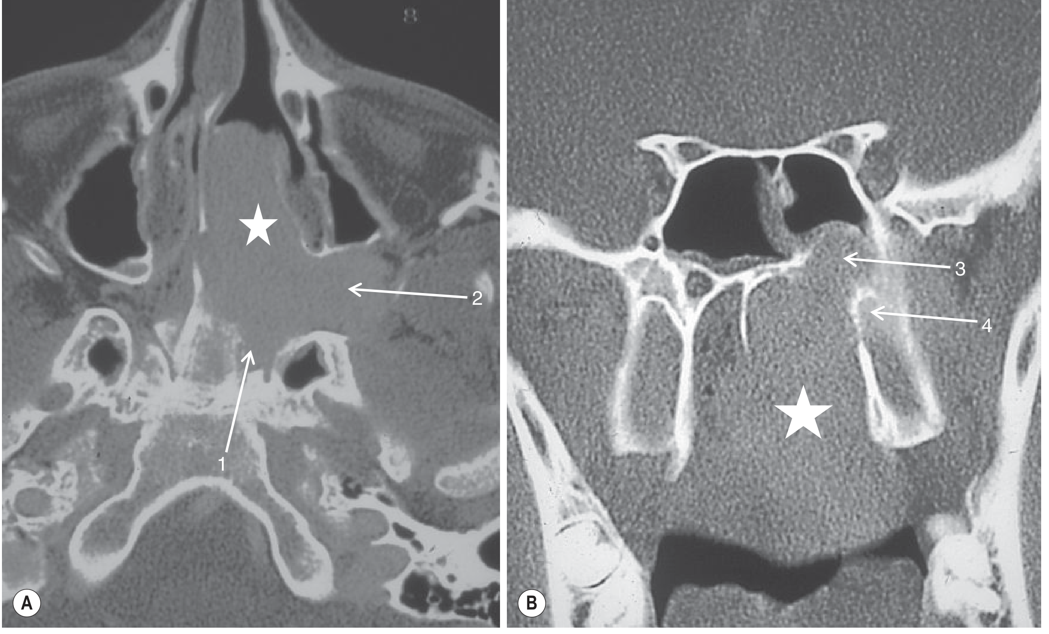
CT of JNA: (A) Axial - mass fills nasal cavity (white star), erodes sphenoid/vidian canal aperture (1), widens pterygopalatine fossa (2). (B) Coronal - vidian canal erosion (3), medial pterygoid plate erosion (4).
Part 3: Classic CT Signs - Memorize These
| Sign | What it means |
|---|---|
| Holman-Miller sign (Antral sign) | Anterior bowing of the posterior wall of the maxillary sinus - pathognomonic |
| Widening of pterygopalatine fossa | The primary epicenter - always present |
| Erosion of medial pterygoid plate | Early, consistent finding |
| Erosion of sphenopalatine foramen | Site of tumor origin |
| Vidian canal erosion | Tumor tracking along vidian nerve to skull base |
| Intense post-contrast enhancement | Due to high vascularity - hallmark finding |
| Permeative skull base erosion | Advanced disease (Stage III) |
Part 4: Full Clinical Profile (What the Examiner Expects You to Know)
Definition
JNA (Juvenile Nasopharyngeal Angiofibroma) is a benign, locally aggressive, highly vascular fibrovascular tumor of the nasopharynx. It is histologically benign but behaves aggressively, eroding bone and invading adjacent spaces.
Epidemiology
- Almost exclusively in adolescent males (ages 10-25 years)
- Accounts for only 0.05% of all head and neck tumors but is the most common benign nasopharyngeal tumor
- No reliable racial predilection, but higher reported incidence in India and Egypt
Origin
- Arises at the sphenopalatine foramen / base of the pterygoid process (where the pterygoid process meets the body of the sphenoid)
- Primary epicenter: pterygopalatine fossa (PPF)
Pathology
- Histology: Vascular spaces (endothelium-lined, NO smooth muscle layer) embedded in a fibrous stroma
- The vessels lack a muscular coat, which is why they cannot contract - hence the profuse, uncontrollable bleeding
- The stroma contains stellate fibroblasts and collagen
- Considered by many to be a vascular malformation/hamartoma rather than a true neoplasm
- Androgen-dependent tumor - explains why it occurs in pubescent males and may involute after puberty
Clinical Features
- Nasal obstruction - unilateral, progressive (most common early symptom)
- Epistaxis - recurrent, profuse, often intractable (hallmark symptom)
- Advanced cases: cheek swelling (ITF), proptosis/diplopia (orbital), headache (intracranial)
- Endoscopy: smooth, hypervascularized, reddish-purple mass behind the middle turbinate, laterally displacing it
Important: Never biopsy in the clinic - risk of catastrophic hemorrhage. Diagnosis is made on imaging.
Part 5: Staging Systems (Examiner Favorite)
Radkowski Staging (Most Commonly Used in Exams)
| Stage | Description |
|---|---|
| Ia | Limited to nasal cavity and nasopharynx |
| Ib | Extension into one or more sinuses |
| IIa | Minimal extension into pterygopalatine fossa |
| IIb | Full occupation of pterygopalatine fossa; orbital erosion |
| IIc | Infratemporal fossa extension; posterior to pterygoid plates |
| IIIa | Minimal intracranial (middle fossa or pterygoid plates) |
| IIIb | Intracranial extension with/without cavernous sinus involvement |
Andrews-Fisch Staging (Also Common)
| Stage | Description |
|---|---|
| I | Nasal cavity, nasopharynx - no bone destruction |
| II | Pterygomaxillary fossa, sinuses - bone destruction |
| III | Infratemporal fossa, orbit, parasellar region - lateral to cavernous sinus |
| IV | Massive intracranial; cavernous sinus, optic chiasm, pituitary |
Exam tip: Know Radkowski well - it's the one most examiners quiz. Mention that the pterygopalatine fossa is the key landmark between Stage I and Stage II.
Part 6: Imaging - What Each Modality Shows
CT (What you're being tested on)
- Gold standard for bone detail - shows erosion of pterygoid plates, skull base, vidian canal
- Post-contrast: intense homogeneous enhancement
- Shows: tumor extent, PPF widening, Holman-Miller sign, sinus involvement
- Limitation: poor soft tissue differentiation from retained secretions vs. tumor
MRI
- Best for soft tissue extent - differentiates tumor from retained secretions in sinuses
- T1: intermediate signal with flow voids (characteristic of high vascularity)
- T2: heterogeneous with flow voids
- T1 post-contrast: intense enhancement
- Best for orbital, intracranial, and cavernous sinus involvement
- Flow voids on both T1 and T2 = salt-and-pepper appearance (characteristic)
- MRI is preferred for staging intracranial extension
Plain X-ray (PNS views)
- Waters' view: soft tissue mass in nasopharynx, anterior bowing of posterior maxillary sinus wall
- Rarely used today but may be asked about
Digital Subtraction Angiography (DSA)
- Shows the vascular supply in detail
- Blood supply: primarily internal maxillary artery (branch of ECA) and ascending pharyngeal artery
- Advanced lesions: ICA supply (up to 35% of cases)
- DSA is done prior to embolization, not primarily for diagnosis
Part 7: Differential Diagnosis of Unilateral Nasal Mass (Examiner Will Ask)
| Condition | Key Distinguishing Features |
|---|---|
| JNA | Young male, PPF widening, intense enhancement, Holman-Miller sign |
| Inverted papilloma | Older male, arises from lateral nasal wall, calcifications, thickened bone |
| Antrochoanal polyp | Soft tissue, no enhancement, no bone erosion, from maxillary sinus |
| Nasopharyngeal carcinoma | Older patient, lymphadenopathy, bone destruction, EBV-related |
| Rhabdomyosarcoma | Children, rapidly destructive, heterogeneous enhancement |
| Chordoma | Midline, clivus destruction, low T1/high T2 |
| Olfactory neuroblastoma | Cribriform plate involvement, older patients |
| Lymphoma | Homogeneous, minimal bone erosion, bilateral |
Part 8: Treatment (Examiner Will Definitely Ask)
Preoperative Embolization (Standard of Care)
- Done 24-48 hours before surgery using polyvinyl alcohol (PVA) particles
- Dramatically reduces intraoperative hemorrhage
- DSA first - maps vasculature, identifies ICA feeders
- If ICA feeders present: direct tumor puncture with liquid embolic agents (Onyx/NBCA) may be used
Surgery - Primary Treatment
- Endoscopic resection - now standard for Stage I, II, and selected Stage III
- Open approaches for advanced lesions:
- Transpalatal approach (Stage I/II)
- Lateral rhinotomy / midfacial degloving (Stage II/III)
- Infratemporal fossa approach (Stage III with ITF extension)
- Combined craniofacial (Stage IV/intracranial)
Radiotherapy
- Reserved for surgically inaccessible lesions (cavernous sinus, intracranial)
- Also used for recurrences not amenable to surgery
- Risk: radiation-induced malignancy in young patients
Hormonal therapy
- Anti-androgens (flutamide) - limited historical use, not standard
Observation
- Residual disease after surgery may be monitored with serial MRI if in a pubescent patient, as spontaneous involution is possible
Recurrence
- Rate: 6-24%, higher in advanced stages
- Monitor with MRI
- Re-embolization and re-surgery is the standard approach
Part 9: Likely Examiner Questions and How to Answer
Q: What is the most pathognomonic finding of JNA on CT?
Widening of the pterygopalatine fossa with anterior bowing of the posterior wall of the maxillary sinus (Holman-Miller sign/antral sign).
Q: Why does this tumor occur only in males?
It is androgen-dependent. Androgen receptors are expressed in the fibrous stroma, and testosterone stimulation at puberty is thought to drive its development. It may involute spontaneously after puberty.
Q: Why should you NOT biopsy this lesion?
The vascular spaces in JNA lack a smooth muscle layer and cannot contract. Biopsy causes profuse, uncontrollable hemorrhage. Diagnosis is made on clinical + imaging grounds.
Q: What is the site of origin of JNA?
The sphenopalatine foramen / base of the pterygoid process (junction of the pterygoid process and the body of the sphenoid). The pterygopalatine fossa is always the primary epicenter.
Q: What is the blood supply?
Primarily from the internal maxillary artery (branch of external carotid artery) and ascending pharyngeal artery. In advanced lesions, the internal carotid artery also contributes (found in up to 35% of cases). This is important before embolization.
Q: What does "flow voids" on MRI mean in JNA?
Signal voids on T1 and T2 weighted sequences due to rapid blood flow through large intralesional vascular channels. This gives the "salt-and-pepper" or "honeycomb" appearance on MRI and confirms high vascularity.
Q: What is the Holman-Miller sign?
Anterior bowing of the posterior wall of the maxillary sinus on CT or plain X-ray (Waters' view), caused by the expanding JNA mass pressing forward from the pterygopalatine fossa. It is pathognomonic of JNA.
Q: How do you differentiate JNA from inverted papilloma on CT?
JNA: young male, PPF widening, intense contrast enhancement, Holman-Miller sign, no calcification. Inverted papilloma: older male, arises from lateral nasal wall/middle meatus, may have calcifications, thickened (sclerotic) bony margins, less intense enhancement.
Q: What is the role of preoperative embolization?
Performed 24-48 hours before surgery using PVA particles via DSA. Reduces intraoperative blood loss dramatically, improves visibility of tumor margins, and makes complete resection easier. It does NOT improve long-term recurrence rates.
Q: What is the most common surgical approach for early-stage JNA today?
Endoscopic endonasal resection. It is now the standard approach for Stage I, II, and even selected Stage III lesions, replacing older open approaches.
Q: Can JNA become malignant?
Rarely. Malignant transformation to fibrosarcoma or angiofibrosarcoma has been rarely reported, usually following radiotherapy. The lesion is otherwise histologically benign.
Part 10: The Perfect Examiner Script (Put It Together)
"This is a contrast-enhanced CT of the paranasal sinuses and skull base in the [axial/coronal] plane. I can see a well-defined, lobulated soft tissue mass in the posterior nasal cavity and nasopharynx on the right side, showing intense post-contrast enhancement, consistent with a highly vascular lesion.The key findings are: widening of the right pterygopalatine fossa, erosion of the base of the medial pterygoid plate, erosion of the sphenopalatine foramen, and anterior bowing of the posterior wall of the right maxillary sinus - which is the Holman-Miller sign.The mass appears to extend into the right pterygopalatine fossa and there is [describe extent]. There is no evidence of intracranial extension / orbital involvement [or state what is present].Based on these findings - a unilateral, highly enhancing nasopharyngeal mass with pathognomonic pterygopalatine fossa widening and the antral sign in what appears to be a young male patient - the most likely diagnosis is Juvenile Nasopharyngeal Angiofibroma, Radkowski Stage [IIb, for example].I would recommend complementary MRI with contrast for precise soft tissue staging, and digital subtraction angiography prior to preoperative embolization and surgical resection."
Sources: Cummings Otolaryngology Head and Neck Surgery (JUVENILE ANGIOFIBROMA section, p.988-991); Grainger & Allison's Diagnostic Radiology (Angiofibroma section, p.1605); Scott-Brown's Otorhinolaryngology Head & Neck Surgery Vol. 1; StatPearls - Nasopharyngeal Angiofibroma (NCBI Bookshelf).
This is a shared conversation. Sign in to Orris to start your own chat.