
Cytochrome p 450 is required for hydroxylation
The cytochrome P450 (CYP) enzyme system is a superfamily of heme-containing monooxygenases that catalyze hydroxylation reactions using molecular oxygen and NADPH. The general reaction is: RH + O₂ + NADPH + H⁺ → ROH + H₂O + NADP⁺.
Searching sources...
Reading File
Finding Sources
Searching the Web
cytochrome P450 hydroxylation mechanism biochemistry
Reading File
Reading File
Loading Image
Loading Image
Reading File
Here is a thorough breakdown of Cytochrome P450 and its role in hydroxylation, drawn from Harper's Illustrated Biochemistry, 32nd Ed.
Cytochrome P450 (CYP) and Hydroxylation
What Is Cytochrome P450?
Cytochrome P450 is a large superfamily of heme-containing monooxygenases with over 50 genes identified in the human genome (at least 57 CYP genes). They are found mainly in the smooth endoplasmic reticulum of liver cells and enterocytes, and also in mitochondria of steroidogenic tissues (adrenal cortex, testis, ovary, placenta).
The name comes from the characteristic absorption peak at 450 nm when the reduced enzyme is bound to carbon monoxide (CO) - visible in the cycle diagram above where CO is shown as an inhibitor.
The Core Reaction
$$\text{RH} + \text{O}_2 + \text{NADPH} + \text{H}^+ \rightarrow \text{R-OH} + \text{H}_2\text{O} + \text{NADP}^+$$
- One atom of O₂ is incorporated into the substrate as a hydroxyl group (-OH)
- The other oxygen atom is reduced to water
- This is why CYP enzymes are called monooxygenases (only one oxygen atom goes into the substrate)
The Hydroxylase Cycle (Step-by-Step)
The diagram above (Figure 12-6) shows the complete cycle. Here is what each step represents:
| Step | What Happens |
|---|---|
| 1. Substrate binding | Substrate (RH) binds to oxidized P450 (Fe³⁺) |
| 2. First reduction | NADPH donates an electron via NADPH-CYP reductase; Fe³⁺ → Fe²⁺ |
| 3. O₂ binding | Reduced P450 (Fe²⁺) binds molecular O₂ |
| 4. Second reduction | A second electron is added (from NADPH or cytochrome b₅); O₂ is activated |
| 5. Hydroxylation | Activated oxygen attacks the substrate C-H bond; product R-OH is released; Fe³⁺ is restored |
Class I vs. Class II P450 Systems
As shown in the electron transport diagram above:
| Feature | Class I | Class II |
|---|---|---|
| Location | Mitochondria (steroidogenic tissues) | Endoplasmic reticulum (liver) |
| Electron carrier | FAD-reductase + iron-sulfur protein (Fe₂S₂) | CYP450 reductase (FAD + FMN) |
| Electron donor | NADH or NADPH | NADPH |
| Function | Steroid hormone biosynthesis | Drug metabolism, xenobiotics |
Cytochrome b₅ can donate the second electron to P450 in class II systems, making it a cofactor in some drug detoxification reactions.
Nomenclature of CYP Isoforms
The naming follows a systematic pattern:
- CYP = cytochrome P450 root
- Number = family (≥40% amino acid identity; e.g., CYP1, CYP2, CYP3)
- Letter = subfamily (≥55% identity; e.g., CYP1A)
- Number = individual enzyme (e.g., CYP1A1)
Major drug-metabolizing families: CYP1, CYP2 (13 subfamilies), and CYP3 - with overlapping substrate specificities allowing metabolism of a very wide range of xenobiotics.
Physiological Roles of CYP Hydroxylation
CYP enzymes hydroxylate a broad range of substrates:
| Substrate | Reaction | CYP Involved |
|---|---|---|
| Steroids | Cholesterol side chain cleavage (C20, C22), corticosteroid synthesis | CYP11A1, CYP11B1 |
| Vitamin D | 25-hydroxylation (liver) → 25-OH-D₃; then 1α-hydroxylation (kidney) → calcitriol | CYP27A1, CYP27B1 |
| Bile acids | 7α-hydroxylation of cholesterol (first regulatory step) | CYP7A1 |
| Drugs/xenobiotics | Phase 1 metabolism - converts lipophilic drugs to water-soluble hydroxylated metabolites | CYP1A2, CYP2C9, CYP2D6, CYP3A4 |
| Retinoic acid | Hydroxylation for metabolism | CYP26 |
At least half of all common drugs are metabolized by CYP isoforms.
Role in Drug Metabolism (Phase 1)
Xenobiotic metabolism occurs in two phases:
- Phase 1: Hydroxylation by CYP enzymes → introduces -OH group, making the molecule more reactive
- Phase 2: Conjugation of the hydroxylated product with glucuronic acid, sulfate, or glutathione (making it water-soluble for excretion)
Together, Cytochromes P450 and b₅ are responsible for about 75% of drug modification and degradation in the body.
Induction of CYP Enzymes
Most CYP isoforms are inducible - meaning their expression can be upregulated by certain substrates:
- Drugs like phenobarbital cause hypertrophy of the smooth ER and a 3-4 fold increase in CYP levels within days
- This happens mainly by increased transcription but can also involve mRNA stabilization or increased translation
- Clinical importance: Inducing one CYP isoform can accelerate the metabolism of other drugs sharing that isoform - the basis of many drug-drug interactions
Key Exam Point
A classic MCQ tests what is NOT an activity of CYP:
- Activation of vitamin D ✓
- Hydroxylation of steroid hormone precursors ✓
- Hydroxylation of xenobiotics ✓
- Hydroxylation of retinoic acid ✓
- Methylation of xenobiotics ✗ - This is a Phase 2 reaction, NOT performed by CYP enzymes
Source: Harper's Illustrated Biochemistry, 32nd Ed., Chapters 12 & 47
Reduction in the detoxification of xenobiotics.
Reduced detoxification of xenobiotics occurs when CYP450 enzyme activity is impaired - through liver disease, genetic polymorphisms, enzyme inhibition by competing drugs, or nutritional deficiencies (NADPH, iron). This leads to drug accumulation, toxicity, and adverse effects.
Searching sources...
Finding Sources
Searching the Web
reduction detoxification xenobiotics CYP450 impairment causes consequences
Reading File
Finding Sources
Can you entirely explain the detoxification of xenobiotics?
Xenobiotic detoxification is a two-phase process. Phase 1 introduces reactive groups (mainly via CYP450 hydroxylation), and Phase 2 conjugates those groups to make the compound water-soluble for excretion. Glutathione, glucuronic acid, and sulfate are major conjugating agents.
Searching sources...
Reading File
Finding Sources
Searching the Web
xenobiotic detoxification phase 1 phase 2 biochemistry complete overview
Finding Sources
Reading File
Finding Sources
Reading File
Reading File
Reading File
Finding Sources
Reading File
Loading Image
Here is a complete, structured explanation of xenobiotic detoxification, drawn from Harper's Illustrated Biochemistry, 32nd Ed. and Goodman & Gilman's Pharmacological Basis of Therapeutics.
Complete Detoxification of Xenobiotics
What Are Xenobiotics?
Xenobiotics (from Greek xenos = foreign) are chemical compounds foreign to the body. They include:
- Drugs and medicines
- Food additives and preservatives
- Environmental pollutants (e.g., polychlorinated biphenyls/PCBs, pesticides, insecticides)
- Naturally occurring plant compounds (some beneficial as antioxidants, some toxic)
- Chemical carcinogens
Most xenobiotics are lipophilic - they dissolve in fat, are easily absorbed, and cannot be directly excreted in urine. Detoxification converts them to water-soluble compounds that can be excreted. The liver is the primary site.
Overview: Two-Phase System
Lipophilic Xenobiotic
        ↓
  [PHASE 1] - Functionalization
        ↓
  Reactive Intermediate
        ↓
  [PHASE 2] - Conjugation
        ↓
  Water-soluble Conjugate
        ↓
  Excreted in URINE or BILE
PHASE 1 - Functionalization Reactions
Goal: Introduce a reactive functional group (-OH, -COOH, -SH, -NH₂, -O-) onto the xenobiotic molecule to make it a substrate for Phase 2.
Main Enzymes in Phase 1
| Enzyme | Reaction | Key Features |
|---|---|---|
| Cytochrome P450 (CYP) | C and O oxidation, hydroxylation, dealkylation, epoxidation, deamination, desulfuration | Primary Phase 1 enzyme; heme-containing monooxygenase; located in smooth ER and mitochondria of liver |
| Flavin-containing monooxygenases (FMOs) | N, S, and P oxidation | Works alongside CYP; also in smooth ER |
| Epoxide hydrolase (EH) | Hydrolysis of epoxides → dihydrodiols | Protective against carcinogenic epoxides |
| Esterases / Hydrolases | Hydrolysis of esters and amides | Activate many prodrugs |
| Alcohol/Aldehyde dehydrogenases | Reduction of alcohols and aldehydes | Also involved in ethanol metabolism |
The Key CYP Reaction:
$$\text{RH} + \text{O}_2 + \text{NADPH} + \text{H}^+ \rightarrow \text{R-OH} + \text{H}_2\text{O} + \text{NADP}^+$$
One oxygen atom hydroxylates the substrate; the other is reduced to water.
Important Note - Prodrug Activation:
Phase 1 does NOT always detoxify. Sometimes it activates an inert compound:
- Prodrugs - inactive drugs converted to active forms (e.g., cyclophosphamide → active alkylating agent; clopidogrel → active platelet inhibitor)
- Procarcinogens - harmless compounds converted to carcinogens or mutagens (e.g., benzo[a]pyrene activated by CYP to a carcinogenic epoxide)
PHASE 2 - Conjugation Reactions
Goal: Attach a large, polar (hydrophilic) molecule to the Phase 1 product to make it water-soluble and excretable.
Conjugation Reactions and Their Donors
| Enzyme | Conjugating Group Added | Donor Molecule | Substrates |
|---|---|---|---|
| UDP-Glucuronosyltransferases (UGTs) | Glucuronic acid | UDP-glucuronic acid | Most common; acts on -OH, -NH₂, -COOH, -SH groups |
| Sulfotransferases (SULT) | Sulfate | PAPS (active sulfate = adenosine 3'-phosphate-5'-phosphosulfate) | Alcohols, arylamines, phenols, steroids |
| Glutathione-S-Transferases (GSTs) | Glutathione (GSH) | Glutathione (γ-Glu-Cys-Gly) | Electrophilic compounds: R + GSH → R-S-G |
| N-Acetyltransferases (NATs) | Acetyl group | Acetyl-CoA | Isoniazid (INH), sulfonamides; polymorphic - slow vs fast acetylators |
| Methyltransferases (MTs) | Methyl group | S-adenosylmethionine (SAM) | Some xenobiotics |
1. Glucuronidation (most common)
- Catalyzed by UDP-glucuronosyltransferases in the ER and cytosol
- UDP-glucuronic acid donates glucuronate to -OH, -NH₂, -SH, or -COOH groups
- Examples: bilirubin, aniline, benzoic acid, phenol, steroid hormones, meprobamate
2. Sulfation
- Uses PAPS (active sulfate) as donor
- Sulfotransferases conjugate alcohols, phenols, and arylamines
- Also used for steroids, glycosaminoglycans, glycolipids, glycoproteins
3. Glutathione Conjugation
- The tripeptide glutathione (GSH) is central to this pathway
- Reaction: R (electrophile) + GSH → R-S-G
- The liver has extremely high glutathione-S-transferase activity - its entire GSH pool can be depleted within minutes of heavy xenobiotic exposure
- GST enzymes also act as ligandin - they bind carcinogens, bilirubin, and steroid hormones to sequester them and prevent DNA damage
- Glutathione conjugates are exported from the liver, processed by γ-glutamyltranspeptidase (GGT) and dipeptidases, taken up by the kidney, and N-acetylated to form mercapturic acids which are excreted in urine
4. Acetylation (polymorphic)
- Reaction: X + Acetyl-CoA → Acetyl-X + CoA
- Catalyzed by acetyltransferases in hepatic cytosol
- Key clinical example: Isoniazid (INH) for tuberculosis
- Slow acetylators - drug persists longer → greater risk of toxicity (peripheral neuropathy)
- Fast acetylators - drug cleared quickly → may need higher doses
5. Methylation
- Uses S-adenosylmethionine (SAM) as methyl donor
- Less common; some xenobiotics only
Epoxide Hydrolase - A Protective Enzyme
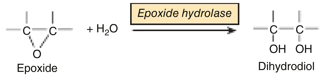
When CYP acts on procarcinogens, it can generate epoxides - highly reactive, potentially mutagenic intermediates. Epoxide hydrolase (in the ER membrane) converts these dangerous epoxides to much less reactive dihydrodiols, providing a protective mechanism.
Adverse Consequences When Detoxification Fails
When xenobiotic metabolism goes wrong, three major categories of harm result:
1. Cytotoxicity (Cell Death)
- Reactive metabolites covalently bind to macromolecules (DNA, RNA, proteins)
- This triggers the DNA repair mechanism - poly(ADP-ribose polymerase) transfers ADP-ribose units to DNA-binding proteins, consuming large amounts of NAD
- Severe DNA damage depletes NAD → impairs ATP synthesis → cell death
2. Immunological / Autoimmune Effects
- A reactive xenobiotic metabolite binds to a protein as a hapten, altering its antigenicity
- The immune system generates antibodies against the modified protein
- These antibodies cross-react with the unmodified protein → potential autoimmune disease
3. Chemical Carcinogenesis
- Some chemicals are indirect carcinogens (procarcinogens) - they need CYP activation to become carcinogenic
- Example: benzo[a]pyrene (in cigarette smoke) → CYP converts it to a reactive epoxide → epoxide attacks DNA → mutation → cancer
- Epoxide hydrolase can partially counter this by detoxifying the epoxide intermediate
- Glutathione S-transferase can also sequester carcinogens, preventing them from reaching DNA
Summary Flow Chart
XENOBIOTIC (lipophilic, foreign compound)
          ↓
     LIVER (main site)
          ↓
   ┌─── PHASE 1 ──────────────────────────┐
   │  CYP450 → hydroxylation, epoxidation │
   │  FMOs → N/S/P oxidation             │
   │  Esterases → hydrolysis             │
   │  Epoxide hydrolase → dihydrodiols   │
   └──────────────────────────────────────┘
          ↓ (reactive intermediate with -OH, -NH₂, etc.)
   ┌─── PHASE 2 ──────────────────────────┐
   │  + Glucuronic acid (UGT/UDP-GlcUA)  │
   │  + Sulfate (SULT/PAPS)              │
   │  + Glutathione (GST/GSH)            │
   │  + Acetyl group (NAT/Acetyl-CoA)    │
   │  + Methyl group (MT/SAM)            │
   └──────────────────────────────────────┘
          ↓
  WATER-SOLUBLE CONJUGATE
          ↓
  Excreted in URINE (kidney) or BILE (intestine)
Key Clinical Points to Remember
| Concept | Detail |
|---|---|
| Most common Phase 2 reaction | Glucuronidation |
| Active sulfate donor | PAPS |
| Glutathione function | Conjugation + intracellular reducing agent |
| Isoniazid metabolism | Acetylation - slow vs fast acetylator polymorphism |
| CYP inducers | Phenobarbital, rifampicin - cause 3-4x increase in CYP → accelerate drug clearance |
| CYP inhibitors | Ketoconazole, erythromycin - block metabolism → drug toxicity |
| Benzo[a]pyrene | Procarcinogen activated by CYP to epoxide carcinogen |
| GGT elevation | Indicates hepatobiliary disease (released from liver ER during damage) |
Sources: Harper's Illustrated Biochemistry, 32nd Ed., Chapter 47; Goodman & Gilman's Pharmacological Basis of Therapeutics, Table 5-1
This is a shared conversation. Sign in to Orris to start your own chat.