
Hand write notes on topic vessels of upper limb
Reading File
Finding Sources
Finding Sources
Finding Sources
Reading File
Reading File
Reading File
Finding Sources
Finding Sources
Reading File
Reading File
Reading File
Now I have comprehensive material. Let me also fetch the key anatomical images.
Loading Image
Loading Image
Now let me also get a veins image:
Finding Sources
Reading File
Loading Image
I now have all the material needed to write comprehensive handwritten-style notes. Let me compile everything.
🩺 Vessels of the Upper Limb - Handwritten Notes
Source: Color Atlas of Human Anatomy Vol. 2 (Internal Organs) & Gray's Anatomy for Students
ARTERIAL SUPPLY - Overview
The main arterial axis of the upper limb runs continuously:
Subclavian A. → Axillary A. → Brachial A. → Radial A. + Ulnar A. → Palmar Arches
1. SUBCLAVIAN ARTERY
Origin:
- Right side: from the brachiocephalic trunk
- Left side: directly from the aortic arch
Three parts (divided by the anterior scalene muscle):
- Part I - medial to anterior scalene
- Part II - posterior to anterior scalene
- Part III - lateral to anterior scalene → becomes axillary A. at the inferior border of rib I
Branches (mnemonic: VITamin C & D):
| Branch | Supply |
|---|---|
| Vertebral artery | Ascending through foramina of C6-C1, enters cranium via foramen magnum; joins opposite side to form basilar A. |
| Internal thoracic artery | Descends 1 cm lateral to sternum; gives anterior intercostal branches; terminal branches = musculophrenic + superior epigastric arteries |
| Thyrocervical trunk | → Inferior thyroid a., suprascapular a., transverse cervical a., ascending cervical a. |
| Costocervical trunk | → Deep cervical a., supreme (highest) intercostal a. |
| Dorsal scapular (posterior scapular) a. | May arise from subclavian directly; passes to levator scapulae |
2. AXILLARY ARTERY
Extent: Inferior border of rib I → inferior border of pectoralis major (or latissimus dorsi tendon)
Divided by pectoralis minor into 3 parts:
Part 1 (1 branch):
- Superior thoracic artery → 1st & 2nd intercostal spaces, pectorals, serratus anterior (superior)
Part 2 (2 branches):
- Thoracoacromial artery → divides into pectoral, acromial, deltoid, clavicular branches; forms acromial anastomosis near acromion
- Lateral thoracic artery → serratus anterior, lateral chest wall; larger in women (supplies mammary glands)
Part 3 (3 branches):
- Subscapular artery (largest axillary branch)
- → Circumflex scapular a.: passes through triangular space (between teres major/minor), reaches infraspinous fossa; anastomoses with suprascapular a.
- → Thoracodorsal a.: accompanies thoracodorsal nerve to latissimus dorsi; also supplies teres major, subscapularis, serratus anterior
- Anterior circumflex humeral artery → passes anteriorly around surgical neck of humerus
- Posterior circumflex humeral artery (larger) → passes posteriorly through quadrangular space (lateral space between teres major/minor); supplies shoulder joint and surrounding muscles
Scapular anastomosis: suprascapular a. + circumflex scapular a. + posterior scapular a. form a rich network. Even if axillary A. is ligated proximal to the subscapular branch, the arm rarely becomes ischemic.
3. BRACHIAL ARTERY
Extent: Inferior border of pectoralis major → bifurcation into radial + ulnar arteries at the radial neck (just below the elbow)
Course: Runs in the medial bicipital groove; pulse palpable here and used for BP measurement
Branches:
| Branch | Notes |
|---|---|
| Deep artery of arm (profunda brachii) | Arises at lower border of teres major; passes posteriorly with radial nerve around the humeral shaft in the spiral groove; branches: radial collateral a. + medial collateral a. → to cubital anastomosis |
| Superior ulnar collateral a. | Runs alongside ulnar nerve; to cubital anastomosis |
| Inferior ulnar collateral a. | Arises above medial epicondyle near olecranon fossa; to cubital anastomosis |
| Nutrient artery of humerus |
Cubital anastomosis: Arterial plexus around the elbow formed by descending branches (profunda brachii, brachial artery) and ascending recurrent branches (radial recurrent, ulnar recurrent, interosseous recurrent). Permits safe ligation of brachial A. distal to the profunda brachii origin. Also permits radial A. harvest as a graft (collateral flow via ulnar A. recurrents).
4. RADIAL ARTERY
Origin: Terminal branch of brachial A. (smaller of the two terminal branches, despite its name)
Course:
- Proximally: between pronator teres and brachioradialis
- Distally: between tendons of brachioradialis and flexor carpi radialis → radial pulse palpable here
- Turns posteriorly → passes between 1st and 2nd metacarpal heads → enters palm of hand (deep)
Branches:
- Radial recurrent a. → ascends to cubital anastomosis
- Superficial palmar branch → joins ulnar A. to complete superficial palmar arch
- Palmar carpal branch → palmar carpal arch (wrist plexus)
- Posterior carpal branch → posterior carpal arch
- Princeps pollicis a. → thumb (flexor surface)
- Radialis indicis a. → radial side of index finger (may arise from princeps pollicis)
- Deep palmar arch → main terminal branch; lies on metacarpal bases deep to long flexor tendons; anastomoses with deep palmar branch of ulnar A.
5. ULNAR ARTERY
Origin: Larger terminal branch of brachial A.
Course:
- Initially runs deep to pronator teres
- Then accompanies flexor carpi ulnaris
- Enters hand lateral to pisiform
Branches:
- Ulnar recurrent a. (anterior + posterior) → cubital anastomosis
- Common interosseous a. → divides into:
- Anterior interosseous a. → runs on interosseous membrane (anterior surface)
- Posterior interosseous a. → pierces interosseous membrane (posterior surface); gives recurrent interosseous a. to cubital anastomosis
- Palmar carpal branch → palmar carpal arch
- Posterior carpal branch → posterior carpal arch
- Deep palmar branch → deep palmar arch
- Superficial palmar arch → main terminal branch (see below)
6. VASCULAR ARCHES OF THE HAND
Superficial Palmar Arch
- Formed by: Terminal branch of ulnar A. + superficial palmar branch of radial A.
- Mainly fed by: Ulnar artery
- Lies between: Palmar aponeurosis and long flexor tendons
- Gives: 3 common palmar digital arteries → each divides into 2 proper palmar digital arteries (supplies adjacent sides of fingers)
Deep Palmar Arch
- Formed by: Terminal branch of radial A. + deep palmar branch of ulnar A.
- Mainly fed by: Radial artery
- Lies on: Bases of metacarpals, beneath long flexor tendons
- Gives: 3-4 palmar metacarpal arteries → interdigital spaces; also perforating branches to dorsum
Posterior Carpal Arch (Dorsal)
- Formed by posterior carpal branches of radial + ulnar arteries
- Gives 4 posterior (dorsal) metacarpal arteries → 2 posterior digital arteries each
Allen's Test checks the patency of the superficial palmar arch and is done before radial A. cannulation or harvest.
VEINS OF THE UPPER LIMB
Divided into deep and superficial systems:
Deep Veins
All deep veins are paired companion veins (venae comitantes) that accompany the arteries:
| Vein | Notes |
|---|---|
| Subclavian vein | Continuation of axillary vein; runs between sternocleidomastoid and anterior scalene; joins internal jugular vein behind sternoclavicular joint → forms brachiocephalic vein |
| Axillary vein | Companion vein of axillary artery; collects blood from subscapular, circumflex humeral, lateral thoracic, thoraco-epigastric, and areolar plexus veins |
| Brachial veins (paired) | Accompany brachial artery; unite proximally to form axillary vein |
| Radial veins (paired) | Companion veins of radial artery |
| Ulnar veins (paired) | Accompany ulnar artery in ulnar neurovascular bundle |
| Anterior & posterior interosseous veins | Accompany interosseous arteries |
| Deep venous palmar arch + palmar metacarpal veins | Deep hand drainage |
Clinical note: The subclavian vein (actually first part of axillary vein) is used for central venous access. Infraclavicular needle entry aimed superomedially. Risk: pneumothorax, brachial plexus injury, subclavian artery puncture.
Superficial Veins
Lie in subcutaneous tissue above muscle fascia (epifascial). Originate mainly from the posterior (dorsal) venous network of the hand (a well-developed plexus on the dorsum).
Three main superficial veins:
1. Cephalic Vein
- Arises from the superficial posterior (dorsal) venous network of the hand
- Passes to the flexor (anterior) side of the wrist
- Ascends on the radial side of the forearm
- Travels in the lateral bicipital groove of the arm
- Pierces the clavipectoral fascia in the deltopectoral (clavipectoral) triangle
- Opens into the axillary vein
2. Basilic Vein
- Arises above the distal ulna
- Ascends on the ulnar side of the forearm
- Pierces the muscle fascia at the middle of the arm
- Enters the medial bicipital groove
- Opens into one of the brachial veins
3. Median Cubital Vein
- Connects cephalic and basilic veins across the cubital fossa
- Passes from inferolateral to superomedial
- Also communicates with deep veins
- Clinically important: Most commonly used site for venipuncture and IV cannulation
Clinical note: Subcutaneous veins of the hand and elbow are used for IV access, blood sampling, and long-term catheterization (Hickman lines, PICC lines).
CLINICAL CORRELATIONS
| Scenario | Key Anatomy |
|---|---|
| Rib I fracture | Fixes subclavian A. to rib; rapid deceleration can damage distal subclavian/proximal axillary A. Rarely causes ischemia due to scapular anastomosis |
| Anterior shoulder dislocation | Humeral head may compress axillary A. + brachial plexus |
| Radial A. harvest (CABG graft) | Safe due to cubital anastomosis - ulnar A. + recurrent branches provide collateral flow |
| Supracondylar fracture | Risk of brachial A. injury - check radial pulse |
| BP measurement | Brachial A. compressed against humerus in medial bicipital groove |
| Radial pulse | Palpated between brachioradialis and flexor carpi radialis tendons at wrist |
QUICK SUMMARY FLOWCHART
Aorta (left) / Brachiocephalic trunk (right)
         ↓
  SUBCLAVIAN ARTERY
  Branches: Vertebral, Internal thoracic, Thyrocervical trunk,
            Costocervical trunk, Posterior scapular
         ↓ (at inferior border of rib I)
  AXILLARY ARTERY (3 parts, pec minor divides)
  Part 1: Superior thoracic
  Part 2: Thoracoacromial, Lateral thoracic
  Part 3: Subscapular, Ant. + Post. circumflex humeral
         ↓ (at inferior border of pec major)
  BRACHIAL ARTERY
  Branches: Profunda brachii, Sup. + Inf. ulnar collateral
         ↓ (at radial neck, cubital fossa)
         ├── RADIAL ARTERY ──────────────────────────┐
         │   Branches: Radial recurrent,              │
         │   Superficial palmar branch, Deep palmar arch│
         │                                            │
         └── ULNAR ARTERY ──────────────────────────┤
             Branches: Ulnar recurrent,              │
             Common interosseous, Superficial palmar arch
                                                     │
                    ┌────────────────────────────────┘
                    ↓
        HAND: Deep Palmar Arch (mainly radial)
              Superficial Palmar Arch (mainly ulnar)
              Posterior Carpal Arch
              Palmar + Dorsal digital arteries
Sources: Color Atlas of Human Anatomy Vol. 2 - Internal Organs (Thieme); Gray's Anatomy for Students
Give the above diagrams with labeling
Here are all three diagrams from the textbook with their full labeling explained:
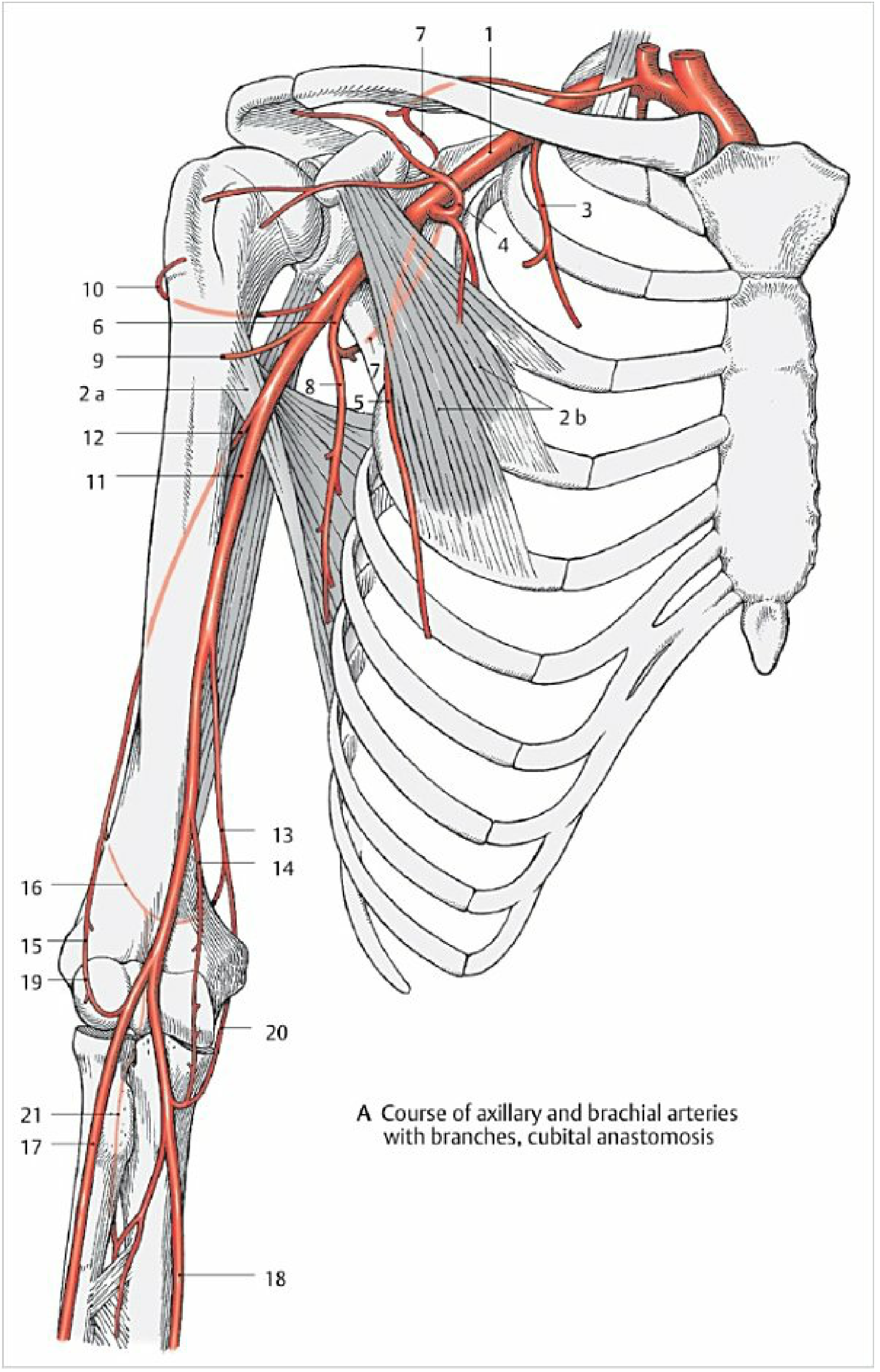
Diagram 1 - Axillary & Brachial Arteries (Fig. 2.25)
Label Key:
| No. | Structure |
|---|---|
| 1 | Axillary artery |
| 2a | Latissimus dorsi (muscle landmark) |
| 2b | Pectoralis minor (muscle landmark) |
| 3 | Superior thoracic artery |
| 4 | Thoracoacromial artery |
| 5 | Lateral thoracic artery |
| 6 | Subscapular artery |
| 7 | Circumflex scapular artery (through triangular space) |
| 8 | Thoracodorsal artery |
| 9 | Anterior circumflex humeral artery |
| 10 | Posterior circumflex humeral artery (through quadrangular space) |
| 11 | Brachial artery |
| 12 | Deep artery of arm (profunda brachii) - with radial & medial collateral branches |
| 13 | Superior ulnar collateral artery |
| 14 | Inferior ulnar collateral artery |
| 15 | Radial recurrent artery |
| 16 | Anterior ulnar recurrent artery |
| 17 | Posterior ulnar recurrent artery |
| 18 | Interosseous recurrent artery |
| 19 | Radial collateral artery |
| 20 | Middle collateral artery |
| 21 | Radial artery (proximal) |
Note: Labels 13-20 together form the cubital anastomosis - the arterial plexus encircling the elbow joint.
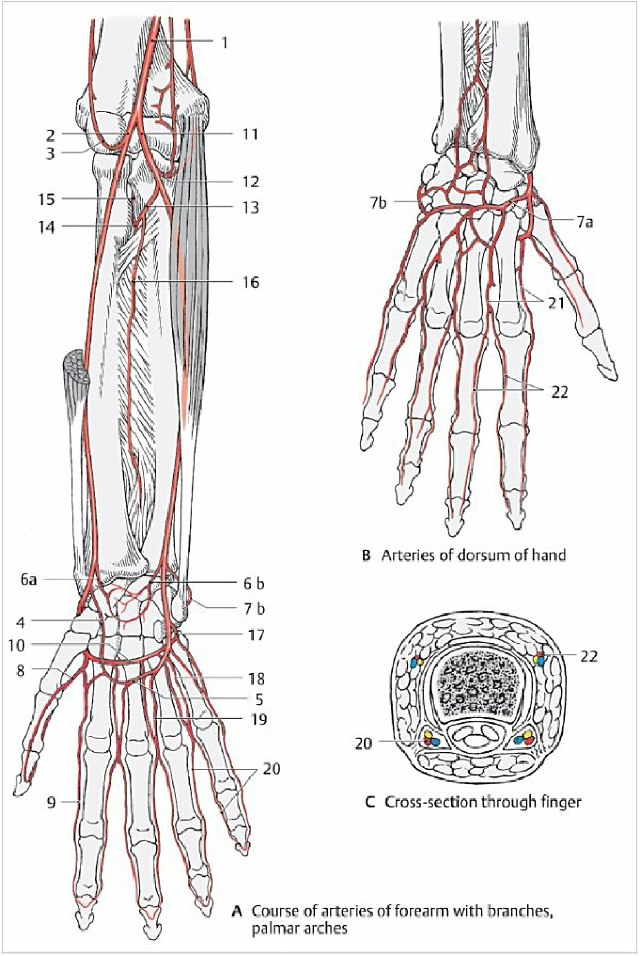
Diagram 2 - Radial & Ulnar Arteries, Palmar Arches (Fig. 2.26)
Panel A - Arteries of Forearm & Palm (Anterior view):
| No. | Structure |
|---|---|
| 1 | Brachial artery (terminal portion) |
| 2 | Radial artery |
| 3 | Radial recurrent artery |
| 4 | Superficial palmar branch of radial artery |
| 5 | Superficial palmar arch (mainly fed by ulnar A.) |
| 6a | Palmar carpal branch of radial artery |
| 6b | Palmar carpal branch of ulnar artery |
| 7a | Posterior carpal branch of radial artery |
| 7b | Posterior carpal branch of ulnar artery |
| 8 | Princeps pollicis artery (to thumb) |
| 9 | Radialis indicis artery (to radial side of index finger) |
| 10 | Deep palmar arch (mainly fed by radial A.) |
| 11 | Ulnar artery |
| 12 | Ulnar recurrent artery |
| 13 | Common interosseous artery |
| 14 | Anterior interosseous artery |
| 15 | Posterior interosseous artery |
| 16 | Recurrent interosseous artery |
| 17 | Deep palmar branch of ulnar artery |
| 18 | Palmar metacarpal arteries (3-4 branches from deep palmar arch) |
| 19 | Common palmar digital arteries (3 branches from superficial palmar arch) |
| 20 | Proper palmar digital arteries |
Panel B - Arteries of Dorsum of Hand:
| No. | Structure |
|---|---|
| 7a | Posterior carpal branch of radial artery |
| 7b | Posterior carpal branch of ulnar artery |
| 21 | Posterior (dorsal) metacarpal arteries (4 vessels) |
| 22 | Posterior (dorsal) digital arteries (2 per metacarpal) |
Panel C - Cross-section through Finger:
- Shows paired proper palmar digital arteries (yellow/red dots - ventral)
- Shows paired proper dorsal digital arteries (blue dots - dorsal)
- Central white = phalanx bone
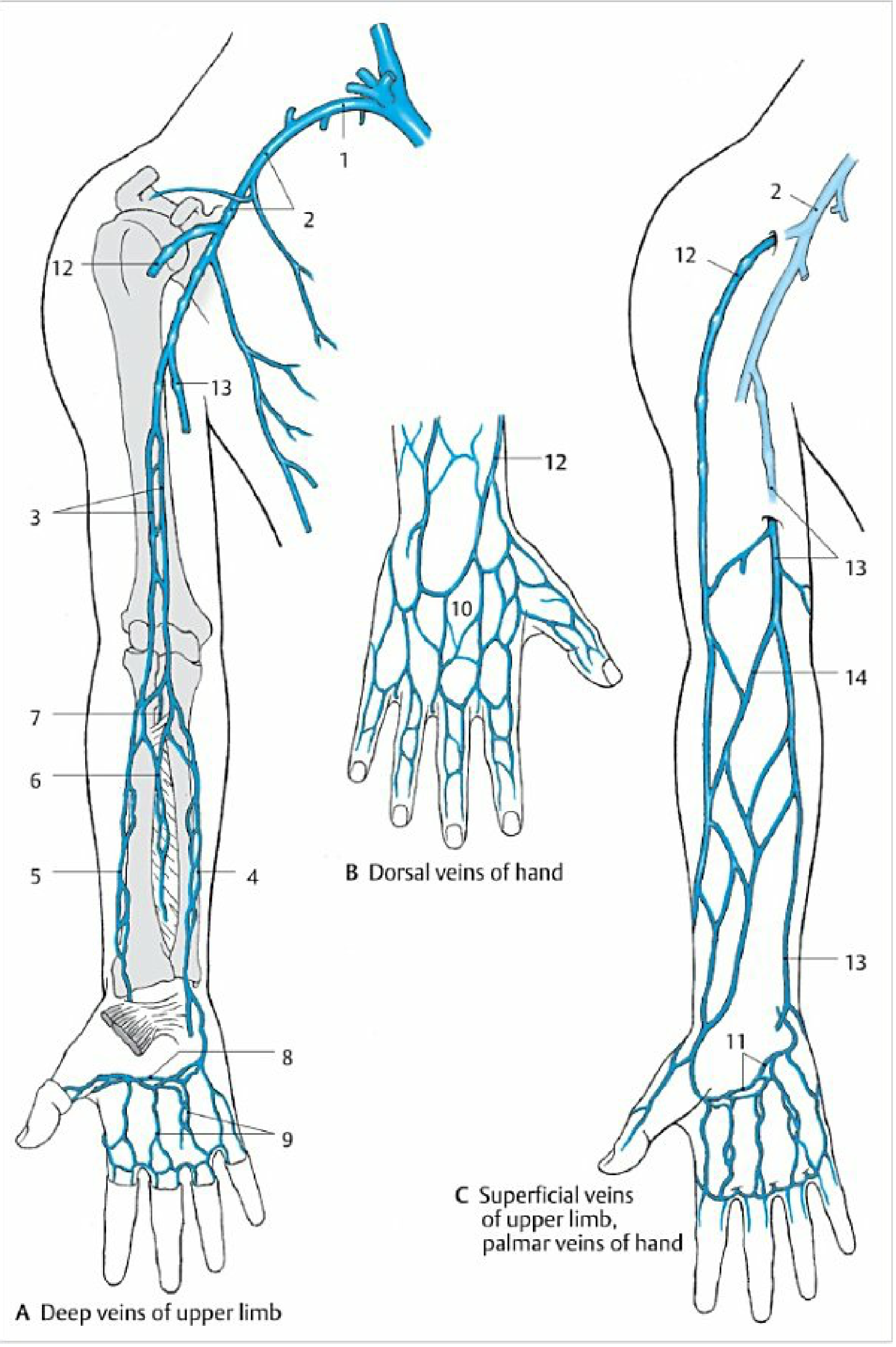
Diagram 3 - Veins of the Upper Limb (Fig. 2.34)
Panel A - Deep Veins of Upper Limb:
| No. | Structure |
|---|---|
| 1 | Subclavian vein → draining into brachiocephalic vein |
| 2 | Axillary vein (companion vein of axillary artery) |
| 3 | Brachial veins (paired, companion veins of brachial artery) |
| 4 | Radial veins (paired companion veins) |
| 5 | Ulnar veins (paired companion veins) |
| 6 | Anterior interosseous veins |
| 7 | Posterior interosseous veins |
| 8 | Deep venous palmar arch |
| 9 | Palmar metacarpal veins |
Panel B - Dorsal (Posterior) Venous Network of Hand:
| No. | Structure |
|---|---|
| 10 | Posterior (dorsal) venous network of hand - the well-developed plexus on the dorsum from which cephalic and basilic veins originate |
| 12 | Cephalic vein (arising from dorsal network, radial side) |
Panel C - Superficial Veins (Anterior view):
| No. | Structure |
|---|---|
| 2 | Axillary vein (receiving cephalic vein superiorly) |
| 11 | Superficial palmar venous arch |
| 12 | Cephalic vein - ascends on radial side of forearm and arm in lateral bicipital groove; pierces clavipectoral fascia → drains into axillary vein |
| 13 | Basilic vein - ascends on ulnar side of forearm; pierces deep fascia at mid-arm; enters medial bicipital groove → drains into brachial vein |
| 14 | Median cubital vein - connects cephalic (lateral) to basilic (medial) across the cubital fossa; runs from inferolateral to superomedial; also communicates with deep veins |
Summary Diagram - Relationship of the Three Superficial Veins
Cephalic V.                    Basilic V.
(radial/lateral side)          (ulnar/medial side)
      |                               |
      |         Median Cubital V.     |
      |___________(cubital fossa)_____|
      |
Ascends in lateral              Ascends in medial
bicipital groove                bicipital groove
      |                               |
Pierces clavipectoral           Pierces deep fascia
fascia (deltopectoral           at mid-arm level
triangle)                              |
      |                               |
Drains into                     Drains into
AXILLARY VEIN                   BRACHIAL VEIN
Venipuncture tip: The median cubital vein is preferred for blood draws because it is large, superficial, relatively fixed (supported by bicipital aponeurosis), and separated from the brachial artery and median nerve by the bicipital aponeurosis - reducing the risk of accidental arterial puncture.
This is a shared conversation. Sign in to Orris to start your own chat.