
Dic pathology
I'll look up DIC pathology from the medical library right away.
Searching sources...
Reading File
Finding Sources
Finding Sources
Searching Images
disseminated intravascular coagulation DIC pathology microthrombi fibrin
Now let me read the most comprehensive sections - Henry's Clinical Diagnosis (lab-focused), Goldman-Cecil (clinical), and Rosen's Emergency Medicine:
Reading File
Reading File
Reading File
Now let me get the dedicated Goldman-Cecil DIC chapter and Andrews' dermatology DIC section for clinical and morphological details:
Reading File
Reading File
I now have a rich set of information. Let me fetch the DIC pathophysiology flowchart image and compose the full answer:
Loading Image
Loading Image
Here is a comprehensive breakdown of DIC pathology, drawn from multiple authoritative textbooks.
Disseminated Intravascular Coagulation (DIC) - Pathology
Definition
DIC is a clinicopathologic syndrome in which dysregulated, simultaneous activation of both the coagulation cascade and the fibrinolytic system results in the paradoxical combination of widespread microvascular thrombosis and hemorrhagic diathesis. It is always secondary to an underlying disease - it is never a primary disorder.
- Henry's Clinical Diagnosis and Management by Laboratory Methods
Pathophysiology
The central event is uncontrolled thrombin generation, triggered by massive exposure of tissue factor (TF) to circulating blood.
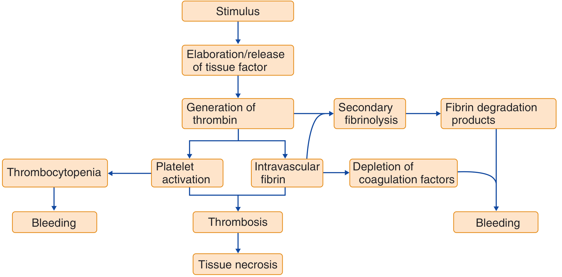
Step-by-step cascade:
- Trigger/stimulus - an underlying insult (sepsis, trauma, malignancy, obstetric catastrophe) causes elaboration and release of tissue factor into the circulation.
- Thrombin generation - TF-VIIa complex activates the extrinsic pathway, producing massive, unregulated amounts of thrombin that overwhelm natural inhibitors (antithrombin III, protein C, TFPI).
- Intravascular fibrin deposition - thrombin cleaves fibrinogen to fibrin, depositing fibrin thrombi in small vessels throughout multiple organs → tissue ischemia and end-organ damage.
- Platelet activation and consumption - thrombin activates platelets; platelet aggregation and trapping in microthrombi deplete circulating platelets → thrombocytopenia.
- Consumption of coagulation factors - fibrinogen and factors V, VIII, and XIII are consumed faster than they can be replaced → coagulopathy.
- Secondary fibrinolysis - plasmin is activated to lyse fibrin; this generates fibrin degradation products (FDPs) and D-dimers that further impair platelet function and inhibit fibrin polymerization.
- Inhibitor depletion - antithrombin III, protein C, and TFPI levels all fall, removing the natural brake on coagulation.
Net result: a vicious cycle of simultaneous thrombosis and bleeding.
- Rosen's Emergency Medicine, Concepts and Clinical Practice
Causes / Precipitating Conditions
| Category | Examples |
|---|---|
| Sepsis | Gram-negative (most common), gram-positive bacteria, fungi |
| Obstetric | Abruptio placentae, amniotic fluid embolism, placenta previa, HELLP syndrome, retained dead fetus |
| Malignancy | Acute promyelocytic leukemia (APL), solid tumors (Trousseau syndrome) |
| Massive tissue injury | Major trauma, burns, crush injury |
| Surgery | Aortic arch, pulmonary artery operations with hypothermia; release of thromboplastin |
| Others | Transfusion reactions, antiphospholipid antibody syndrome, SLE, envenomation, G-CSF therapy, kaposiform hemangioendothelioma (Kasabach-Merritt syndrome in children) |
- Henry's Clinical Diagnosis and Management; Andrews' Diseases of the Skin
Clinical Presentations / Phenotypes
DIC is not one single phenotype. Three major patterns exist:
1. Acute Hemorrhagic DIC ("Fibrinolytic Phenotype")
- Seen with: major trauma, abruptio placentae, APL
- Dominant feature: fulminant, uncontrolled fibrinolysis → life-threatening bleeding
- Widespread ecchymoses, oozing from venipuncture sites, wounds, mucosal surfaces
2. Thrombotic DIC
- Seen with: solid tumors, chronic underlying disease (Trousseau syndrome)
- Dominant feature: microvascular and large-vessel thrombosis → organ ischemia
- Acrocyanosis, digital gangrene (especially in patients on vasopressors), ischemic organ dysfunction
3. Chronic / Compensated DIC
- Often asymptomatic; detected only on lab abnormalities
- Seen in malignancy, liver disease, retained dead fetus
Skin manifestations (up to 2/3 of patients):
- Petechiae, ecchymoses, ischemic necrosis, hemorrhagic bullae
- Purpura fulminans - symmetric peripheral gangrene; a life-threatening manifestation
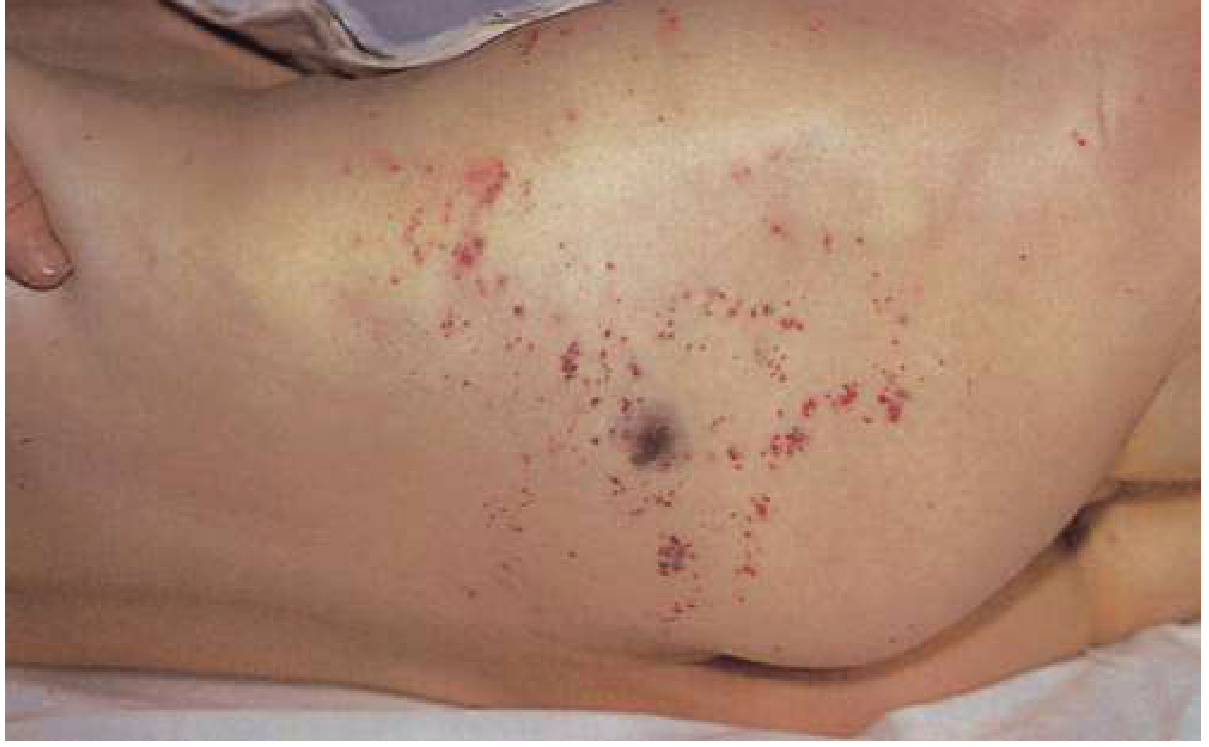
Characteristic skin hemorrhage in DIC from staphylococcal septicemia - ranging from small purpuric lesions to larger ecchymoses.
- Goldman-Cecil Medicine; Andrews' Diseases of the Skin
Laboratory Diagnosis
The combination of findings below confirms fulminant DIC; no single test is diagnostic:
| Lab Test | Finding in DIC |
|---|---|
| Platelet count | Low (thrombocytopenia) |
| PT / aPTT | Prolonged (factor consumption + inhibition) |
| Thrombin time | Prolonged |
| Fibrinogen | Decreased (consumed; note: normal in 57% of cases) |
| D-dimer | Elevated (secondary fibrinolysis marker) |
| FDPs | Elevated |
| Peripheral smear | Schistocytes / helmet cells (microangiopathic hemolytic anemia) - absence does not exclude DIC |
ISTH Scoring System (for Overt DIC):
| Parameter | Score |
|---|---|
| Platelet >100k = 0 / <100k = 1 / <50k = 2 | 0-2 |
| D-dimer <0.4 μg/mL = 0 / 0.4-4.0 = 2 / >4.0 = 3 | 0-3 |
| PT prolonged <3s = 0 / 3-6s = 1 / >6s = 2 | 0-2 |
| Fibrinogen >100 mg/dL = 0 / <100 = 1 | 0-1 |
Score ≥ 5 = compatible with overt DIC (repeat daily). Score < 5 = suggestive of non-overt DIC (repeat in 1-2 days).
- Henry's Clinical Diagnosis and Management by Laboratory Methods
Differential Diagnosis
| Condition | Key Distinguishing Features |
|---|---|
| Severe liver disease | Clinical jaundice + splenomegaly; all factors produced/catabolized by liver are low |
| Primary fibrinolysis | Rare; affects fibrinogen/fibrin specifically; platelets, factor V, and factor VIII usually preserved in low-normal range |
| TTP/HUS | Normal coagulation times, very low platelets, no FDP elevation; ADAMTS13 activity reduced |
Management Principles
-
Treat the underlying cause - paramount; many episodes self-resolve once the trigger is removed (e.g., antibiotics for sepsis, delivery in obstetric DIC).
-
Hemorrhagic DIC - replacement therapy:
- Platelets (for thrombocytopenia)
- Fresh frozen plasma (FFP) to replenish clotting factors
- Cryoprecipitate (for fibrinogen)
-
Thrombotic DIC - heparin is considered when fibrin deposition and thrombosis dominate (e.g., purpura fulminans, Trousseau syndrome); LMWH for thromboprophylaxis until bleeding or platelets < 30 × 10⁹/L.
-
Adjuncts: Protein C concentrate in severe sepsis-DIC; recombinant thrombomodulin and tranexamic acid have been studied but data remain limited.
- Rosen's Emergency Medicine; Andrews' Diseases of the Skin; Goldman-Cecil Medicine
Key Takeaways
- DIC = simultaneous thrombosis + consumption coagulopathy + secondary fibrinolysis
- The central mediator is unregulated thrombin generated via tissue factor
- Always secondary - identify and treat the underlying cause first
- Lab hallmark: low platelets + elevated D-dimer + prolonged PT/aPTT + low fibrinogen
- Two phenotypes: hemorrhagic (acute, fibrinolytic) vs. thrombotic (chronic, associated with malignancy)
- Purpura fulminans is the most severe cutaneous manifestation
This is a shared conversation. Sign in to Orris to start your own chat.