
Give me important history points,clinical features,investigations and Management of upper GIT bleeding
Reading File
Now let me search the medical library for upper GIT bleeding content in parallel.
Finding Sources
Finding Sources
Searching PubMed
"upper gastrointestinal bleeding" AND management
Excellent results. Now let me read the most comprehensive sources in parallel - Sleisenger & Fordtran (the top gastroenterology text), Tintinalli's Emergency Medicine, Frameworks for Internal Medicine, and Fischer's Surgery.
Reading File
Reading File
Reading File
Reading File
Now let me get the detailed management sections from Sleisenger & Fordtran and Tintinalli's, including their treatment algorithms:
Reading File
Reading File
Excellent - very rich content. Let me get the remaining management sections from Tintinalli's (treatment details) and Sleisenger's (medical treatment), and also fetch the classification diagram:
Reading File
Reading File
Loading Image
I now have comprehensive content from multiple authoritative textbooks. Let me compile the complete, well-structured answer.
Upper Gastrointestinal (GIT) Bleeding - Complete Clinical Review
Definition: Upper GI bleeding (UGIB) is any hemorrhage originating proximal to the ligament of Treitz (esophagus, stomach, duodenum).
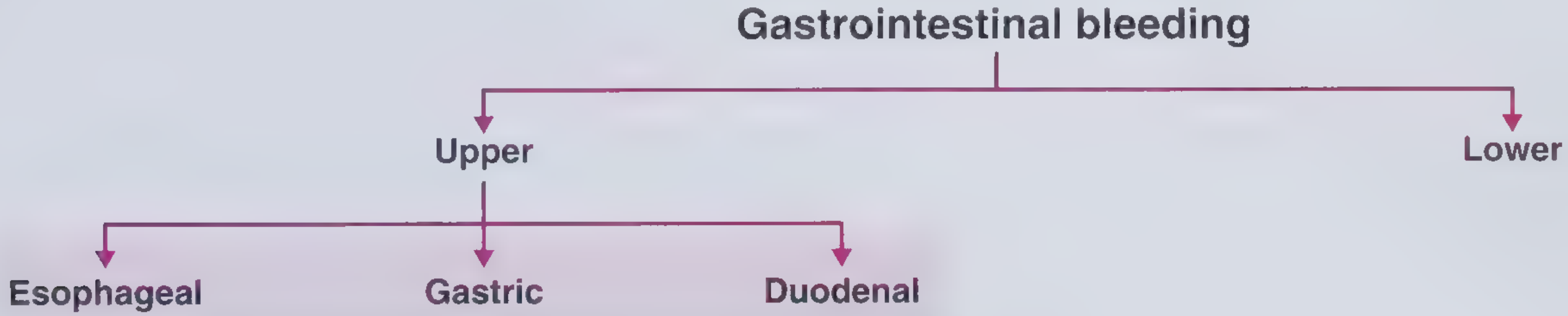
CAUSES / ETIOLOGY
| Cause | Frequency |
|---|---|
| Peptic ulcer disease (gastric + duodenal) | ~35-50% |
| Esophageal / gastric varices | ~22% |
| Portal hypertension-related lesions | ~5% |
| Esophagitis | ~5% |
| Mallory-Weiss tear | ~4% |
| Angioectasia / Angiodysplasia | ~4% |
| Dieulafoy lesion | ~3% |
| UGI neoplasm | ~3% |
| Erosive gastritis/duodenitis | ~1-2% |
| Aortoenteric fistula (post-aortic graft) | Rare but lethal |
(Source: Sleisenger and Fordtran's Gastrointestinal and Liver Disease, UCLA CURE Database, n=968)
IMPORTANT HISTORY POINTS
Presenting symptoms:
- Hematemesis - bright red blood in vomitus = active or recent bleeding
- Coffee-ground emesis - digested blood, suggests slower or stopped bleeding
- Melena - black, tarry, malodorous stools - strongly suggests UGIB (especially in patients <50 years)
- Hematochezia - bright red blood per rectum - can arise from UGIB in ~14% of cases when bleeding is massive and rapid
Key history questions to ask:
- Prior GI bleeding episodes - up to 60% of recurrent UGIBs arise from the same lesion
- History of peptic ulcer disease or H. pylori infection
- Alcohol use and liver disease - cirrhotic patients have a 30% chance of variceal bleeding; ~60% rebleed within 12 months
- NSAID / aspirin / anticoagulant use - most patients presenting with UGIB used aspirin or an NSAID in the preceding week; half use these OTC
- Retching and vomiting before hematemesis - classic for Mallory-Weiss tear (also seen with DKA, chemotherapy, alcohol binge)
- History of aortic graft surgery - "herald bleed" (self-limited initial hematemesis) preceding massive hemorrhage = aortoenteric fistula
- Iron or bismuth ingestion - can simulate melena (stool guaiac will be negative)
- Red-dye liquids or beets - can simulate hematochezia
- Smoking, alcohol, GERD - risk factors for esophageal cancer
- Known varices, prior TIPS, banding - high-risk endoscopic history
- Syncope, weakness, angina, confusion - may be the only presenting feature without overt bleeding
CLINICAL FEATURES
Symptoms
- Hematemesis (bright red or coffee-ground)
- Melena
- Hematochezia (in massive UGIB)
- Syncope, dizziness, weakness
- Angina or palpitations (demand ischemia from blood loss)
Signs - Vital Signs
- Tachycardia (often first sign)
- Hypotension / orthostatic hypotension
- Decreased pulse pressure
- Tachypnea
- Paradoxical bradycardia can occur even with significant hemorrhage
- Younger patients without comorbidities may tolerate substantial volume loss with minimal vital sign changes
Signs - Physical Examination
- Pallor, cool clammy extremities (hypovolemia)
- Jaundice, spider angiomas, palmar erythema, splenomegaly, ascites - suggest liver disease and variceal bleeding
- Epigastric tenderness - suggests peptic ulcer disease
- Digital rectal exam - assess stool color (melena vs. hematochezia) and confirm rectal bleeding
Postural (Orthostatic) Vital Signs
- Drop in systolic BP >10 mmHg OR rise in HR >20 bpm on standing suggests >15% volume loss
INVESTIGATIONS
Bedside / Initial Assessment
- Nasogastric aspirate (NG tube) - red blood or coffee-ground material confirms UGIB; useful when diagnosis is not clinically apparent (no strong evidence it provokes variceal bleeding)
- Stool guaiac / FOBT - confirms blood in stool; differentiates true melena from iron/bismuth ingestion
Laboratory Studies
| Test | What to look for |
|---|---|
| FBC / CBC | Haemoglobin, haematocrit (may be normal initially before hemodilution) |
| Blood group & cross-match | Prepare for transfusion |
| Coagulation screen (PT, INR, aPTT) | INR >1.5 = high risk (part of AIMS65); guides FFP use |
| Platelet count | <50,000 = needs correction before/during procedures |
| Urea / BUN and creatinine | Elevated BUN:creatinine ratio is a biochemical clue to UGIB (digested blood = urea source); typical ratio >20:1 |
| LFTs, albumin | Albumin <3.0 g/dL = poor prognosis (part of AIMS65) |
| Serum electrolytes | Baseline |
| Blood glucose |
Scoring Systems (Risk Stratification)
Pre-endoscopy - Glasgow-Blatchford Score (GBS):
Uses: blood pressure, BUN, haemoglobin, heart rate, syncope, melena, liver disease, heart failure
- Predicts need for clinical intervention (transfusion, endoscopy, surgery)
- Score 0 = very low risk; can consider outpatient management
AIMS65 Score (pre-endoscopy):
- Albumin <3.0 g/dL
- INR >1.5
- Mental status alteration
- Systolic BP <90 mmHg
- Age >65 years
- Score ≥2 = higher mortality, longer stay, higher cost
Complete Rockall Score (post-endoscopy):
Combines: age + shock + comorbidities + endoscopic findings (SRH)
- Score 0-2 = low risk, consider early discharge
- Correlates well with mortality
Imaging and Endoscopy
-
Upper GI Endoscopy (EGD) - gold standard - diagnostic AND therapeutic
- Perform within 24 hours of presentation in most patients with overt bleeding after adequate resuscitation
- Identifies stigmata of recent haemorrhage (SRH): active spurting, visible vessel, adherent clot, flat spot, clean base
- Can be performed urgently (<12 hours) in haemodynamically unstable or high-risk patients
- Side-viewing duodenoscope for suspected duodenal/ampullary bleeding not seen on standard scope
-
CT angiography (CTA) - when endoscopy is non-diagnostic or not feasible; detects bleeding >0.3-0.5 mL/min
-
Mesenteric angiography - for active bleeding; can be therapeutic (coil embolization)
-
Scintigraphy (tagged RBC scan) - detects slow intermittent bleeding (>0.1 mL/min)
MANAGEMENT
Step 1: Immediate Resuscitation ("ABC" First)
- Airway - consider prophylactic endotracheal intubation if massive active bleeding, altered consciousness, or aspiration risk; use smaller induction agent doses to minimize peri-intubation hypotension
- IV access - two large-bore IVs (16-gauge or larger)
- IV fluids - crystalloid resuscitation while awaiting blood products
- Oxygen - supplemental O2 in all patients
Step 2: Blood Transfusion
| Trigger | Threshold |
|---|---|
| Most patients | Transfuse if Hb ≤ 7 g/dL |
| Elderly / comorbidities (cardiac disease) | Transfuse if Hb ≤ 9 g/dL |
| Massive bleeding | Activate massive transfusion protocol (MTP) - balanced ratio of pRBC : FFP : platelets |
- Correct coagulopathy: FFP if INR elevated; platelets if <50,000; vitamin K if on warfarin
- Correct thrombocytopenia aggressively in active bleeding
Step 3: Pharmacological Treatment
| Drug | Dose | Indication |
|---|---|---|
| PPI (IV pantoprazole / omeprazole) | 80 mg bolus then 8 mg/hr infusion | Non-variceal UGIB (peptic ulcer) - reduces rebleeding rate |
| Somatostatin analogues (Octreotide) | 50 mcg IV bolus then 50 mcg/hr | Variceal bleeding - reduces portal pressure |
| Vasopressin / Terlipressin | Terlipressin 2 mg IV q4h | Variceal bleeding |
| Prokinetics (Erythromycin) | 250 mg IV 30-60 min before endoscopy | Promotes gastric emptying to improve endoscopic visualization |
| Metoclopramide | 10 mg IV | Alternative prokinetic |
| Antibiotics (Ceftriaxone / Norfloxacin) | Ceftriaxone 1 g IV daily | Prophylaxis in cirrhotic patients with variceal bleeding (reduces infection and rebleeding) |
| Tranexamic acid | Under study; current meta-analyses show no mortality benefit in GI bleeding | Not currently routinely recommended |
Step 4: Endoscopic Therapy (Definitive for Most)
Endoscopic hemostasis modalities:
- Injection therapy - adrenaline/epinephrine injection (local vasoconstriction + tamponade), tissue adhesives (e.g., cyanoacrylate for gastric varices)
- Mechanical therapy - hemostatic clips, band ligation (variceal banding), suturing devices
- Thermal therapy:
- Contact: electrocoagulation, thermocoagulation (heater probe)
- Non-contact: argon plasma coagulation (APC), Nd:YAG laser photocoagulation
- Combination therapy (e.g., epinephrine + thermal/clip) is superior to monotherapy for high-risk lesions
For varices specifically:
- Esophageal varices - endoscopic band ligation (EBL) is first-line
- Gastric varices - cyanoacrylate glue injection or thrombin
Step 5: Management of Refractory Bleeding
| Situation | Intervention |
|---|---|
| Endoscopy fails to control variceal bleeding | Balloon tamponade (Sengstaken-Blakemore tube) as bridge; then TIPS (Transjugular Intrahepatic Portosystemic Shunt) |
| Refractory non-variceal bleeding | Repeat endoscopy, then interventional radiology (angioembolization) |
| All else fails | Surgery (underrunning of ulcer, partial gastrectomy) |
Step 6: Secondary Prevention
- H. pylori eradication - test-and-treat in all peptic ulcer bleeders
- Stop NSAIDs - if not possible, add PPI prophylaxis
- PPI long-term - for high-risk ulcers or ongoing NSAID use
- Beta-blockers (propranolol/carvedilol) + EBL - for variceal bleed secondary prevention
- TIPS - for refractory or recurrent variceal bleeding in cirrhosis
RISK STRATIFICATION SUMMARY
| Very Low Risk | High Risk |
|---|---|
| Age <60 | Advanced age |
| No major comorbidities | Significant comorbidities |
| No red hematemesis | Red hematemesis |
| No hematochezia | Hematochezia or melena |
| Negative NG aspirate | Positive NG aspirate |
| Haemodynamically stable | Haemodynamically unstable |
| Normal labs | Abnormal labs (low Hb, elevated INR, low albumin) |
KEY PROGNOSTIC FACTS
- Bleeding is self-limited in 80% of UGIB patients even without specific therapy
- Of the remaining 20% who rebleed, mortality is 30-40%
- Overall UGIB mortality is 5-10% (unchanged since the 1970s despite advances, due to older and sicker patient population)
- Variceal bleeding carries 20% mortality per episode; ~60% rebleed within 12 months
- Peptic ulcer most common cause (~35-50%); varices carry highest per-episode mortality
RECENT EVIDENCE UPDATES
A 2025 meta-analysis (PMID 40029534) found tranexamic acid shows no significant mortality benefit in acute GI bleeding, meaning it is not currently recommended as routine therapy - contradicting earlier enthusiasm. A 2024 systematic review (PMID 38818853) compared endoscopic vs. medical management for peptic ulcers with adherent clots, highlighting that endoscopic removal of adherent clots with hemostasis reduces rebleeding.
Sources: Sleisenger and Fordtran's Gastrointestinal and Liver Disease; Tintinalli's Emergency Medicine (9e); Frameworks for Internal Medicine; Fischer's Mastery of Surgery (8e)
Give its differentials and points to rule out them
The differentials for upper GIT bleeding are essentially the causes within the upper GIT itself (each presenting with hematemesis/melena) that need to be distinguished from one another, plus conditions that mimic UGIB. Let me pull detailed distinguishing features from the textbooks.
Searching sources...
Finding Sources
Finding Sources
Finding Sources
Finding Sources
Finding Sources
Finding Sources
Finding Sources
Reading File
Reading File
Reading File
Reading File
Now let me get detailed content on peptic ulcer features, varices clinical features, esophagitis, aortoenteric fistula, and pseudo-bleeding mimics (epistaxis, haemoptysis):
Finding Sources
Finding Sources
Finding Sources
Reading File
Reading File
Loading Image
Excellent - comprehensive data collected from multiple authoritative sources. Here is the complete structured answer:
Differentials of Upper GIT Bleeding and How to Rule Each Out
The differentials fall into two major categories:
- True UGIB causes - conditions within the upper GIT that genuinely bleed (need to be distinguished from each other)
- Pseudo-UGIB / Mimics - conditions that simulate UGIB but do not originate there
CATEGORY 1: TRUE UGIB DIFFERENTIALS
1. Peptic Ulcer Disease (PUD) - Most Common (~35-50%)
Suggestive (points IN):
- Chronic, recurring epigastric/burning pain - often relieved by food or antacids (duodenal ulcer), or worsened by food (gastric ulcer)
- History of NSAID, aspirin, or steroid use - most patients used NSAIDs the week before
- History of H. pylori infection or previous ulcer
- Tobacco smoking history
- Middle-aged to elderly patient
- Melena more common than hematemesis (unless actively bleeding)
- Coffee-ground emesis typical
To rule out / rule in:
- EGD (gold standard) - shows discrete ulcer crater with clean base, flat spot, adherent clot, visible vessel, or active bleeding
- H. pylori testing (CLO test at endoscopy, urea breath test, stool antigen)
- Elevated BUN:Creatinine ratio supports upper GI source
- Absence of liver disease signs argues against varices
2. Esophageal / Gastric Varices (Second Most Common, Highest Mortality per Episode)
Suggestive (points IN):
- Known cirrhosis / chronic liver disease - alcoholic, viral hepatitis, NAFLD
- Physical signs of portal hypertension: jaundice, spider angiomas, palmar erythema, caput medusae, splenomegaly, ascites, hepatomegaly or small shrunken liver
- Sudden, massive, painless hematemesis of bright red blood - often torrential
- Altered sensorium (hepatic encephalopathy)
- No preceding epigastric pain (unlike PUD)
- History of alcohol binge; prior known varices; prior banding/sclerotherapy
- Coagulopathy (raised INR, low platelets from hypersplenism)
To rule out / rule in:
- EGD - shows dilated tortuous submucosal veins at lower esophagus or gastric fundus; red-wale signs indicate high bleed risk
- LFTs, albumin, INR - deranged in liver disease
- Platelets typically low (hypersplenism)
- Abdominal USS/Doppler - portal vein diameter >13 mm, reversal of flow, splenomegaly
- MELD or Child-Pugh score to quantify liver disease severity
- No ulcer crater on endoscopy distinguishes from PUD
- Note: cirrhotic patients bleed from non-variceal sources ~40% of the time, so EGD remains mandatory
3. Mallory-Weiss Tear
Suggestive (points IN):
- Classic triad: repeated forceful vomiting → THEN hematemesis (the nonbloody vomiting comes first)
- History of alcohol binge, DKA, chemotherapy, bowel preparation
- Can also occur with Valsalva (coughing, seizures, lifting)
- Usually younger patients
- Bleeding often self-limiting and less severe than varices
- No prior GI history
- Sometimes hiatal hernia is present
To rule out / rule in:
- EGD - shows longitudinal mucosal laceration at the gastroesophageal junction, often extending into a hiatal hernia; best seen on retroflexed view
- Single or occasionally multiple tears
- Tear heals spontaneously within ~48 hours in most cases
- Absence of cirrhosis signs distinguishes from variceal bleeding
- No chronic pain history distinguishes from PUD
4. Dieulafoy Lesion
Suggestive (points IN):
- Intermittent, recurrent, massive hematemesis or melena with absolutely NO precipitating factor and NO history of liver disease or NSAID use
- Patient has often had multiple previous negative endoscopies ("serial negative workups")
- Classically in the proximal stomach within 6 cm of the GEJ, but can be anywhere
- No epigastric pain; no alcohol history; no cirrhosis
- Any age, but more common in older males with cardiovascular comorbidities
- Bleeding can be profuse and life-threatening from normal-looking mucosa
To rule out / rule in:
- EGD - profuse bleeding from an area of apparently normal mucosa is the hallmark
- When not bleeding: only a subtle vascular protuberance (1-3 mm) visible; overlying mucosa appears normal
- Doppler endoscopic probe (DEP) can detect submucosal arterial blood flow even when the lesion is not visually apparent
- Mark the site with submucosal ink tattoo if found - aids relocation during rebleed
- Angiography can demonstrate the culprit vessel if endoscopy fails
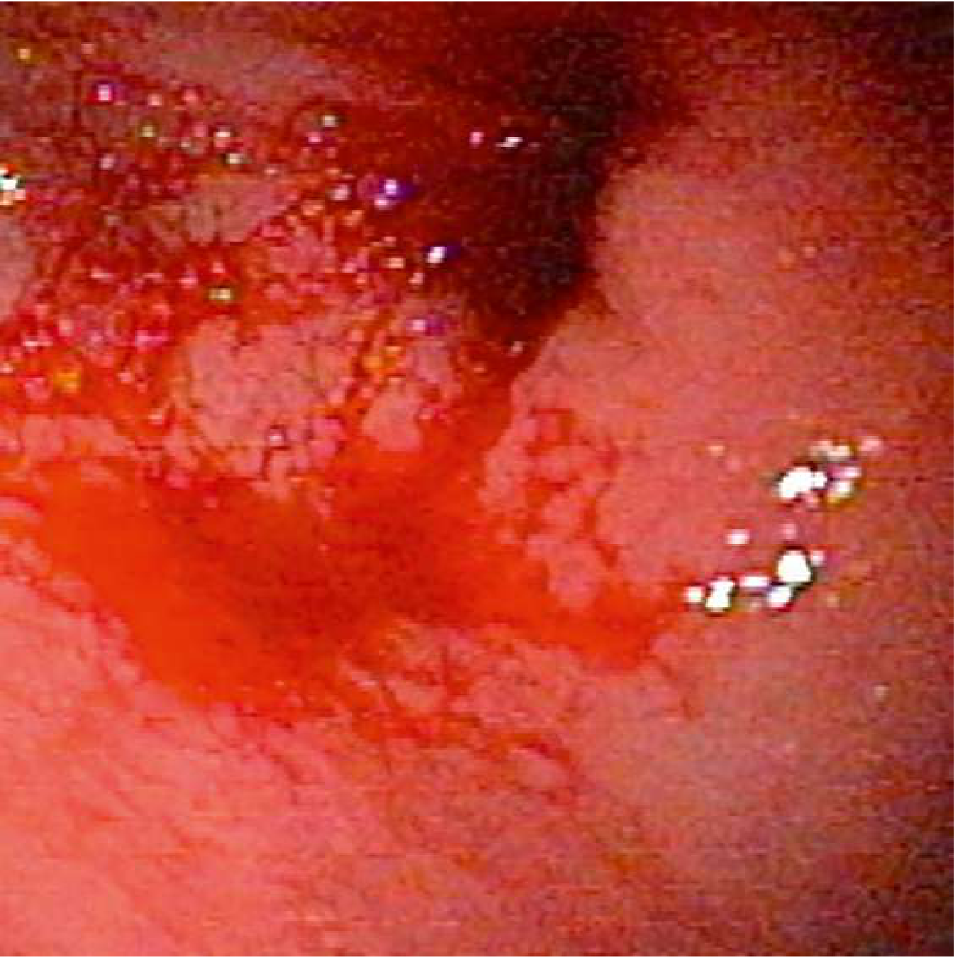
5. Erosive Gastritis / Duodenitis / Esophagitis
Suggestive (points IN):
- Coffee-ground emesis more than frank hematemesis (usually oozing rather than arterial)
- History of alcohol, aspirin, NSAIDs (stress-related mucosal disease)
- Critical illness - sepsis, trauma, mechanical ventilation (stress ulcers)
- Dysphagia or odynophagia with heartburn suggests erosive esophagitis
- Immunocompromised patients (HIV, transplant): esophagitis from Candida, HSV, CMV
- Pill-induced esophagitis: doxycycline, bisphosphonates, potassium, NSAIDs
- Rarely causes massive bleeding; more often occult or minor
To rule out / rule in:
- EGD - multiple superficial erosions without a discrete deep ulcer crater (distinguishes from PUD)
- No vascular lesion (distinguishes from Dieulafoy and varices)
- Biopsy for infection in immunocompromised patients (Candida plaques, viral inclusions)
6. Upper GIT Neoplasm (Gastric / Esophageal Cancer, GIST)
Suggestive (points IN):
- Older patient (>50), progressive symptoms over weeks to months
- Weight loss, anorexia, dysphagia (esophageal cancer), early satiety
- Chronic, unremitting, occult bleeding leading to iron-deficiency anaemia - rarely torrential
- History of smoking, alcohol, GERD (esophageal SCC/adenocarcinoma)
- Family history of gastric cancer; H. pylori-related
- Palpable epigastric mass, left supraclavicular lymph node (Virchow's node), Sister Mary Joseph nodule
- GISTs (gastrointestinal stromal tumors) may present with massive UGIB in 20-30% of cases
To rule out / rule in:
- EGD with biopsy - irregular, firm, ulcerated mass; rolled edges in gastric cancer
- CT chest/abdomen/pelvis - staging, lymph nodes, metastases
- Endoscopic ultrasound (EUS) - depth of invasion, GIST characterisation
- Absence of discrete ulcer with surrounding normal mucosa distinguishes from PUD
- Elevated tumour markers (CEA, CA 19-9) in advanced disease
7. Aortoenteric Fistula (Rare but Lethal - Must Not Miss)
Suggestive (points IN):
- History of prior aortic surgery (AAA repair, aortic graft) - typically 3-5 years prior
- Or known abdominal aortic aneurysm (AAA)
- Classic "herald bleed" - initial self-limited, mild hematemesis or hematochezia hours to months before catastrophic exsanguination
- Pulsatile abdominal mass on exam
- Most commonly involves the 3rd portion of the duodenum
To rule out / rule in:
- Urgent CT angiography with IV contrast or MR angiography is first-line in stable patients - shows inflammation around graft, may demonstrate the fistula
- EGD / push enteroscopy to directly visualize the 3rd part of duodenum for compression, blood, or graft material
- Endoscopic therapy plays NO role - surgical vascular intervention is required
- Do NOT delay with endoscopy alone if clinical suspicion is high
8. Angioectasia / Hereditary Haemorrhagic Telangiectasia (HHT / Osler-Weber-Rendu)
Suggestive (points IN):
- Recurrent episodes of obscure GI bleeding with iron-deficiency anaemia
- HHT: visible telangiectasias on lips, tongue, buccal mucosa, fingers - autosomal dominant family history; frequent epistaxis since childhood
- Patients with CREST syndrome, scleroderma, Turner syndrome, or radiation injury
- Often elderly patients with no liver disease or NSAID history
- Chronic, indolent course; rarely catastrophic
To rule out / rule in:
- EGD shows flat, reddish, spider-like vascular lesions in stomach/duodenum (vs. normal mucosa around a Dieulafoy, vs. varices at GEJ)
- HHT: genetic testing for ENG or ALK-1 mutations
- Clinical criteria for HHT (Curacao criteria): epistaxis + telangiectasias + visceral lesions + positive family history
CATEGORY 2: PSEUDO-UGIB MIMICS (Rule These Out First)
These conditions simulate hematemesis or melena without true GI bleeding:
| Condition | How It Mimics | How to Rule Out |
|---|---|---|
| Haemoptysis (coughed blood) | Blood in mouth, may be swallowed then vomited | Blood is frothy/bright red, mixed with sputum, preceded by cough; pH is alkaline; respiratory symptoms present; chest X-ray/CT abnormal; negative NG aspirate |
| Epistaxis (swallowed) | Blood from posterior nasal bleed swallowed then vomited | Examine the nasopharynx - active bleeding point; blood dripping into posterior pharynx; no GI symptoms; elevated BUN:Cr ratio absent |
| Ingestion of iron supplements / bismuth | Black stools mimicking melena | Stool is guaiac-negative; history of iron/bismuth intake; no anaemia progression; no haemodynamic instability |
| Foods/dyes - beets, red Kool-Aid, food colouring | Red/maroon stools or vomit | Guaiac test negative; dietary history; no haemodynamic compromise |
| Oropharyngeal / dental bleeding (swallowed) | Blood in vomit | Examine the oral cavity carefully; guaiac test may be positive but very low BUN:Cr ratio |
QUICK-REFERENCE DIFFERENTIATING TABLE
| Feature | PUD | Varices | Mallory-Weiss | Dieulafoy | Neoplasm | Aortoenteric Fistula |
|---|---|---|---|---|---|---|
| Age | Any | Any (cirrhotic) | Any (young-mid) | Older males | >50 | Older (post-surgical) |
| Pain | Chronic epigastric | None | None | None | Weight loss/dysphagia | None (until catastrophe) |
| Precipitant | NSAIDs, H. pylori | Alcohol/liver disease | Retching/vomiting | None | None | Prior aortic graft |
| Character | Coffee-ground / melena | Massive bright red | Hematemesis after retching | Intermittent massive | Occult/chronic | "Herald" then massive |
| Liver signs | Absent | Present | Absent | Absent | Absent | Absent |
| Endoscopy | Ulcer crater | Variceal columns | GEJ laceration | Bleeding from normal mucosa | Mass/ulcer | 3rd duodenum (graft) |
| Key investigation | EGD + H. pylori | EGD + LFTs + USS | EGD (retroflexed view) | EGD + DEP probe | EGD + biopsy + CT | CT angiography FIRST |
Sources: Sleisenger and Fordtran's Gastrointestinal and Liver Disease; Goldman-Cecil Medicine (22e); Bailey and Love's Surgery (28e); Fischer's Mastery of Surgery (8e); Tintinalli's Emergency Medicine (9e)
This is a shared conversation. Sign in to Orris to start your own chat.