
What is MI
Myocardial infarction (MI), or "heart attack," occurs when blood flow to part of the heart muscle is blocked long enough to cause cell death. It is most commonly caused by rupture of an atherosclerotic plaque in a coronary artery, leading to thrombus formation and occlusion.
Searching sources...
Reading File
Searching the Web
myocardial infarction MI overview pathophysiology 2025
Finding Sources
Reading File
Reading File
Reading File
Finding Sources
Reading File
Reading File
Reading File
Here is a thorough overview of Myocardial Infarction (MI) drawn from authoritative medical textbooks:
Myocardial Infarction (MI) - "Heart Attack"
Definition
Myocardial infarction (MI) is necrosis (death) of heart muscle resulting from ischemia - an inadequate supply of oxygenated blood to the myocardium. The 2018 joint U.S./European Cardiology task force defines it as:
"The presence of acute myocardial injury detected by abnormal cardiac biomarkers in the setting of evidence of acute myocardial ischemia."
- Robbins & Kumar Basic Pathology, p. 354
Causes & Pathogenesis
The vast majority (~90%) of MIs are caused by acute thrombosis within a coronary artery, triggered by rupture or erosion of an atherosclerotic plaque. The sequence of events:
- An atheromatous plaque is disrupted by endothelial injury, intraplaque hemorrhage, or mechanical forces, exposing subendothelial collagen and necrotic plaque contents.
- Platelets adhere, aggregate, and activate, releasing thromboxane A2, ADP, and serotonin - causing further platelet aggregation and vasospasm.
- Coagulation is activated via tissue factor, building a growing thrombus.
- Within minutes, the thrombus completely occludes the coronary artery.
In ~10% of MIs, no occlusive atherosclerosis is found. These cases involve:
-
Coronary artery vasospasm
-
Embolization from mural thrombi (e.g., atrial fibrillation) or valve vegetations
-
Small vessel disease: vasculitis, amyloid deposition, sickle cell stasis
-
Robbins & Kumar Basic Pathology, p. 354
Classification: Types of MI
| Type | Mechanism |
|---|---|
| Type 1 | Spontaneous ischemia from atherosclerotic plaque rupture/erosion + thrombus |
| Type 2 | Oxygen supply-demand imbalance WITHOUT plaque rupture (e.g., severe tachycardia, anemia, hypotension) |
By ECG Pattern (Clinical Subtypes)
| Subtype | ECG Finding | Extent |
|---|---|---|
| STEMI | ST-segment elevation in ≥2 contiguous leads | Transmural (full-thickness) |
| NSTEMI | No ST elevation; elevated troponin | Subendocardial |
| Unstable Angina (UA) | ECG changes but NO troponin rise | No necrosis (ischemia only) |
- STEMI criteria: new ST elevation ≥0.1 mV at J-point in ≥2 contiguous leads (≥0.2 mV in V2-V3 in men ≥40 yrs)
- Frameworks for Internal Medicine; Robbins Pathology
Myocardial Response to Ischemia
Within seconds: aerobic metabolism ceases → ATP drops → lactic acid accumulates.
Within minutes: loss of contractility.
At 20-40 minutes: irreversible cell death (coagulative necrosis) begins.
-
The subendocardium is most vulnerable - it has the highest oxygen demand and its blood vessels are compressed by systolic contraction.
-
Necrosis starts in the subendocardium and expands outward in a "wavefront" pattern.
-
A full transmural infarct achieves maximum extent in 3-6 hours without intervention.
-
Cardiac muscle needs ~1.3 mL O2/100g/min to survive; normal delivery is ~8 mL O2/100g/min. Even 15-30% of normal flow can prevent death.
-
Guyton and Hall Textbook of Medical Physiology, p. 271
Locations of MI (by Vessel)
| Artery Occluded | Area Infarcted | % of MIs |
|---|---|---|
| LAD (proximal) | Anterior LV wall, anterior 2/3 of septum, apex | 40-50% |
| RCA (proximal) | Right ventricle, posterior LV | 30-40% |
| LCX (proximal) | Lateral left ventricle | 15-20% |
- Robbins & Kumar Basic Pathology, p. 355
Patterns of Infarction
- Transmural infarction: Full-thickness necrosis; caused by epicardial vessel occlusion with thrombosis. Produces Q waves on ECG (STEMI).
- Subendocardial infarction: Restricted to the inner <1/2 of ventricular wall; corresponds to NSTEMI. More common when there is diffuse severe atherosclerosis without complete occlusion.
Causes of Death After MI
- Decreased cardiac output and cardiogenic shock (systolic stretch - ischemic muscle bulges outward instead of contracting)
- Pulmonary edema from damming of blood in pulmonary vessels
- Ventricular fibrillation - the most common cause of death (80-90% of cardiac deaths post-MI)
- Cardiac rupture (less common)
- Guyton and Hall Textbook of Medical Physiology, p. 271
Epidemiology (2025 ACC/AHA Data)
- ~805,000 MIs per year in the U.S. (605,000 first-time; 200,000 recurrent)
- Economic burden: ~$84.9 billion annually
- ~10% of MIs occur before age 40; ~45% before age 65
- Men are at greater risk, but the gap narrows with age (women are relatively protected pre-menopause)
- Mortality has declined thanks to early reperfusion and evidence-based prevention
Key Biomarker: Troponin
The earliest detectable sign of myocyte necrosis is disruption of the sarcolemmal membrane, allowing intracellular proteins - especially cardiac troponin I and T - to leak into the bloodstream. This is the basis of the troponin blood test used to diagnose MI.
Summary
MI = blocked coronary artery → ischemia → myocardial cell death. The key points are:
- Most are caused by plaque rupture + thrombus
- Time is muscle - intervention within 3-6 hours limits infarct size
- Classified as STEMI (transmural, needs immediate reperfusion) vs. NSTEMI (subendocardial)
- Diagnosed by troponin rise + ECG changes + symptoms
- The #1 cause of death post-MI is ventricular fibrillation
Stages of hair fall Medications and procedure for hair transplant
Hair loss progresses through recognized stages (the Norwood scale for men, Ludwig scale for women), and treatment ranges from medications like minoxidil and finasteride to surgical hair transplant procedures (FUT and FUE).
Searching sources...
Reading File
Searching the Web
stages of hair loss Norwood Ludwig scale classification 2025
Searching the Web
hair transplant procedure FUE FUT medications 2025
Finding Sources
Finding Sources
Reading File
Finding Sources
Reading File
Reading File
Reading File
Reading Web Page
https://www.finishingtouchesgroup.com/blogs/news/norwood-lud…
Reading File
Reading File
I now have comprehensive information from the dermatology textbook and web sources. Here is the full answer:
Hair Loss: Stages, Medications & Hair Transplant
Part 1 - The Hair Cycle (Biology of Hair Loss)
Before understanding stages, it helps to know the normal hair cycle:
| Phase | Duration | What Happens |
|---|---|---|
| Anagen (growth) | 2-7 years | Active hair shaft production |
| Catagen (transition) | 2-3 weeks | Follicle shrinks, growth stops |
| Telogen (resting) | 3 months | Hair rests, then sheds |
| Exogen | Days-weeks | Old hair falls out, new cycle begins |
In androgenetic alopecia (AGA), DHT (dihydrotestosterone) progressively miniaturizes terminal pigmented anagen hairs into fine, hypopigmented vellus hairs - eventually silencing the follicle altogether.
- Dermatology 2-Volume Set 5e, p. 3158
Part 2 - Stages of Hair Loss
In Men: The Norwood (Hamilton-Norwood) Scale
The most widely used classification - 7 stages of increasing baldness:
| Stage | Description |
|---|---|
| Stage 1 | No significant hair loss; hairline intact |
| Stage 2 | Slight recession at temples - "M" shape (mature hairline) |
| Stage 3 | Deeper temple recession; first stage of clinically significant baldness. Crown thinning may begin (Stage 3 Vertex) |
| Stage 4 | More pronounced hairline recession + distinct bald spot on crown; strip of hair separates the two areas |
| Stage 5 | The two bald areas (frontal + crown) continue to enlarge and approach each other; narrow bridge of hair between them |
| Stage 6 | Bridge disappears; frontal and crown regions merge into one large bald area; hair remaining only on sides |
| Stage 7 | Most advanced - only a thin horseshoe band of hair remains around the sides and back of the head |
Here is the Norwood grading scale illustration from Dermatology textbook:
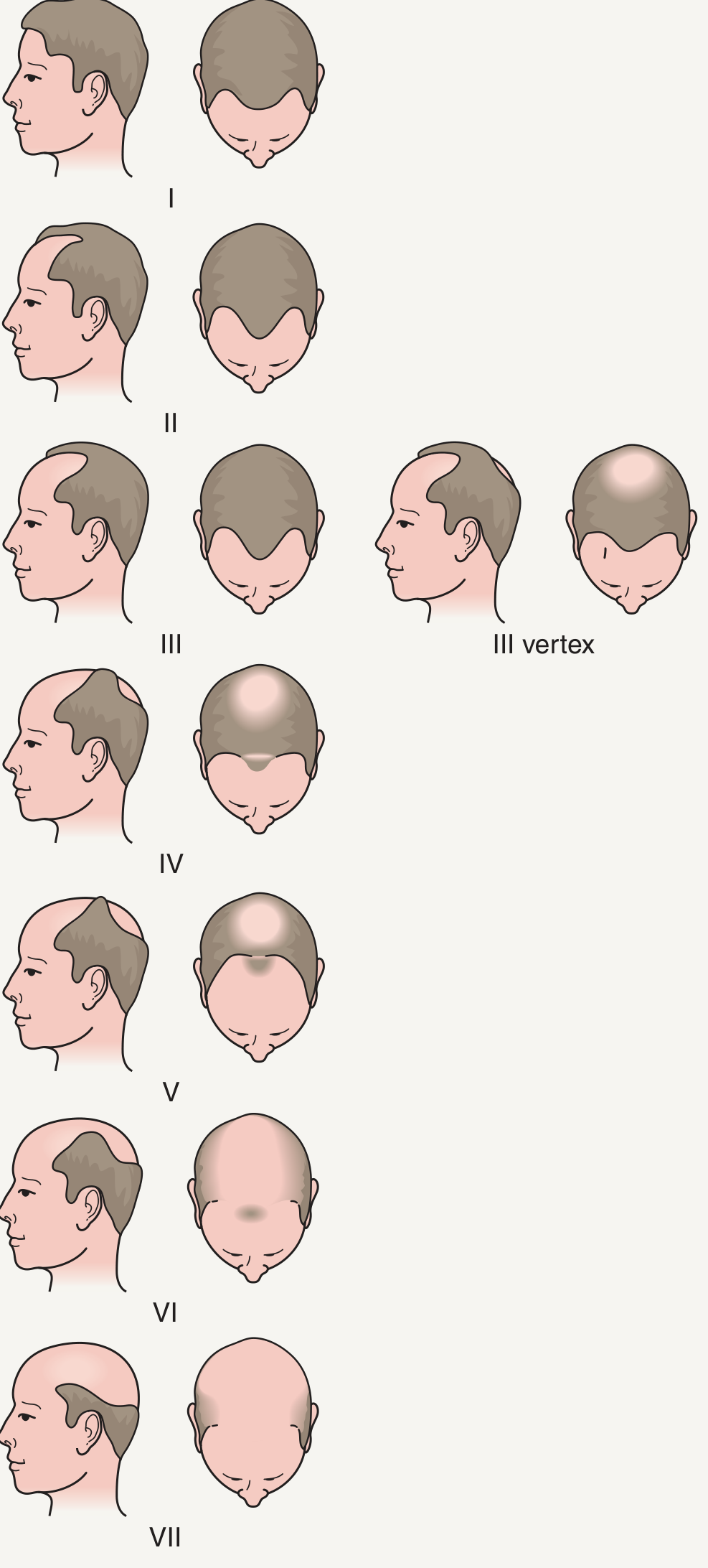
- Dermatology 2-Volume Set 5e, Fig. 157.2
In Women: The Ludwig Scale
Female pattern hair loss (FPHL) typically presents as diffuse thinning over the crown (not a receding hairline), graded in 3 stages:
| Stage | Description |
|---|---|
| Ludwig I | Mild - slight widening of the central part; thinning on top, often unnoticed |
| Ludwig II | Moderate - noticeable widening and volume loss; significant thinning of crown |
| Ludwig III | Severe - near-complete loss of hair on top of scalp; frontal hairline usually preserved |
A "Christmas tree pattern" of midline parting widening is a characteristic clue in women. The Sinclair scale (5 stages) is also used: Stage 1 = normal, Stage 2 = widening of central part, Stage 3 = volume loss lateral to part, Stage 4 = anterior bald spot, Stage 5 = advanced.
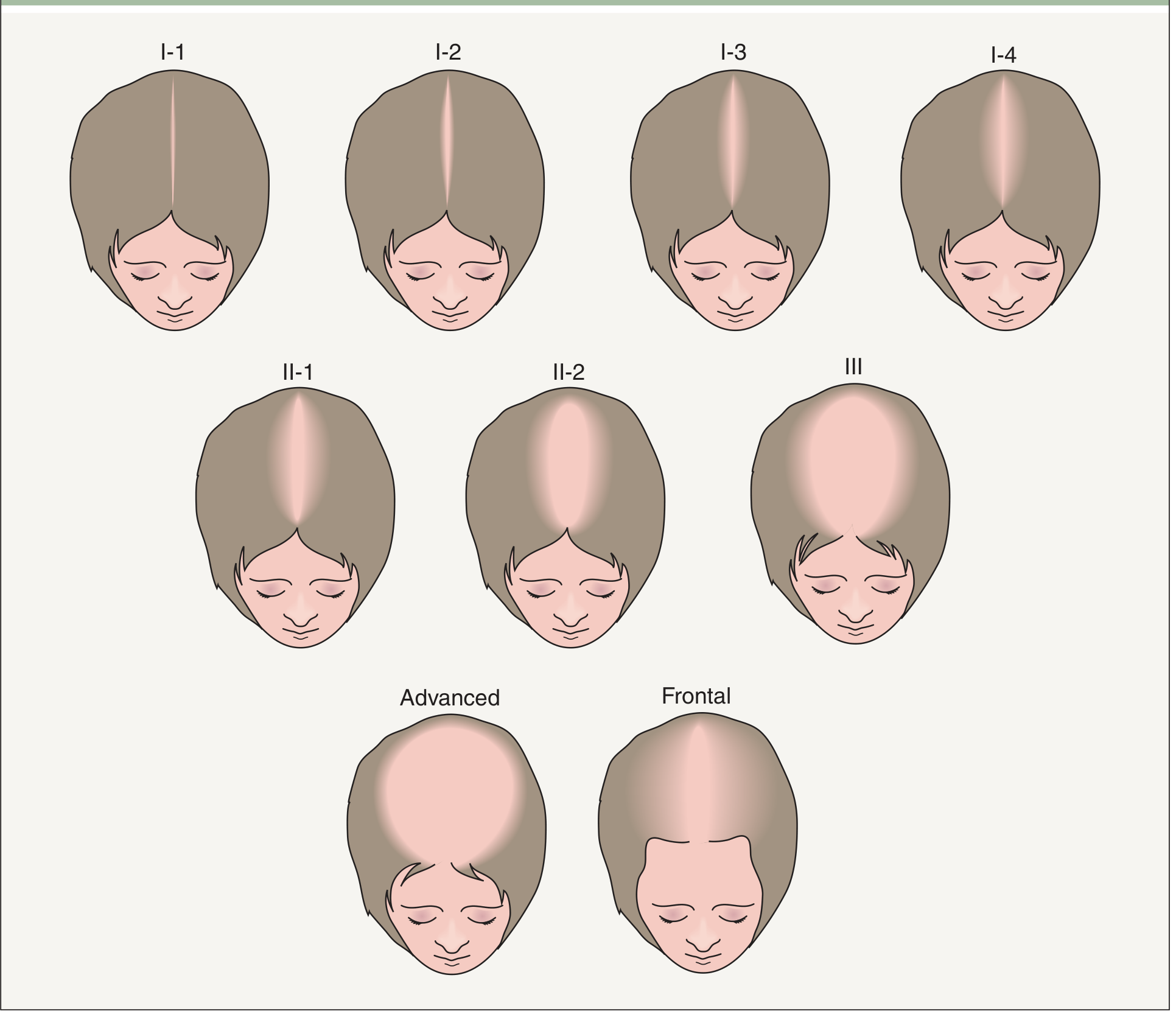
- Dermatology 2-Volume Set 5e, Fig. 157.3
Part 3 - Medications for Hair Loss
1. Minoxidil (Topical / Oral)
- Mechanism: Vasodilation + angiogenic properties; prolongs anagen phase; increases dermal papilla cell proliferation; acts on potassium channels
- FDA-approved forms:
- 5% solution/foam - men and women
- 2% solution - women
- Oral low-dose minoxidil: 0.25-2.5 mg/day (women); up to 5 mg/day (men) - effective and usually well tolerated
- Important: Stopping minoxidil causes an acute telogen efflux 3-4 months after discontinuation
- Must be used continuously - effects reverse on stopping
2. Finasteride (Oral) - Men
- Mechanism: Inhibits 5α-reductase, blocking conversion of testosterone to DHT
- Dose: 1 mg/day orally
- FDA-approved for men
- Stopping finasteride causes gradual hair loss, returning to pretreatment state within ~1 year
- Topical finasteride spray is equally effective with less systemic DHT suppression
- Side effect: "Post-finasteride syndrome" - persistent sexual dysfunction and depression (reported in some men)
3. Dutasteride (Oral)
- Inhibits both isoforms of 5α-reductase (vs. finasteride which inhibits only type II)
- Dose: 0.5 mg/day
- More effective than finasteride with a similar safety profile
- Approved in some countries (not FDA-approved in the US for hair loss)
4. Other Medical Options
| Treatment | Details |
|---|---|
| Spironolactone | Anti-androgen used in women with FPHL; 50-200 mg/day |
| Platelet-Rich Plasma (PRP) | Injections into the scalp; stimulates follicles; used as adjunct |
| Low-Level Laser Therapy (LLLT) | Red light stimulates follicular activity; non-invasive |
| Ketoconazole shampoo | Anti-fungal with mild anti-androgenic effect; used as adjunct |
Treatment should be continued for at least 6 months before assessing efficacy. - Goldman-Cecil Medicine
Part 4 - Hair Transplant: Procedure
Hair transplantation moves follicular unit grafts from a donor region (back/sides of scalp - not affected by AGA) to balding recipient areas.
Who Is a Good Candidate?
- Age >25 years preferred (younger patients have unpredictable future hair loss)
- Donor hair density >80 follicular units/cm² = excellent; <40 = poor candidate
- Hair shaft caliber >70 microns gives denser coverage
- Norwood Stages 3-6 (frontal baldness) show the most dramatic results
The Two Main Techniques
1. FUT - Follicular Unit Transplantation (Strip Method)
Steps:
- A thin strip of scalp is surgically excised from the mid-occipital (back) donor area
- The strip is divided by technicians into slivers, then further cut into individual follicular units (1-4 hairs each)
- Grafts are stored in chilled saline
- The surgeon creates small recipient sites (0.5-1 mm) using a blade or needle in balding areas at 35-40 follicular units/cm²
- Technicians insert the grafts into recipient sites
- Donor wound is sutured - leaves a linear scar (concealed by surrounding hair)
Pros: Higher graft yield per session
Cons: Permanent linear scar - not ideal for very short haircuts
Cons: Permanent linear scar - not ideal for very short haircuts
2. FUE - Follicular Unit Extraction
Steps:
- Individual follicular units are punched out directly from the donor scalp using a small (0.8-1 mm) circular punch
- Each follicular unit is extracted one by one
- Grafts are implanted into recipient sites (same as FUT)
- No strip removed = no linear scar - leaves small "white dot" scars instead
Pros: No linear scar; faster recovery; ideal for patients who want short hair
Cons: Slightly lower graft yield; more time-consuming per graft
2025 Update: Robotic-assisted FUE (e.g., ARTAS system) improves precision and reduces follicle transection rates
Cons: Slightly lower graft yield; more time-consuming per graft
2025 Update: Robotic-assisted FUE (e.g., ARTAS system) improves precision and reduces follicle transection rates
- Dermatology 2-Volume Set 5e, pp. 3162-3165
Important Surgical Principles
- Frontal scalp is prioritized over vertex (crown): frontal transplants give maximum cosmetic impact and are more stable long-term
- Crown transplants carry risk: progressive 360° hair loss can create a peripheral ring of bald scalp around grafts that looks unnatural over time
- A typical session takes 4+ hours; 2-4 sessions may be needed months apart
- Conservative hairline design is essential - must look natural over decades
Post-Transplant Medications
After surgery, the following are prescribed:
| Medication | Purpose |
|---|---|
| Pain medications | Post-operative comfort |
| Antibiotics | Prevent infection |
- Anti-inflammatory steroids | Reduce swelling | | Minoxidil | Enhances and hastens growth of transplanted hairs during recovery | | Finasteride | Minimizes ongoing native hair loss; improves long-term transplant results |
The transplanted hair falls out 2-3 weeks after the procedure (shock loss) - this is normal. New hair growth begins at 3-6 months, with full results at 12-18 months.
Key Points to Remember
-
AGA is ongoing and progressive - transplantation does not stop future loss
-
Medications (finasteride, minoxidil, PRP, LLLT) must continue alongside transplant to preserve surrounding hair
-
The surgeon must always plan for future hair loss when designing the hairline
-
Hair color, shaft caliber, and donor density all affect the final cosmetic result
-
Dermatology 2-Volume Set 5e; Goldman-Cecil Medicine; Healthline Hair Transplant Guide
Explain briefly about finasteride Composition mechanisam action side effects
Finasteride is a 5α-reductase inhibitor used for male pattern baldness (1 mg) and benign prostatic hyperplasia (5 mg). It blocks DHT production, which is responsible for follicle miniaturization and prostate growth. Side effects include sexual dysfunction and, rarely, persistent post-finasteride syndrome.
Searching sources...
Finding Sources
Searching the Web
finasteride composition mechanism action side effects pharmacology 2025
Reading File
Reading File
Reading File
Finding Sources
Finding Sources
Reading File
Finding Sources
Finding Sources
I have enough information from the textbooks to deliver a thorough answer. Here it is:
Finasteride - Complete Overview
1. Composition & Drug Class
| Property | Details |
|---|---|
| Drug class | 5α-reductase inhibitor (5-ARI) / Antiandrogen |
| Chemical name | N-(1,1-Dimethylethyl)-3-oxo-4-aza-5α-androst-1-ene-17β-carboxamide |
| Brand names | Propecia (1 mg - hair loss) / Proscar (5 mg - BPH) |
| Route | Oral tablet |
| Formulation | 1 mg tablet (androgenetic alopecia); 5 mg tablet (BPH) |
| Structure | Synthetic steroidal compound - azasteroid derivative of testosterone |
2. Mechanism of Action
Finasteride works by competitively inhibiting the enzyme 5α-reductase (specifically type II and type III isoenzymes), which normally converts testosterone into the far more potent androgen dihydrotestosterone (DHT).
Testosterone  ──[5α-reductase]──►  DHT
                  ↑
             BLOCKED by Finasteride
What DHT does (and why blocking it helps):
- In the scalp: DHT binds to androgen receptors in hair follicles of genetically predisposed individuals, causing progressive miniaturization of terminal hairs into thin vellus hairs → eventual baldness
- In the prostate: DHT drives prostate cell growth → benign prostatic hyperplasia (BPH) and enlarged prostate volume
Quantitative effects of finasteride:
-
Reduces serum DHT by ~70%
-
Reduces intraprostatic DHT by ~90%
-
Does NOT reduce testosterone to castration levels (type I 5α-reductase in skin and liver continues to produce some DHT)
-
Reduces serum PSA by ~50% (important: must double PSA value to correct for this when screening for prostate cancer)
-
Campbell Walsh Wein Urology; Katzung's Pharmacology 16th Ed.
3. Indications & Dosing
| Indication | Dose | Duration |
|---|---|---|
| Androgenetic alopecia (male pattern baldness) | 1 mg/day orally | Minimum 3-6 months to assess; indefinite for benefit |
| Benign prostatic hyperplasia (BPH) | 5 mg/day orally | Long-term |
- Finasteride and minoxidil are the only two FDA-approved medications for male pattern hair loss
- Must be used continuously - stopping leads to reversal of benefit within ~12 months for hair loss
4. Pharmacokinetics
| Parameter | Details |
|---|---|
| Absorption | Well absorbed orally (~65% bioavailability); unaffected by food |
| Protein binding | ~90% bound to plasma proteins |
| Metabolism | Hepatic (CYP3A4); converted to inactive metabolites |
| Half-life | ~5-6 hours (6-8 hours in elderly) |
| Excretion | Feces (~57%) and urine (~39%) |
5. Side Effects
Sexual Side Effects (Most Common)
| Side Effect | Frequency |
|---|---|
| Decreased libido | ~1.8% |
| Erectile dysfunction | ~1.3% |
| Ejaculatory disorder (decreased volume) | ~1.2% |
| Orgasm disorders | Reported |
In most men, these resolve with continued therapy or upon discontinuation.
Post-Finasteride Syndrome (PFS) - Serious Concern
A subset of men report persistent sexual, neurological, and psychological symptoms even after stopping the drug:
- Persistent erectile dysfunction
- Decreased libido
- Depression and anxiety
- Brain fog (cognitive impairment)
- Fatigue and insomnia
- Testicular pain
- Suicidal ideation (rare)
The FDA (2025) has also issued alerts about compounded topical finasteride - skin absorption is significant, and adverse events similar to oral use have been reported despite topical application.
Other Side Effects
| Side Effect | Notes |
|---|---|
| Orthostatic hypotension | Dizziness on standing |
| Breast tenderness / gynecomastia | Uncommon |
| Mood changes / depression | Reported, especially with 5 mg dose |
| PSA reduction | Must double PSA to avoid masking prostate cancer |
| Possible increased high-grade prostate cancer risk | Seen in PCPT trial (though possibly a detection artifact due to prostate volume reduction) |
- Katzung's Basic and Clinical Pharmacology 16th Ed.; Lippincott Illustrated Reviews Pharmacology
6. Contraindications
| Contraindication | Reason |
|---|---|
| Pregnancy (absolute) | Even handling crushed tablets is dangerous - causes hypospadias (abnormal urethral opening) in male fetuses |
| Women of childbearing age | Teratogenic to male fetus |
| Children | Not indicated |
| Hypersensitivity to finasteride or any excipient | Standard contraindication |
7. Drug Interactions
- No major pharmacokinetic drug interactions identified
- Caution with other CYP3A4 inhibitors (may increase finasteride levels)
- No interaction with antihypertensives, digoxin, warfarin, or theophylline (studied in trials)
8. Key Points Summary
| Class | 5α-reductase inhibitor |
| Target | Type II & III 5α-reductase |
| Effect | ↓ DHT by ~70% (serum), ~90% (prostate) |
| Hair dose | 1 mg/day |
| BPH dose | 5 mg/day |
| Onset of hair benefit | 3-6 months |
| Main side effects | Sexual dysfunction, post-finasteride syndrome |
| Absolute contraindication | Pregnancy (male fetal teratogen) |
| Must continue | Yes - effects reverse on stopping |
- Sources: Katzung's Basic and Clinical Pharmacology 16th Ed., p. 1690; Lippincott Illustrated Reviews Pharmacology, p. 1513; Campbell Walsh Wein Urology; FDA Topical Finasteride Alert
This is a shared conversation. Sign in to Orris to start your own chat.