
Diabetic foot
Reading File
Searching sources...
Finding Sources
Searching PubMed
"diabetic foot"[MeSH Terms] AND management
Good results. Now let me read from the most comprehensive surgical/clinical sources in parallel.
Reading File
Reading File
Reading File
Excellent content. Let me now read the remaining truncated Bailey & Love content, the Goldman-Cecil classification/management content, and the Current Surgical Therapy content in parallel.
Reading File
Reading File
Reading File
Good. Now let me get the Current Surgical Therapy content on evaluation/ischemia/infection that was truncated, and fetch the clinical image from Bailey & Love:
Reading File
Loading Image
I now have comprehensive content from multiple authoritative sources. Let me compile the full answer.
Diabetic Foot
Overview
Diabetic foot disease is one of the most serious complications of diabetes mellitus, accounting for approximately 20% of all diabetic hospitalizations. The annual incidence of foot complications is 1-2% per year. About 1 in 4 patients with diabetes will develop a foot complication during their lifetime, and 15-20% of diabetic foot ulcers (DFUs) result in lower extremity amputation (LEA). Approximately 85% of all lower extremity amputations are preceded by a DFU.
- Current Surgical Therapy 14e, p. 1186
- Bailey and Love's Short Practice of Surgery 28th Ed, p. 628
Pathophysiology: The Triad
DFUs are multifactorial, driven by a triad of three interacting mechanisms:
1. Peripheral Neuropathy
- Sensory neuropathy: Loss of protective sensation (LOPS) - patients don't notice pressure, heat, or small wounds
- Motor neuropathy: Muscle atrophy causing structural deformity (claw toe, hammertoe), which creates abnormal pressure points
- Autonomic neuropathy: Decreased sweating → dry skin → cracks, fissures, and ulceration
The most common cause of injury is pressure on plantar bony prominences. Sensory loss is assessed with a 10-g monofilament plus at least one other modality (pinprick, temperature, vibration, or proprioception).
2. Ischemia / Peripheral Arterial Disease (PAD)
- Macro-vascular: large-vessel atherosclerosis - diabetics are prone to medial calcinosis ("lead-pipe" calcification), making ABI unreliable
- Micro-vascular: impaired tissue perfusion and impaired healing
- PAD worsens both ulcer healing and post-operative wound closure
3. Infection
- Begins as bacterial invasion into compromised tissue
- Rapidly spreads to deep structures including bone (osteomyelitis)
- Immune defects and hyperglycemia further impair the response
Altered foot mechanics (Charcot foot) from repeated undetected fractures can destroy normal foot architecture completely.
- Goldman-Cecil Medicine, p. 1425
Classification Systems
WIfI Classification (Wound, Ischemia, foot Infection)
Developed by the Society for Vascular Surgery, this is now the preferred system. Each domain is graded 0-3 (none to severe), giving 64 permutations that predict:
- Risk of major LEA at 1 year
- Theoretical benefit of revascularization
WIfI grade also correlates with wound healing time and 1-year amputation-free survival.
Wound Grades (WIfI)
| Grade | Description |
|---|---|
| 0 | No wound / pre-ulceration |
| 1 | Shallow ulcer, no exposed bone (except distal phalanx), no gangrene |
| 2 | Ulcer with exposed bone, digit gangrene, or shallow heel ulcer |
| 3 | Extensive ulcer/gangrene, full-thickness heel ulcer |
Other Systems
-
Wagner Classification - widely used but does not account for ischemia fully
-
University of Texas Classification - grades wound depth + infection + ischemia
-
SINBAD Score - used in epidemiological comparisons
-
Current Surgical Therapy 14e, p. 1186
Clinical Presentation
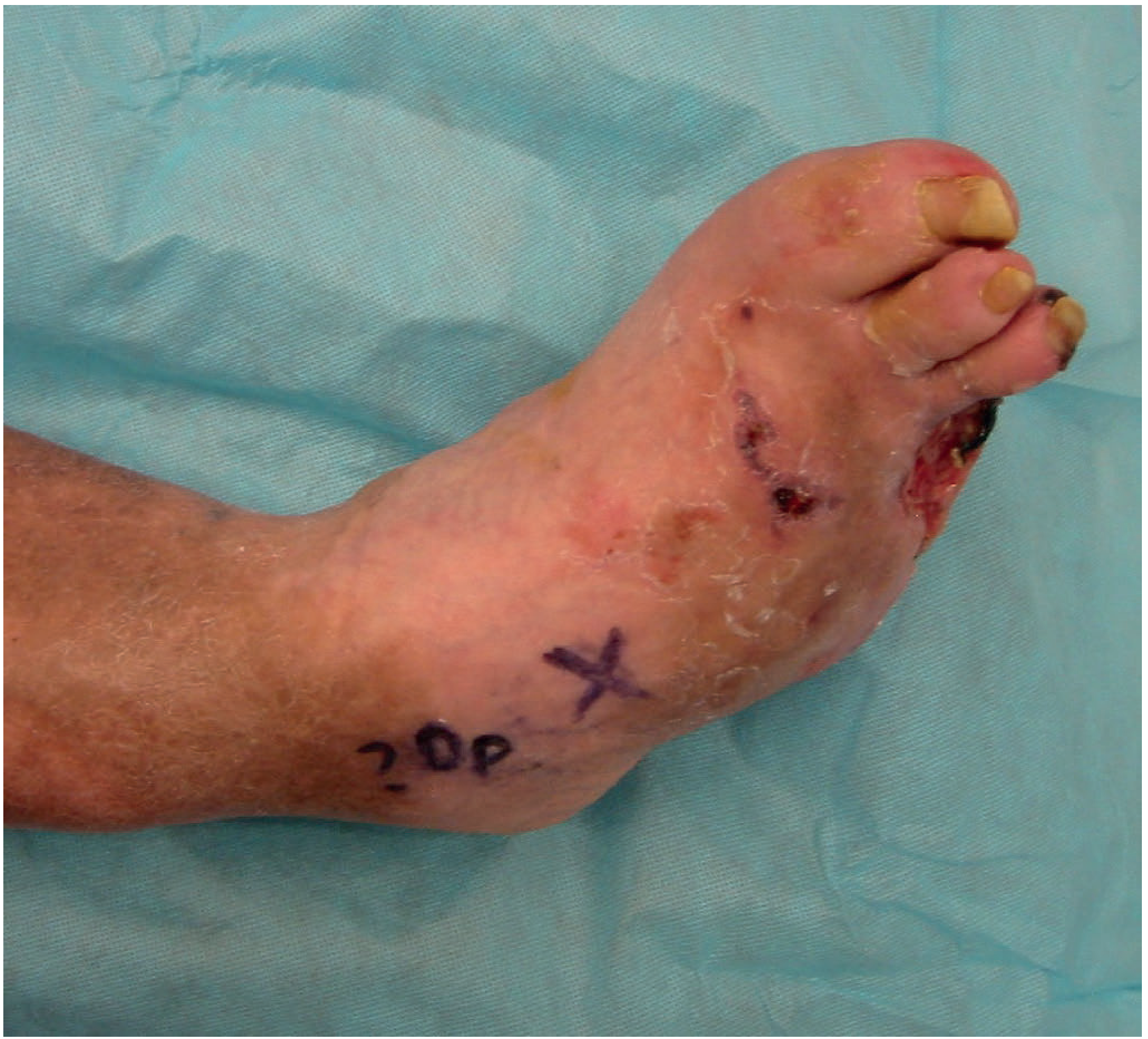
- Ulceration typically on the plantar surface under metatarsal heads, or the calcaneum, surrounded by callus
- Spreading cellulitis, fevers, or critical ischemia indicate severe infection
- Signs of local inflammation: erythema, warmth, swelling, crepitation (gas in tissues)
- Patients may be asymptomatic due to neuropathy - infection can fester for weeks undetected
Investigations
| Investigation | Finding / Role |
|---|---|
| Blood tests | Often unhelpful - inflammatory markers may be normal or only mildly raised |
| Plain X-ray (3-view) | First-line; may show osteomyelitis (can be normal early), gas in soft tissue, foreign bodies, Charcot deformity |
| MRI | Most sensitive modality for bone involvement / osteomyelitis |
| Probe-to-bone test | Combined with elevated CRP and abnormal X-ray confirms osteomyelitis |
| Wound swabs | Superficial swabs are unreliable for deep infection pathogens |
| Bone biopsy | Gold standard for culture in complex/severe infection |
| ABI / Toe pressures | Assess for PAD; ABI may be falsely elevated due to calcification - use toe-brachial index or TcPO2 instead |
- Bailey and Love's 28th Ed, p. 628
- Current Surgical Therapy 14e, p. 1186
Microbiology
-
Mild infections: Gram-positive cocci - S. aureus, beta-haemolytic streptococci
-
Moderate-severe infections: Polymicrobial - aerobic Gram-positives + Gram-negative bacilli + anaerobes
-
Pseudomonas aeruginosa is over-represented; empiric therapy for severe infections must include Pseudomonas coverage
-
Anaerobes: Consider metronidazole especially for abscesses and devitalized tissue
-
Ulcer swabs/sinus tract cultures are not reliable - they do not reflect deep pathogen burden
-
Bailey and Love's 28th Ed, p. 628
Management
1. Antibiotic Therapy
| Severity | Treatment |
|---|---|
| Mild | Oral: TMP-SMX 800/160 mg BD, or clindamycin 300 mg QID, or cephalexin 500 mg QID |
| Moderate-Severe | IV: Piperacillin-tazobactam 3.375 g IV q8h + vancomycin 15 mg/kg IV q12h |
Tissue culture (surgical) should guide definitive therapy. Osteomyelitis typically requires 4-6 weeks of antibiotics (even with disease-free margins at resection, based on practical experience).
2. Wound Care
- Debridement: Callus debridement first to assess wound depth; surgical debridement for collections, necrotic areas, or osteomyelitis
- Pressure off-loading: Total-contact casts, special shoes, orthotics - essential for plantar ulcer healing
- Strict non-weight-bearing regimen
- Advanced wound dressings with healing accelerators
- Negative-pressure wound therapy (NPWT) - evidence from recent meta-analysis (PMID 39241769) supports efficacy in DFUs
3. Surgical Intervention
- Drainage of abscesses
- Debridement of devitalized tissue
- Ray amputation (partial or full) for infected toes/metatarsals
- Wound closure is staged - infected wounds left open initially
- For wounds with exposed tendon/bone or unable to close: Integra dermal regeneration template, followed by split-thickness skin grafting (12-month healing rate ~79%, 18-month ~93%)
- Amputation - not a simple option; wound healing is unpredictable; ensure all infected material is excised with tension-free closure
4. Revascularization
- Mandatory vascular assessment in all patients with poor peripheral pulses
- Angioplasty or bypass surgery for PAD to improve distal vascularity before or alongside foot surgery
- WIfI classification predicts the benefit of revascularization
5. Glycemic Control
- Tight glycemic control important for healing
- HbA1c ≤8% (rather than the traditional ≤7%) has been shown to be sufficient for avoiding major amputation in patients with DFUs
- Endocrinology should be integrated into the multidisciplinary team
6. Other Adjuncts
-
Hyperbaric oxygen - some evidence for complicated infections, particularly anaerobic
-
Bioengineered skin substitutes
-
Autologous leukocytes and platelet-rich plasma for non-healing ulcers
-
Smart wearable technology - emerging role in prevention and monitoring (systematic review, PMID 40682082)
-
Rosen's Emergency Medicine, p. 2540
-
Goldman-Cecil Medicine, p. 1425-1426
Osteomyelitis in Diabetic Foot
The most important risk factor for osteomyelitis is the presence of a foot ulcer. Key points:
- Probe-to-bone test + elevated inflammatory markers + abnormal X-ray = confirmed diagnosis
- MRI is the most sensitive investigation
- Bone biopsy (surgical) is preferred over swabs for culture
- Treatment: surgical resection of infected bone + prolonged antibiotics (4-6 weeks post-resection)
- Distinguishing superficial osteitis (from soft tissue loss/vascular compromise) from deep osteomyelitis is important - the former may require only optimization of glycaemic control, vascular supply, and footwear
Multidisciplinary Team (MDT) Approach
MDT care is now the standard of care for diabetic foot wounds. A comprehensive MDT reduces LEA rates through early recognition and management. Core members include:
- Vascular surgeon
- Surgical podiatrist
- Wound care specialist
- Endocrinologist
- Infectious disease specialist (as needed)
- Plastic/orthopaedic surgery (as needed)
MDT management improves wound healing, lowers major amputation rates, reduces readmissions, and is cost-effective.
- Current Surgical Therapy 14e, p. 1190
Prevention
-
Routine foot examination at every medical visit for patients with sensory loss
-
Daily self-inspection for cracks, fissures, ulcers, or inflammation
-
Never walk barefoot (even at home)
-
Wear closed protective footwear; avoid sandals outdoors
-
Avoid heating pads / hot water bottles on feet (thermal injury risk due to LOPS)
-
Refer to foot care specialist for: sensory loss, foot deformity, extensive callus, non-healing ulcers
-
Custom therapeutic footwear and periodic foot inspection are part of optimal DFU management
-
Goldman-Cecil Medicine, p. 1426
Charcot Foot (Neuropathic Arthropathy)
Altered foot mechanics from repeated undetected fractures lead to destruction of normal foot architecture - the classic Charcot foot deformity. This significantly increases susceptibility to ulceration from abnormal pressure points and is identified on plain X-ray.
Recent Evidence:
- Advanced wound dressings vs standard care for DFUs: Italian guideline meta-analysis (PMID 38864979) - different advanced dressings show varied efficacy; the guideline recommends tailored selection
- NPWT in DFUs: Systematic review/meta-analysis (PMID 39241769) - supports NPWT as an effective adjunct in DFU management
This is a shared conversation. Sign in to Orris to start your own chat.