
Does m2 and m4 increase receptor increase calcium levels or decrease
M2 and M4 muscarinic receptors are Gi-coupled receptors. When activated, they inhibit adenylyl cyclase (reducing cAMP) and do NOT directly increase calcium - in fact, they generally decrease intracellular calcium signaling compared to the Gq-coupled M1/M3/M5 receptors.
Searching sources...
Reading File
Finding Sources
Finding Sources
The textbooks confirm the answer clearly. Here is the full breakdown:
M2 and M4 Muscarinic Receptors: Effect on Calcium
M2 and M4 receptors DECREASE (inhibit) calcium signaling - they do NOT increase it.
Here is how the different muscarinic subtypes compare:
Excitatory Receptors (M1, M3, M5) - INCREASE calcium
- Coupled to Gq protein
- Activate phospholipase C → generate IP3 and DAG
- IP3 triggers release of calcium from the endoplasmic reticulum → increases intracellular calcium
Inhibitory Receptors (M2, M4) - DECREASE/inhibit
- Coupled to Gi protein (inhibitory G protein, pertussis toxin-sensitive)
- Inhibit adenylyl cyclase → decrease cAMP
- Do not generate IP3, so they do not mobilize intracellular calcium
- They actually attenuate calcium-dependent signaling pathways
Summary Table
| Receptor | G Protein | Effect on cAMP | Effect on Calcium |
|---|---|---|---|
| M1, M3, M5 | Gq | No direct effect | ↑ Increase (via IP3) |
| M2, M4 | Gi | ↓ Decrease (inhibit adenylyl cyclase) | ↓ Decrease / No increase |
Clinical relevance
- In the heart, M2 receptors (predominant subtype) slow the rate via Gi → reduced cAMP → reduced Ca²⁺ influx through L-type channels → negative chronotropy/dromotropy
- In the bladder detrusor, M3 (not M2/M4) mediates the main contractile response because it is the calcium-mobilizing subtype
Sources: Katzung's Basic and Clinical Pharmacology, 16th Ed. | Campbell-Walsh Urology
Give very very mbbs detalied comparision btw sympathetic ns and parasympathetic ns
Searching sources...
Finding Sources
Finding Sources
Finding Sources
Reading File
Reading File
Finding Sources
Reading File
Finding Sources
Reading File
Finding Sources
Reading File
Finding Sources
Reading File
Finding Sources
Reading File
Finding Sources
Finding Sources
Loading Image
Loading Image
Sympathetic vs Parasympathetic Nervous System - Complete MBBS Comparison
1. OVERVIEW / NICKNAME
| Feature | Sympathetic (SNS) | Parasympathetic (PSNS) |
|---|---|---|
| Nickname | "Fight or Flight" / "Thoracolumbar" | "Rest and Digest" / "Craniosacral" |
| Walter Cannon's term | "Preparation for flight or fight" | "Vegetative nervous system" |
| General function | Emergency, catabolic, energy expenditure | Conservation, anabolic, housekeeping |
| Discharge pattern | Mass discharge (whole system at once) | Discrete, organ-specific responses |
2. ANATOMICAL ORIGIN (Outflow)
| Feature | Sympathetic | Parasympathetic |
|---|---|---|
| Origin in CNS | Thoracic T1-L2 (or T1-L3) spinal cord | Brainstem (CN III, VII, IX, X) + Sacral S2-S4 |
| Cell body location (preganglionic) | Intermediolateral cell column (lateral horn) of spinal cord | Cranial nerve nuclei in brainstem + sacral parasympathetic nuclei |
| Alternate name | Thoracolumbar division | Craniosacral division |
Mnemonic - PSNS cranial nerves: "Some Say Marry Money But My Brother Says Big Brains Matter More" (or simply: CN III, VII, IX, X)
3. GANGLIA - LOCATION AND DISTANCE
| Feature | Sympathetic | Parasympathetic |
|---|---|---|
| Ganglia location | Near the spinal cord (paravertebral or prevertebral) - far from target organ | Near/in the wall of target organ (terminal ganglia) - close to effector |
| Paravertebral ganglia | Sympathetic chain/trunk on either side of vertebral column | None |
| Prevertebral ganglia | Celiac, superior mesenteric, inferior mesenteric, aorticorenal ganglia | None |
| Terminal (intramural) ganglia | None | Present (in or near organ wall) |
| Named ganglia (head/neck for PSNS) | Superior, middle (often absent), inferior (stellate) cervical ganglia | Ciliary, pterygopalatine, otic, submandibular ganglia |
Sympathetic chain allows: Upper thoracic cord (T1-T3) to reach head and neck via cervical chain ganglia.
4. PRE- AND POSTGANGLIONIC FIBERS
| Feature | Sympathetic | Parasympathetic |
|---|---|---|
| Preganglionic fiber | Short (ganglion near cord) | Long (ganglion near organ) |
| Postganglionic fiber | Long (travels far to organ) | Short (just the last bit to organ) |
| Preganglionic fiber type | Lightly myelinated (B fibers) | Lightly myelinated (B fibers) |
| Postganglionic fiber type | Unmyelinated (C fibers) | Unmyelinated (C fibers) |
| Preganglionic branching | High (1 pre → many post neurons) = diffuse response | Low (1 pre → few post neurons) = discrete response |
| Ratio (pre:post) | 1:20 or more | 1:1 to 1:3 |
Clinical implication: SNS produces widespread simultaneous responses; PSNS produces targeted responses.
5. NEUROTRANSMITTERS
| Site | Sympathetic | Parasympathetic |
|---|---|---|
| Preganglionic → Ganglion | Acetylcholine (ACh) - acts on Nicotinic receptors (Nn) | Acetylcholine (ACh) - acts on Nicotinic receptors (Nn) |
| Postganglionic → Target organ | Norepinephrine (NE) - acts on Adrenergic receptors | Acetylcholine (ACh) - acts on Muscarinic receptors |
| Exception 1 - Sweat glands | Acetylcholine (cholinergic sympathetic!) on M3 receptors | - |
| Exception 2 - Adrenal medulla | Pre-ganglionic ACh → Chromaffin cells release Epinephrine (80%) + NE (20%) directly into blood | - |
| Exception 3 - Blood vessels (some) | Vasodilator fibers use ACh | - |
Key fact: The adrenal medulla = modified sympathetic ganglion. Chromaffin cells = modified postganglionic neurons that secrete into blood instead of a synapse.
6. RECEPTORS
Adrenergic Receptors (Sympathetic targets)
| Receptor | Location | Effect when activated |
|---|---|---|
| α1 | Vascular smooth muscle, iris (dilator), bladder sphincter, prostate | Vasoconstriction, mydriasis, sphincter contraction |
| α2 | Presynaptic (autoreceptors), platelets, fat cells | Inhibit NE release, platelet aggregation, fat inhibition |
| β1 | Heart (SA node, AV node, ventricles) | ↑HR (chronotropy), ↑conduction (dromotropy), ↑contractility (inotropy) |
| β2 | Bronchial smooth muscle, vascular smooth muscle (skeletal muscle), uterus, bladder detrusor | Bronchodilation, vasodilation, uterine relaxation |
| β3 | Adipose tissue, bladder detrusor | Lipolysis, detrusor relaxation |
Cholinergic Receptors (Parasympathetic targets + autonomic ganglia)
| Receptor | Location | Effect |
|---|---|---|
| Nicotinic (Nm) | NMJ (skeletal muscle) | Muscle contraction |
| Nicotinic (Nn) | All autonomic ganglia, adrenal medulla | Ganglionic transmission |
| Muscarinic M1 | CNS, gastric parietal cells | ↑Gastric acid, CNS excitation |
| Muscarinic M2 | Heart (SA, AV node) | ↓HR, ↓conduction (Gi - inhibit cAMP) |
| Muscarinic M3 | Smooth muscle, glands, endothelium | Contraction of smooth muscle, gland secretion, vasodilation (via NO) |
| Muscarinic M4 | CNS, lung | Inhibitory (Gi) |
| Muscarinic M5 | CNS | Excitatory (Gq) |
7. ORGAN-BY-ORGAN EFFECTS
Diagrammatic Overview
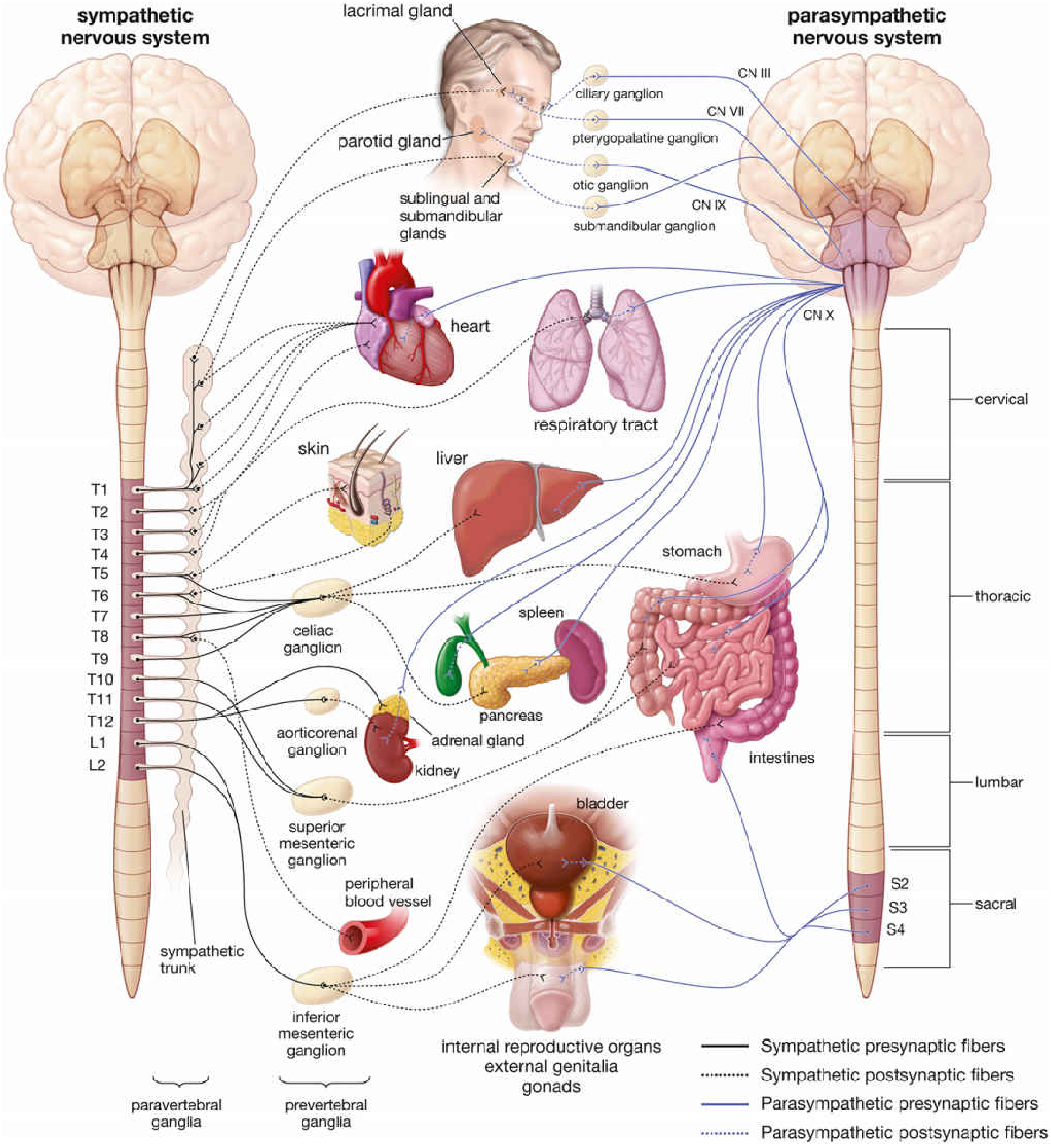
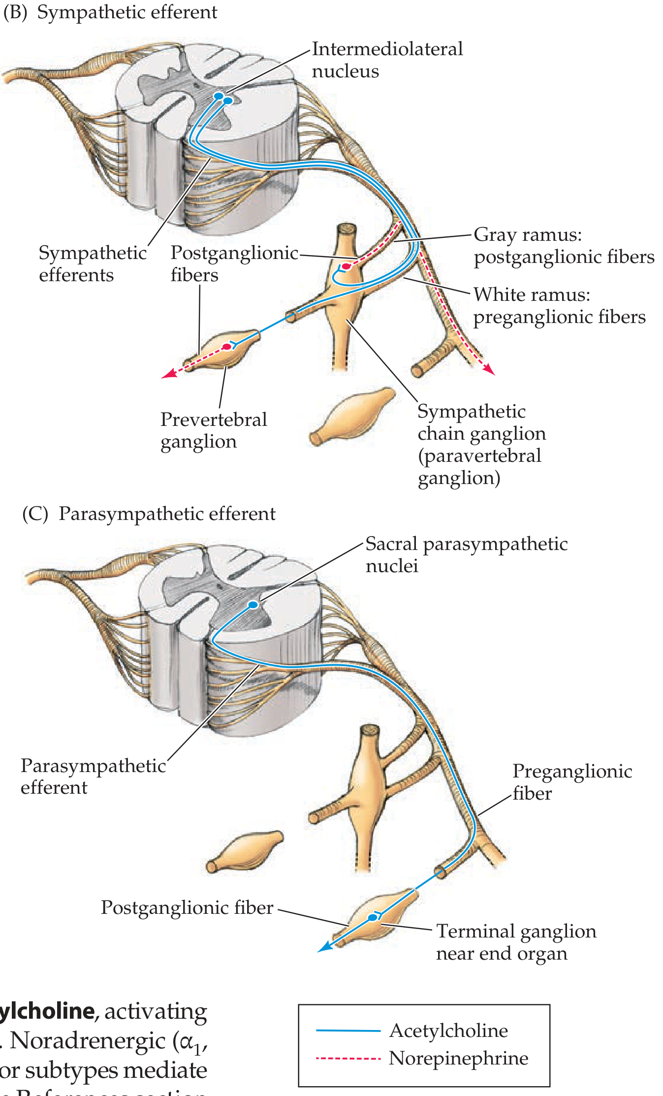
Detailed Organ Effects Table
| Organ / System | Sympathetic Effect | Receptor | Parasympathetic Effect | Receptor |
|---|---|---|---|---|
| Heart - Rate | ↑ Heart rate (tachycardia) | β1 | ↓ Heart rate (bradycardia) | M2 |
| Heart - Contractility | ↑ Force of contraction | β1 | ↓ Slightly | M2 |
| Heart - Conduction (AV node) | ↑ Conduction velocity | β1 | ↓ Conduction (AV block possible) | M2 |
| Blood vessels (most) | Vasoconstriction | α1 | Minimal innervation (vasodilation via NO) | M3 |
| Blood vessels (skeletal muscle) | Vasodilation | β2 | None | |
| Bronchi / Bronchioles | Bronchodilation, ↓ secretions | β2 | Bronchoconstriction, ↑ secretions | M3 |
| Eye - Pupil | Mydriasis (dilates) - radial/dilator muscle contracts | α1 | Miosis (constricts) - sphincter pupillae contracts | M3 |
| Eye - Ciliary muscle | Relaxation → far vision | β | Contraction → near vision (accommodation) | M3 |
| Salivary glands | Thick, viscous saliva (↓ volume) | α1, β | Watery, copious saliva (↑ volume) | M3 |
| Lacrimal glands | No significant innervation | - | ↑ Tear secretion | M3 |
| GI - Motility | ↓ Peristalsis, ↓ tone | α2, β2 | ↑ Peristalsis, ↑ tone | M3 |
| GI - Sphincters | Contracts (closes) | α1 | Relaxes (opens) | M3 |
| GI - Secretions | ↓ | α2 | ↑ (gastric acid, enzymes) | M1, M3 |
| Liver | Glycogenolysis, gluconeogenesis (↑ blood glucose) | β2, α1 | Glycogen synthesis | M |
| Pancreas | ↓ Insulin secretion | α2 | ↑ Insulin and enzyme secretion | M3 |
| Bladder (Detrusor) | Relaxation (allows filling) | β2, β3 | Contraction (micturition) | M3 |
| Bladder (Internal sphincter) | Contracts (retention) | α1 | Relaxes (permits micturition) | M3 |
| Uterus (pregnant) | Contraction | α1 | Variable | |
| Uterus (non-pregnant) | Relaxation | β2 | Variable | |
| Male genitalia | Ejaculation | α1 | Erection (↑ blood flow via NO) | M |
| Kidney (renin) | ↑ Renin secretion | β1 | ↓ Renin | - |
| Skin - Sweat glands | ↑ Sweating (cholinergic!) | M3 | None | |
| Skin - Piloerection | Hair stands up (goosebumps) | α1 | None | |
| Adrenal medulla | ↑ Epi + NE release (via ACh on Nn) | Nn | None | |
| Metabolic | ↑ Glycogenolysis, ↑ lipolysis, ↑ glucose, ↑ FFA | β | Anabolic/storage | - |
Memory trick for erection/ejaculation: "Point and Shoot" - Parasympathetic = erection (Point), Sympathetic = ejaculation (Shoot)
8. STRUCTURES WITH ONLY ONE DIVISION
| Structure | Innervated only by... |
|---|---|
| Blood vessels (most) | Sympathetic only |
| Sweat glands | Sympathetic only (but cholinergic!) |
| Piloerector muscles | Sympathetic only |
| Adrenal medulla | Sympathetic only (preganglionic directly) |
| Lacrimal gland | Parasympathetic only |
| Ciliary muscle | Parasympathetic only |
| Nasopharyngeal glands | Parasympathetic only |
9. NEUROTRANSMITTER SYNTHESIS, STORAGE AND TERMINATION
Norepinephrine (Sympathetic)
- Synthesis: Tyrosine → DOPA → Dopamine → NE (in vesicles) → Epi (adrenal medulla only, via PNMT enzyme)
- Termination: Mainly reuptake into presynaptic terminal (uptake-1 = NET transporter); also COMT (extraneuronal), MAO (intraneuronal)
- Drugs blocking reuptake: Cocaine, tricyclic antidepressants
Acetylcholine (Parasympathetic)
- Synthesis: Choline + Acetyl-CoA → ACh (via choline acetyltransferase, ChAT)
- Termination: Rapid hydrolysis by Acetylcholinesterase (AChE) in synaptic cleft
- Drugs blocking AChE: Neostigmine, physostigmine, organophosphates
10. SECOND MESSENGER SYSTEMS
| Receptor | G Protein | Second Messenger | Effect |
|---|---|---|---|
| α1 adrenergic | Gq | ↑ IP3/DAG → ↑ Ca²⁺ | Contraction, secretion |
| α2 adrenergic | Gi | ↓ cAMP | Inhibition, NE autofeedback |
| β1, β2, β3 adrenergic | Gs | ↑ cAMP → PKA | Relaxation (smooth muscle), ↑HR/contractility (heart) |
| M1, M3, M5 muscarinic | Gq | ↑ IP3/DAG → ↑ Ca²⁺ | Smooth muscle contraction, secretion |
| M2, M4 muscarinic | Gi | ↓ cAMP | ↓ HR, inhibition |
| Nicotinic (Nn, Nm) | None (ion channel) | Na⁺ influx → depolarization | Fast excitation |
11. COMPARISON TABLE - STRUCTURAL SUMMARY
| Feature | Sympathetic | Parasympathetic |
|---|---|---|
| Origin | T1-L2 (thoracolumbar) | CN III, VII, IX, X + S2-S4 (craniosacral) |
| Preganglionic fiber | Short | Long |
| Postganglionic fiber | Long | Short |
| Ganglion location | Para/prevertebral (near cord) | Terminal (near/in organ) |
| Pre:Post ratio | 1:20+ (divergence) | 1:1-3 (discrete) |
| Preganglionic NT | ACh (Nicotinic Nn) | ACh (Nicotinic Nn) |
| Postganglionic NT | Norepinephrine (adrenergic) | ACh (muscarinic) |
| Postganglionic receptor | Adrenergic (α1, α2, β1, β2, β3) | Muscarinic (M1-M5) |
| Exceptions in NT | Sweat glands = ACh; Adrenal medulla = Epi | None |
| General effect | Catabolic, fight-or-flight | Anabolic, rest-and-digest |
| Distribution | Widespread (entire body) | Selective (mainly head, thorax, abdomen, pelvis) |
12. PHARMACOLOGICAL HANDLE (High-yield for MBBS)
| Drug class | Division | Mechanism | Example | Use |
|---|---|---|---|---|
| Beta-blockers | Block SNS | Block β receptors | Propranolol (β1+β2), Metoprolol (β1) | Hypertension, angina, arrhythmia |
| Alpha-blockers | Block SNS | Block α1 receptors | Prazosin, Tamsulosin | Hypertension, BPH |
| Beta-2 agonists | Mimic SNS | Activate β2 | Salbutamol | Asthma |
| Anticholinergics (antimuscarinics) | Block PSNS | Block M receptors | Atropine | Bradycardia, peptic ulcer, COPD |
| Anticholinesterases | Potentiate PSNS | Block AChE | Neostigmine | Myasthenia gravis, reverse NMJ block |
| Muscarinic agonists | Mimic PSNS | Activate M receptors | Pilocarpine | Glaucoma |
Sources: Neuroanatomy through Clinical Cases 3rd Ed. | Lippincott Illustrated Reviews Pharmacology 7th Ed. | Ganong's Review of Medical Physiology 26th Ed. | Guyton & Hall Textbook of Medical Physiology | Histology - A Text and Atlas (Histology)
How does m3 acting drugs cause hypotension
M3 receptors on vascular endothelium, when activated, trigger nitric oxide (NO) release, which causes smooth muscle relaxation and vasodilation - leading to a drop in blood pressure. This is a paradoxical effect since most smooth muscle M3 activation causes contraction.
Searching sources...
Finding Sources
Finding Sources
Finding Sources
Finding Sources
Reading File
Reading File
Finding Sources
Loading Image
Excellent - the textbooks have given the full picture. Here is the complete answer:
How M3-Acting Drugs Cause Hypotension
The Short Answer - A Receptor Correction First
Important note: Katzung's Pharmacology specifically states the vascular vasodilation by muscarinic agonists is mediated via M2 receptors on endothelium (not M3) in intact vessels. However, M3 receptors are present on endothelial cells in some vascular beds (e.g. skeletal muscle via M1/M3), and the mechanism of NO release is the same. For exam purposes in many MBBS curricula, this effect is attributed to M3. The mechanism is identical regardless of which muscarinic subtype is cited.
Step-by-Step Mechanism
Step 1 - Muscarinic Agonist Activates Endothelial Receptors (M2/M3)
When a muscarinic agonist (e.g. ACh, bethanechol, carbachol, pilocarpine) enters the bloodstream, it binds to muscarinic receptors on the vascular endothelium - the inner lining of blood vessels.
- Receptor subtype: M2 (classic) or M3 (in some beds)
- G protein: Gq → activates phospholipase C → ↑ IP3 → ↑ intracellular Ca²⁺ in endothelial cells
Step 2 - eNOS Activation in Endothelium
The rise in intracellular Ca²⁺ inside the endothelial cell:
- Ca²⁺ binds calmodulin
- Ca²⁺-calmodulin complex activates endothelial Nitric Oxide Synthase (eNOS)
- eNOS converts L-arginine → Nitric Oxide (NO)
NO is also known as EDRF - Endothelium-Derived Relaxing Factor (Furchgott & Zawadzki, 1980 - Nobel Prize discovery)
Step 3 - NO Diffuses to Vascular Smooth Muscle
NO is a lipophilic gas - it diffuses freely from the endothelial cell into the adjacent vascular smooth muscle cell.
Step 4 - Activation of Guanylyl Cyclase → ↑ cGMP
Inside the smooth muscle cell:
- NO activates soluble guanylyl cyclase (sGC)
- sGC converts GTP → cyclic GMP (cGMP)
- ↑ cGMP activates Protein Kinase G (PKG)
Step 5 - Smooth Muscle Relaxation → Vasodilation
PKG causes:
- Phosphorylation of myosin light chain kinase (MLCK) → inactivated
- ↓ intracellular Ca²⁺ (Ca²⁺-ATPase activated, Ca²⁺ channels inhibited)
- Result: smooth muscle RELAXES → vessel DILATES
Step 6 - Vasodilation → ↓ Peripheral Vascular Resistance → Hypotension
Vasodilation reduces total peripheral resistance (TPR).
Since: BP = Cardiac Output × TPR
↓ TPR → ↓ Blood Pressure (Hypotension)
Summary Flowchart
Muscarinic agonist (M2/M3)
        ↓
Endothelial cell: Gq → ↑Ca²⁺ → Ca²⁺-calmodulin → eNOS activated
        ↓
L-arginine → NITRIC OXIDE (NO)
        ↓  (diffuses into vascular smooth muscle)
sGC activated → GTP → ↑ cGMP → PKG activated
        ↓
MLCK inhibited + ↓Ca²⁺ in smooth muscle
        ↓
VASODILATION
        ↓
↓ Total Peripheral Resistance
        ↓
HYPOTENSION
The Critical Paradox - Endothelium Intact vs Absent
This classic experiment (Furchgott & Zawadzki 1980) is a favorite exam topic:
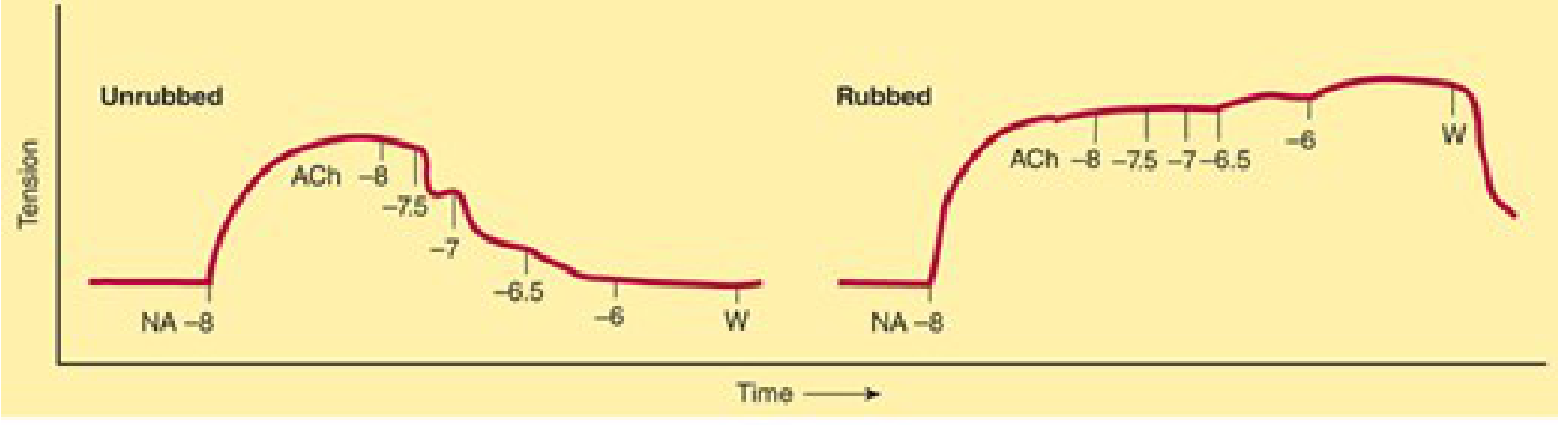
| Condition | Effect of ACh on Blood Vessel |
|---|---|
| Endothelium INTACT (unrubbed) | RELAXATION (vasodilation) - via NO/cGMP |
| Endothelium REMOVED (rubbed) | CONTRACTION (vasoconstriction) - direct M3 on smooth muscle → IP3 → ↑Ca²⁺ |
Key clinical implication: In atherosclerosis, where the endothelium is damaged, ACh (or parasympathetic stimulation) paradoxically causes vasoconstriction instead of vasodilation - a mechanism contributing to angina in diseased coronary arteries.
Additional Mechanisms Contributing to Hypotension
Beyond the NO pathway, muscarinic agonists also lower BP via:
- ↓ Heart rate (M2 on SA node) - bradycardia → ↓ cardiac output
- ↓ AV node conduction (M2) - negative dromotropy
- ↓ Cardiac contractility (M2) - negative inotropy → ↓ cardiac output
- The fall in BP then triggers reflex sympathetic discharge (baroreceptor reflex), which can partially oppose the bradycardia - making the net heart rate effect complex.
Pilocarpine Exception (Exam Favourite)
Pilocarpine IV produces:
- Brief hypotension (M receptor - NO pathway as above)
- Followed by paradoxical hypertension - because it also activates M1 receptors in sympathetic ganglia, causing sympathetic ganglion firing → NE release → vasoconstriction
Sources: Katzung's Basic and Clinical Pharmacology 16th Ed. | Ganong's Review of Medical Physiology 26th Ed. | Barash Clinical Anesthesia 9th Ed.
Myasthenia gravis mbbs 1st year pharmacology
Myasthenia gravis is an autoimmune disease where antibodies destroy nicotinic (Nm) receptors at the NMJ, causing muscle weakness. The pharmacological treatment relies on anticholinesterase drugs (neostigmine, pyridostigmine) that inhibit AChE, allowing ACh to accumulate and overcome the receptor deficit.
Searching sources...
Finding Sources
Finding Sources
Finding Sources
Reading File
Finding Sources
Finding Sources
Finding Sources
Finding Sources
Finding Sources
Finding Sources
Reading File
Finding Sources
I now have all the information needed for a comprehensive MBBS 1st year pharmacology answer. Here it is:
Myasthenia Gravis - MBBS 1st Year Pharmacology
1. DEFINITION
Myasthenia Gravis (MG) is an acquired autoimmune disease of the neuromuscular junction (NMJ) characterized by:
- Production of antibodies against nicotinic acetylcholine receptors (nAChR / Nm receptors) on the postsynaptic (skeletal muscle) membrane
- Progressive loss and destruction of functional AChRs
- Resulting in fatigable skeletal muscle weakness
Name origin: "Myasthenia" = muscle weakness; "Gravis" = severe (Latin)
2. PATHOPHYSIOLOGY - The Core Mechanism
Normal NMJ Transmission (Recap)
Motor nerve terminal
        ↓ Action potential
Vesicles release ACh into synaptic cleft
        ↓
ACh binds Nm (nicotinic) receptors on motor end plate
        ↓
Na⁺ influx → End plate potential (EPP) → Action potential → Muscle contraction
        ↓
ACh hydrolyzed by AChE → choline recycled
What Goes Wrong in MG
- Thymus abnormality (hyperplasia in young women, thymoma in older men) → T-cell sensitization to nAChR α-subunit
- B-cells produce IgG anti-nAChR antibodies (directed mainly at the Main Immunogenic Region / MIR of the α-subunit)
- Antibodies cause:
- Complement activation → destruction of postjunctional folds
- Accelerated receptor internalization/degradation (main mechanism)
- Competitive blockade of ACh binding
- ↓ Surface density of AChRs at the motor end plate
- Miniature End Plate Potential (MEPP) amplitude ↓ (frequency is normal - presynaptic ACh release is intact)
- The EPP no longer reaches threshold → muscle contraction fails
Key electrophysiology: In MG, MEPP frequency = normal (vesicle release intact) but MEPP amplitude = reduced (fewer receptors to respond)
3. CLINICAL FEATURES (Pharmacology-relevant)
| Feature | Detail |
|---|---|
| Hallmark | Fatigable weakness - worse with repetition/end of day, better with rest |
| First muscles affected | Extraocular muscles → ptosis (drooping eyelid), diplopia (double vision) |
| Bulbar involvement | Dysarthria (slurred speech), dysphagia (difficulty swallowing) |
| Generalized form | Neck, shoulder, proximal limb, respiratory muscles |
| Life-threatening | Respiratory muscle paralysis - myasthenic crisis |
| Bimodal age | Young women (20s) with thymic hyperplasia; Older men (60s) with thymoma |
4. PHARMACOLOGICAL BASIS OF TREATMENT
There are two main pharmacological strategies:
| Strategy | Goal | Drugs |
|---|---|---|
| A. Symptomatic | ↑ ACh at NMJ to overcome receptor deficit | Anticholinesterases (AChE inhibitors) |
| B. Disease-modifying | Reduce the autoimmune attack | Corticosteroids, immunosuppressants, IVIG, plasmapheresis |
Plus surgical: Thymectomy (removes source of autosensitization)
5. ANTICHOLINESTERASE DRUGS (1st Year Pharmacology Core)
Mechanism of Action
Normally: ACh → AChE → Choline + Acetate (ACh destroyed)

In MG treatment:
Anticholinesterase drug → INHIBITS AChE
         ↓
ACh accumulates in synaptic cleft
         ↓
More ACh available to compete with antibodies for remaining receptors
         ↓
More receptors activated → muscle contraction restored
Classification of Anticholinesterases Used in MG
| Drug | Type of inhibition | Duration | Route | Special points |
|---|---|---|---|---|
| Neostigmine | Reversible (carbamylation) | 3-4 hours | Oral / IV / IM | Drug of choice for acute/diagnosis; also reverses NMJ block post-surgery |
| Pyridostigmine | Reversible (carbamylation) | 4-6 hours | Oral | Drug of choice for long-term management of MG; longer acting, better tolerated |
| Edrophonium | Reversible (electrostatic - very brief) | 2-10 minutes | IV only | Used for Tensilon test (diagnosis) - not treatment |
| Physostigmine | Reversible (carbamylation) | 3-4 hours | - | Crosses BBB - NOT used in MG (CNS effects) |
| Ambenonium | Reversible | Longer | Oral | Alternative when neostigmine/pyridostigmine cause GI side effects |
Mechanism of Inhibition - How Anticholinesterases Work
- Neostigmine/Pyridostigmine: Carbamylate the serine residue at the esteratic site of AChE → forms carbamyl-enzyme complex → enzyme slowly regenerates over hours (reversible but slow)
- Edrophonium: Binds only by electrostatic interaction to the anionic site of AChE → very brief, easily displaced → lasts only minutes
6. EDROPHONIUM (TENSILON) TEST - Diagnosis of MG
Principle: If weakness is due to MG (too little ACh at NMJ), giving a short-acting AChE inhibitor temporarily improves weakness.
Procedure
- Give edrophonium 2 mg IV initially (test dose)
- If no adverse reaction → give 8 mg IV (total 10 mg)
- Observe for 30-60 seconds
Interpretation
| Result | Meaning |
|---|---|
| Marked improvement in weakness (e.g., ptosis lifts) | Myasthenic crisis - not enough ACh → give more anticholinesterase |
| Worsening weakness (increased paralysis) | Cholinergic crisis - too much ACh (from anticholinesterase overdose) → STOP anticholinesterases, give Atropine |
| No change | Equivocal - consider other diagnosis |
Why edrophonium? Its 2-10 minute duration means any adverse effects are brief and self-limiting - it is safe for diagnostic use.
7. TWO CRISES IN MG - Must Know Distinction
| Feature | Myasthenic Crisis | Cholinergic Crisis |
|---|---|---|
| Cause | Under-treatment (too little AChE inhibitor) or disease exacerbation | Overdose of anticholinesterase |
| Mechanism | ↓↓ ACh at NMJ (antibodies overwhelming) | ↑↑ ACh everywhere → depolarization block at NMJ |
| Muscle weakness | Yes (from under-stimulation) | Yes (from over-stimulation → depolarization block) |
| Muscarinic side effects | Absent | SLUDGE: Salivation, Lacrimation, Urination, Defecation, GI cramps, Emesis; also bradycardia, miosis |
| Edrophonium test | Weakness improves | Weakness worsens |
| Treatment | Increase anticholinesterase dose + ventilatory support | Stop anticholinesterase + Atropine (blocks muscarinic effects) + ventilatory support |
SLUDGE mnemonic = classic muscarinic overdose symptoms from excess ACh
8. DRUGS THAT WORSEN MYASTHENIA GRAVIS (High-yield)
These drugs must be avoided in MG patients:
| Drug Class | Examples | Mechanism of Worsening |
|---|---|---|
| Aminoglycoside antibiotics | Gentamicin, Streptomycin, Neomycin | Inhibit presynaptic ACh release (Ca²⁺ antagonism) |
| Fluoroquinolones | Ciprofloxacin, Azithromycin | Block NMJ |
| Beta-blockers | Propranolol | Reduce NMJ safety margin |
| Calcium channel blockers | Verapamil | ↓ presynaptic ACh release |
| Class Ia/Ib antiarrhythmics | Quinidine, Procainamide, Lidocaine | Membrane stabilization, block NMJ |
| Neuromuscular blockers | Succinylcholine, Vecuronium | Direct NMJ blockers - extreme sensitivity in MG |
| Corticosteroids | Prednisolone | Paradoxically worsen weakness initially before improving |
| Phenytoin, Lithium | CNS drugs | Impair NMJ transmission |
| Magnesium | Mg²⁺ | ↓ presynaptic ACh release |
9. DISEASE-MODIFYING TREATMENTS
Immunosuppressants (Reduce antibody production)
- Corticosteroids (Prednisolone) - 1st line immunotherapy; initial dose can paradoxically worsen weakness (start low, go slow)
- Azathioprine - steroid-sparing agent; delayed onset (months)
- Mycophenolate mofetil, Cyclosporine, Tacrolimus - alternatives
Rapid Short-term Immunotherapy
- Plasmapheresis (plasma exchange) - physically removes anti-AChR antibodies from blood; effect in days, lasts weeks
- IV Immunoglobulins (IVIG) - modulates immune response; similar speed/duration to plasmapheresis
- Both used for myasthenic crisis or pre-operative preparation
Biologic
- Eculizumab (complement inhibitor) - approved for generalized MG; blocks C5 complement activation
Surgical
- Thymectomy - removes thymus (source of autosensitization); improves ~75% of thymoma cases and benefits non-thymoma generalized MG
10. SUMMARY TABLE - Key Drugs at a Glance
| Drug | Class | Use in MG | Mechanism | Key Point |
|---|---|---|---|---|
| Pyridostigmine | AChE inhibitor | Symptomatic (1st line, long-term) | Carbamylation of AChE → ↑ ACh | Drug of choice for maintenance |
| Neostigmine | AChE inhibitor | Symptomatic / IV acute use | Carbamylation of AChE | Does NOT cross BBB |
| Edrophonium | AChE inhibitor | Diagnosis only (Tensilon test) | Electrostatic inhibition | Lasts only 2-10 min |
| Atropine | Muscarinic antagonist | Antidote in cholinergic crisis | Blocks M receptors | Does NOT help muscle weakness (no effect on Nm) |
| Prednisolone | Corticosteroid | Disease-modifying | ↓ anti-AChR antibody production | Can initially worsen MG |
| Azathioprine | Immunosuppressant | Disease-modifying | ↓ lymphocyte proliferation | Delayed onset |
| Eculizumab | Biologic (anti-C5) | Severe generalized MG | ↓ complement destruction of AChR | Expensive, newer |
11. MEMORY AIDS
- Why neostigmine doesn't cross BBB? - It is a quaternary ammonium compound (charged at physiological pH) → cannot cross lipid bilayer of BBB
- Physostigmine DOES cross BBB - it is a tertiary amine (uncharged) → used for atropine/anticholinergic overdose in CNS, NOT for MG
- Pyridostigmine preferred over neostigmine for MG - longer duration (4-6 h vs 3-4 h), less muscarinic side effects
- "Point and Shoot" - remember NMJ = Nm receptor; atropine blocks M (muscarinic) but NOT Nm → atropine treats the SLUDGE symptoms of cholinergic crisis but does NOT reverse the muscle weakness
Sources: Medical Physiology (Boron & Boulpaep) | Morgan & Mikhail's Clinical Anesthesiology 7th Ed. | Katzung's Basic and Clinical Pharmacology 16th Ed.
This is a shared conversation. Sign in to Orris to start your own chat.