
New category Mullerian anomaly category PG exam 30 marks obs gynec
Mullerian anomaly new category classification PG exam obstetrics gynecology 2024 2025
https://www.asrm.org/practice-guidance/practice-committee-do…
Mullerian duct anomaly classification diagram uterus types
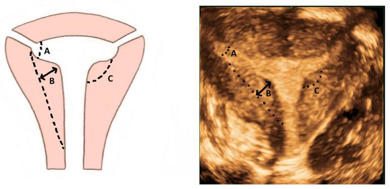
This composite image includes a diagram and a corresponding 3D ultrasound image illustrating the diagnostic criteria for a T-shaped uterus, a type of Mullerian duct anomaly (Class U1). The visualization focuses on the Congenital Uterine Malformation Experts (CUME) criteria in the coronal plane. Both images are annotated with three key measurable parameters: 'A' denotes the T-angle (typically ≤ 40°), formed by the intersection of the lateral borders of the uterine cavity; 'B' indicates the lateral indentation depth (typically ≥ 7 mm), measuring the thickness and inward protrusion of the lateral myometrial walls; and 'C' represents the lateral indentation angle (typically ≤ 130°). The 3D ultrasound demonstrates a constricted, narrow endometrial cavity with thickened lateral walls and a characteristic T-like morphology, contrasting with the normal triangular uterine cavity. This content is intended for medical education in gynecology, reproductive endocrinology, and radiology to improve the objective classification of dysmorphic uteri.
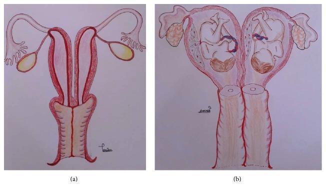
This anatomical schematic diagram illustrates uterus didelphys, a Müllerian duct anomaly, in two states. Image (a) depicts a nongravid (non-pregnant) uterus didelphys, showing two completely separate uterine horns and two distinct cervices. The illustration also shows accompanying fallopian tubes and ovaries. Image (b) presents a gravid (pregnant) uterus didelphys with a dicavitary twin pregnancy. In this state, each independent uterine cavity contains a developing fetus in a cephalic presentation, along with its own placenta and umbilical cord. This comparison highlights the structural duplication of the reproductive tract and the rare clinical scenario of simultaneous gestation in both separate cavities. The diagram is designed for educational use in obstetrics and gynecology to explain congenital uterine malformations and their implications for multifetal pregnancy.
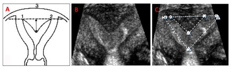
This composite image illustrates the 3-D ultrasound evaluation and measurement of a uterine subseptation, a common Müllerian duct anomaly. Panel A presents a schematic diagram of a subseptate uterus, labeling the left (1) and right (2) tubal ostia and the fundal outer contour (3). A vertical arrow demonstrates the measurement of subseptation length from the inter-ostial line to the apex of the septum. Panel B displays a 3-D ultrasound coronal view rendering, showing the characteristic Y-shaped indentation of the endometrial cavity with a convex fundal external contour. Panel C demonstrates the clinical measurement technique on the ultrasound rendering; dotted lines delineate the cavity width (between the tubal ostia), the subseptation width at its base, and the subseptation length. These standardized measurements are critical for distinguishing between arcuate and septate uteri and for calculating the cavity area, which informs surgical planning for hysteroscopic incision.
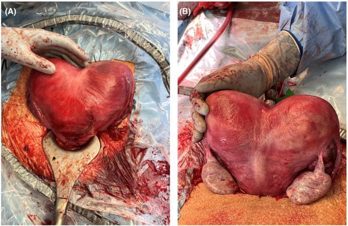
Clinical photograph of a human uterus post-cesarean delivery and hysterotomy repair, demonstrating a bicornuate bicollis anomaly (ASRM classification). The composite image shows (A) an anterior view and (B) a posterior view of the heart-shaped uterus. A deep midline serosal indentation exceeding 1 cm is clearly visible at the fundus, indicative of failed fusion of the Müllerian ducts. The uterine serosa appears erythematous and congested, a physiological finding typical of immediate postpartum and surgical manipulation. In the posterior view (B), both ovaries and fallopian tubes are visible flanking the two distinct uterine horns. The image demonstrates the surgical appearance of a dicavitary uterine anomaly following a twin pregnancy. It serves as an educational reference for Mullerian duct anomalies and their intraoperative presentation during obstetric surgery.
ASRM 2021 Mullerian anomaly nine categories classification table
Clinical photograph of a human uterus post-cesarean delivery and hysterotomy repair, demonstrating a bicornuate bicollis anomaly (ASRM classification). The composite image shows (A) an anterior view and (B) a posterior view of the heart-shaped uterus. A deep midline serosal indentation exceeding 1 cm is clearly visible at the fundus, indicative of failed fusion of the Müllerian ducts. The uterine serosa appears erythematous and congested, a physiological finding typical of immediate postpartum and surgical manipulation. In the posterior view (B), both ovaries and fallopian tubes are visible flanking the two distinct uterine horns. The image demonstrates the surgical appearance of a dicavitary uterine anomaly following a twin pregnancy. It serves as an educational reference for Mullerian duct anomalies and their intraoperative presentation during obstetric surgery.
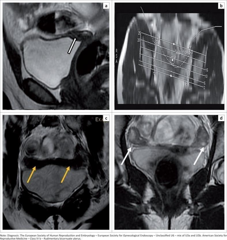
This composite of four magnetic resonance imaging (MRI) panels illustrates the diagnostic evaluation of a Müllerian Duct Anomaly (MDA), specifically a rudimentary bicornuate uterus (ASRM Class IVb). (a) Sagittal T2-weighted image shows a small, rudimentary uterus (white arrow) with lack of normal endometrial-myometrial differentiation. (b) A planning scan demonstrates the technical setup for a T2 CUBE curved Maximum Intensity Projection (MIP) sequence, designed to follow the anatomical curvature of the pelvic structures. (c) The resulting T2 CUBE curved MIP clearly delineates the bicornuate morphology, with yellow arrows highlighting two widely separated uterine horns and an intercornual distance exceeding 4 cm. (d) Coronal T2-weighted image identifies both ovaries (white arrows), appearing as characteristic oval structures lateral to the midline. The imaging illustrates critical diagnostic features for classification under ESHRE-ESGE (Unclassified U6) and ASRM systems, emphasizing the utility of volume sequences and MIP reformats in assessing complex pelvic anatomy and fusion defects.
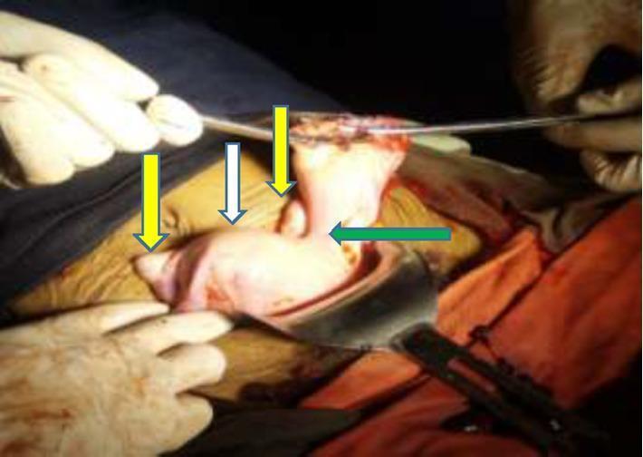
This clinical photograph shows an intraoperative view of a female reproductive system following the resection of a ruptured ectopic pregnancy. The image illustrates a congenital Mullerian duct anomaly, specifically a unicornuate uterus with a non-communicating rudimentary horn. A white arrow identifies the main unicornuate uterine body, while a green arrow points to the left-sided rudimentary horn. The connection between the two structures is visible as a bridge of dense fibro-muscular tissue. Yellow arrows indicate the bilateral ovaries, which appear in their relative anatomical positions. The tissues are fleshy and vascularized, consistent with a surgical environment. This case demonstrates the anatomical relationships associated with Class II Mullerian anomalies according to the American Society for Reproductive Medicine (ASRM) classification, highlighting the risk of obstetric complications such as rudimentary horn pregnancy and rupture.
Mullerian Anomalies - New Classification (ASRM 2021) | PG Exam 30 Marks
Introduction (2 marks)
Classification Systems - Overview (3 marks)
| Classification | Year | No. of Categories | Key Feature |
|---|---|---|---|
| AFS (American Fertility Society) | 1988 | 7 classes (I-VII) | Uterus only, no cervix/vagina |
| ESHRE/ESGE | 2013 | U0-U6 + C0-C4 + V0-V4 | Uterus, cervix, vagina separately |
| ASRM MAC | 2021 | 9 descriptive categories | Uterus + cervix + vagina, no Roman numerals |
ASRM 2021 MAC - 9 NEW CATEGORIES (Exam High Yield) (8 marks)
Category 1 - Mullerian Agenesis
- Previously: AFS Class I (Hypoplasia/Agenesis)
- Complete absence of uterus, cervix, and upper vagina
- Mayer-Rokitansky-Kuster-Hauser (MRKH) syndrome - most important subtype
- 46,XX karyotype, normal secondary sexual characters, primary amenorrhea
- Ovaries normal - so no hormonal deficiency
- Management: Vaginal dilation (Frank's progressive dilation) - first line; surgical creation of neovagina (McIndoe procedure, Vecchietti procedure)
Category 2 - Cervical Agenesis (new separate category)
- Isolated cervical agenesis or stenosis
- Was poorly classified in AFS 1988
- Presents with cryptomenorrhea, cyclic pain, primary amenorrhea
- MRI: normal uterus with absent/stenotic cervix
- Management: Cervical reconstruction or hysterectomy
Category 3 - Unicornuate Uterus
- Previously: AFS Class II
- Failure of one Mullerian duct to develop
- Banana/cigar shaped uterus on imaging
- Rudimentary horn (contralateral) - may be:
- Communicating (with endometrium)
- Non-communicating (with endometrium) - HIGH RISK of rudimentary horn ectopic pregnancy
- Without cavity
- Absent
- Obstetric risk: preterm labor, malpresentation, intrauterine growth restriction (IUGR)
- Associated ipsilateral renal agenesis (renal tract anomalies in ~40%)
Category 4 - Uterus Didelphys
- Previously: AFS Class III
- Complete failure of fusion - two separate uteri, two cervices, often longitudinal vaginal septum
- Bicornis bicollis bipartite appearance
- Best reproductive outcomes among fusion anomalies
- Wunderlich syndrome: didelphys + unilateral obstructed vagina + ipsilateral renal agenesis (triad)
- Obstetric complication: highest rate of preterm birth among MDAs
Category 5 - Bicornuate Uterus
- Previously: AFS Class IV
- Partial fusion failure - heart-shaped uterus with fundal cleft > 1 cm
- Bicornuate unicollis (one cervix) - more common
- Bicornuate bicollis (two cervices) - rare
- Surgical correction (Strassman metroplasty) for recurrent pregnancy loss
- Differentiation from septate uterus is critical (fundal indentation on MRI/3D USG)
Category 6 - Septate Uterus (Most Common MDA)
- Previously: AFS Class V
- Most common MDA (~45%) - but most common TREATABLE cause of recurrent miscarriage
- Failure of resorption of midline septum
- Fundal contour: convex/flat/minor indentation (< 1 cm) externally
- Septum internally: fibrous or fibromuscular - avascular, poor implantation site
- ASRM 2021 key change: Internal indentation > 1 cm (previously > 1.5 cm in older criteria) defines septate uterus
- Complete septate: septum reaches internal or external os
- Partial/subseptate: does not reach os
- Arcuate uterus (ASRM 2021): internal indentation < 1 cm, angle of divergence > 90 degrees - considered normal variant, NOT classified as MDA
- Treatment: Hysteroscopic metroplasty (transcervical resection of septum) - gold standard
Category 7 - Longitudinal Vaginal Septum (LVS)
- NEW standalone category in ASRM 2021 (was not separately classified in AFS 1988)
- May occur with or without other uterine anomalies
- Obstructed hemivagina (Herlyn-Werner-Wunderlich) presents with dysmenorrhea post-menarche
- Treatment: Surgical excision of septum
Category 8 - Transverse Vaginal Septum
- Failure of canalization between Mullerian tubercle and urogenital sinus
- Level: Upper (46%), middle (35%), lower (19%) vagina
- Presents: Cryptomenorrhea, primary amenorrhea, cyclic pain - can mimic imperforate hymen
- Differentiation from imperforate hymen: transverse septum has no bluish bulge, thicker
- Management: Surgical excision + vaginal reconstruction
Category 9 - Complex Anomalies
- Anomalies that do not fit neatly into any single category
- Combinations of above anomalies
- DES (diethylstilbestrol) exposure: T-shaped uterus (ESHRE U1c) - included here
AFS 1988 Classification (Old, Still Tested) (4 marks)
| Class | Type | Description |
|---|---|---|
| I | Hypoplasia/Agenesis | Vaginal/cervical/fundal/tubal/combined |
| II | Unicornuate | With/without rudimentary horn |
| III | Didelphys | Two complete uteri |
| IV | Bicornuate | Partial/complete |
| V | Septate | Partial/complete |
| VI | Arcuate | Minor fundal indentation (now considered normal by ASRM 2021) |
| VII | DES-related | T-shaped uterus |
ESHRE/ESGE 2013 Classification - Key Points (3 marks)
- U0 = Normal
- U1 = Dysmorphic (T-shaped U1a, Infantilis U1b, Others U1c including arcuate)
- U2 = Septate (partial U2a, complete U2b)
- U3 = Bicorporeal/Bicornuate (partial U3a, complete U3b, bicorporeal septate U3c)
- U4 = Hemi-uterus/Unicornuate (with rudimentary cavity U4a, without U4b)
- U5 = Aplastic (with rudimentary cavity U5a, without U5b)
- U6 = Unclassified
Embryology (3 marks)
| Stage | Event | Anomaly if Disrupted |
|---|---|---|
| 6th week | Mullerian ducts form lateral to Wolffian ducts | Agenesis (MRKH) |
| 8-10th week | Caudal migration and fusion of Mullerian ducts | Uterus didelphys, bicornuate |
| 9-11th week | Midline fusion | Septate uterus |
| 11-12th week | Canalization/resorption of septum | Septate, transverse vaginal septum |
| Contact with urogenital sinus | Vaginal formation | Transverse vaginal septum |
- Lateral fusion defects = didelphys, bicornuate (external cleft)
- Vertical fusion defects = transverse vaginal septum
- Resorption defects = septate uterus (no external cleft)
Diagnosis (3 marks)
- Fundal contour assessment (bicornuate vs. septate)
- Complex anomalies
- Soft tissue characterization
| Feature | Bicornuate | Septate |
|---|---|---|
| Fundal cleft (external) | > 1 cm | < 1 cm (convex/flat) |
| Intercornual angle (HSG) | > 105 degrees | < 75 degrees |
| Intercornual distance | > 4 cm | 2-4 cm (normal) |
| Septum tissue | Myometrium (intermediate T2) | Fibrous (T2 hypointense) or muscular |
| Treatment | Strassman metroplasty (laparotomy) | Hysteroscopic resection |
- Sonohysterography (SHG) - outlines cavity well
- Hysteroscopy - direct visualization, therapeutic
- Laparoscopy - external contour, combined with hysteroscopy for definitive diagnosis
Clinical Presentation & Associations (2 marks)
- Primary amenorrhea: MRKH, transverse vaginal septum, cervical agenesis
- Cyclic pain / cryptomenorrhea: obstructed hemivagina, transverse vaginal septum
- Recurrent pregnancy loss (RPL): Septate uterus (most common treatable cause), bicornuate
- Preterm labor / IUGR: All fusion anomalies, especially didelphys
- Malpresentation: Unicornuate, bicornuate
- Infertility: Septate uterus, MRKH
- Renal anomalies: Unicornuate (ipsilateral renal agenesis ~40%), MRKH
Management Summary (2 marks)
| Anomaly | Treatment |
|---|---|
| MRKH | Vaginal dilation (Frank) / McIndoe procedure; surrogacy for fertility |
| Unicornuate + rudimentary horn (with endometrium) | Laparoscopic excision of rudimentary horn |
| Uterus didelphys | Usually no surgery; cerclage if needed |
| Bicornuate | Strassman metroplasty (for RPL) |
| Septate uterus | Hysteroscopic metroplasty (Gold standard) |
| Longitudinal vaginal septum | Surgical excision |
| Transverse vaginal septum | Surgical excision + end-to-end vaginal anastomosis |
High-Yield Exam Points (must-know) (Bonus)
- Most common MDA = Septate uterus (~45%)
- Most common cause of treatable RPL = Septate uterus
- Arcuate uterus = normal variant in ASRM 2021 (no longer a class)
- DES uterus = T-shaped = now Category 9 (Complex) in ASRM 2021
- ASRM 2021 key change: Dropped Roman numeral classes; uses 9 descriptive categories; now includes cervix and vagina; Arcuate removed as a pathological entity
- Longitudinal Vaginal Septum = new standalone Category 7 in ASRM 2021
- MRKH: 46,XX, normal ovaries, normal secondary sex characters, primary amenorrhea, absent uterus/upper vagina
- Best reproductive outcome among uterine anomalies = Didelphys
- Renal agenesis association = Unicornuate uterus (ipsilateral), MRKH (unilateral), Didelphys + obstructed hemivagina (Wunderlich triad)
- Gold standard diagnosis = 3D USG (first-line) + MRI (definitive)
With diagram for exam which can draw
Medical exam diagram showing Mullerian duct anomalies classification with simple line drawings of uterus shapes. Show 9 types in a clean grid layout with labels: 1) Normal Uterus (triangular cavity, convex fundus), 2) Mullerian Agenesis/MRKH (absent uterus, short vagina), 3) Unicornuate Uterus (banana-shaped, one horn, small rudimentary horn on other side), 4) Uterus Didelphys (two complete separate uteri side by side, two cervices), 5) Bicornuate Uterus (heart-shaped, deep fundal cleft >1cm, two horns), 6) Septate Uterus (convex fundus externally, internal septum dividing cavity), 7) Arcuate Uterus (minor internal indentation <1cm, normal variant), 8) Longitudinal Vaginal Septum (normal uterus with vertical septum in vagina), 9) Transverse Vaginal Septum (horizontal bar blocking vaginal canal). Each uterus drawn as simple black outline diagram like a medical textbook hand-drawn style. Include small key differences: Bicornuate vs Septate - external vs internal indentation. White background, clean black lines, suitable for hand-drawing in exam. Title at top: ASRM 2021 Mullerian Anomaly Classification - 9 Categories
Simple medical exam hand-drawn style diagram: Bicornuate uterus versus Septate uterus differentiation. Two large uterus diagrams side by side. LEFT side labeled "BICORNUATE UTERUS": heart-shaped external contour with deep V-shaped fundal cleft greater than 1cm shown with arrow, two separate horns visible externally, internal cavity divided, label showing "External indentation >1cm", "Two horns externally visible", "Intercornual angle >105 degrees on HSG", treatment label "Strassman Metroplasty". RIGHT side labeled "SEPTATE UTERUS (Most Common MDA)": smooth convex outer fundal contour, internal septum shown as shaded wedge dividing cavity labeled "Internal septum only", "Fundus smooth externally", "Intercornual angle <75 degrees on HSG", treatment label "Hysteroscopic Resection - Gold Standard". Arrow in middle pointing to both labeled KEY DIFFERENCE: External fundal contour. Below both: small inset showing HSG appearance of each. Clean black line art on white background, textbook style suitable for drawing in exam hall.
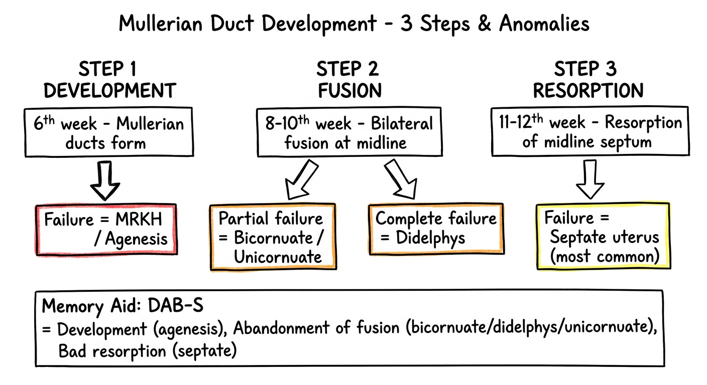
Medical exam flowchart diagram: Embryological basis of Mullerian anomalies. Simple flow diagram with boxes and arrows. Title: "Mullerian Duct Development - 3 Steps & Anomalies". Three columns labeled STEP 1 DEVELOPMENT, STEP 2 FUSION, STEP 3 RESORPTION. Step 1 box: "6th week - Mullerian ducts form" with arrow going down to "Failure = MRKH / Agenesis" shown in red box. Step 2 box: "8-10th week - Bilateral fusion at midline" with arrow to "Partial failure = Bicornuate / Unicornuate" in orange box, "Complete failure = Didelphys" in orange box. Step 3 box: "11-12th week - Resorption of midline septum" with arrow to "Failure = Septate uterus (most common)" in yellow box. Below the flowchart, a simple mnemonic box: "Memory Aid: DAB-S = Development (agenesis), Abandonment of fusion (bicornuate/didelphys/unicornuate), Bad resorption (septate)". Clean black and white with minimal color coding, hand-drawable exam style, white background.
Diagram 1 - ASRM 2021: All 9 Categories
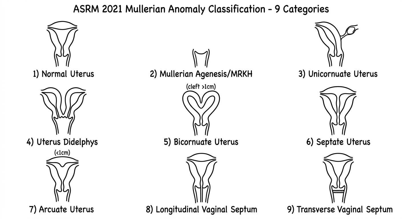
Diagram 2 - Most Important: Bicornuate vs Septate (Most Tested)
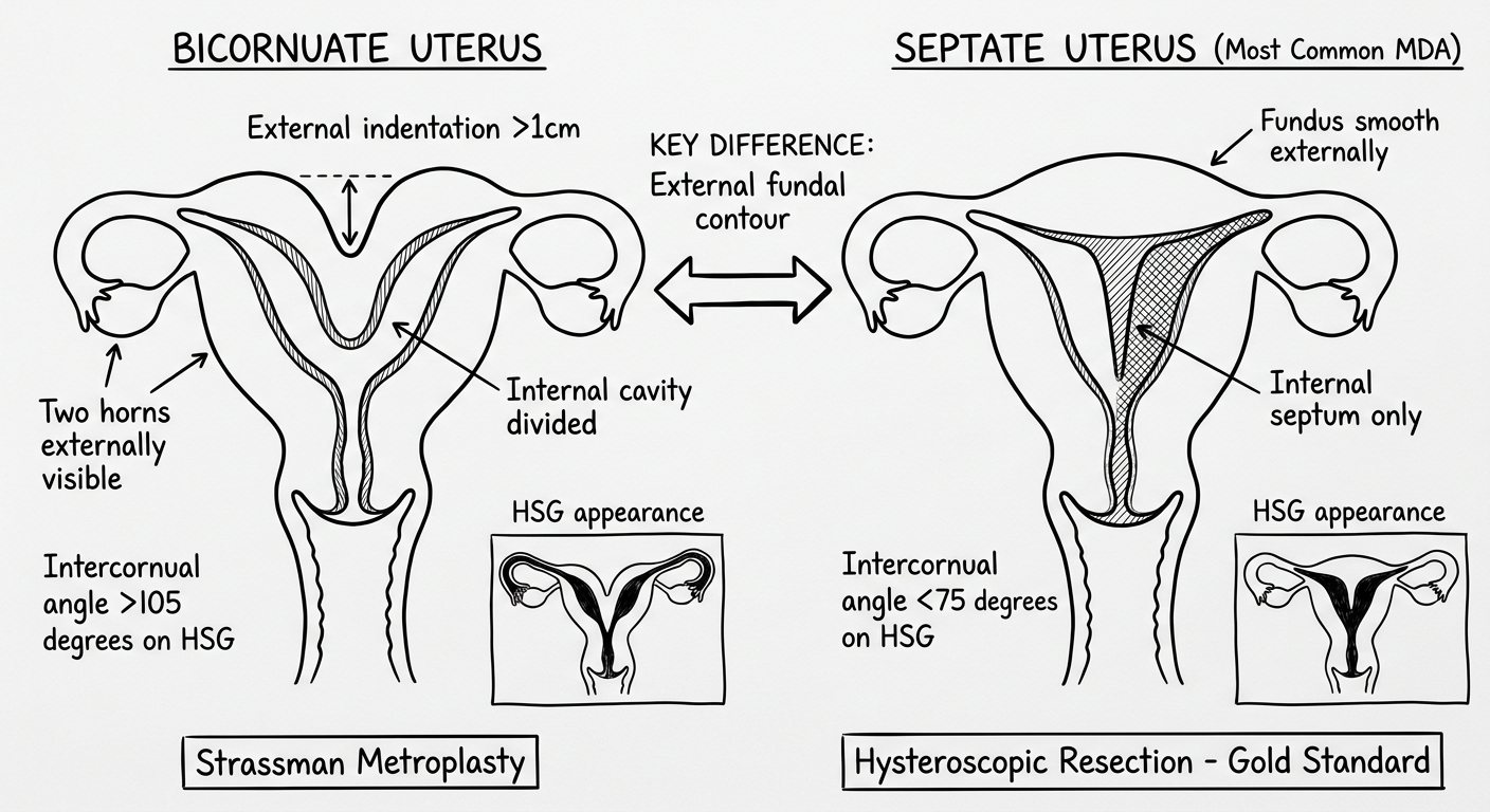
Diagram 3 - Embryological Basis (Flowchart)
How to Draw in Exam (Step-by-Step)
Draw Diagram 1 - ASRM 2021 Nine Types (5 minutes)
Box 1 - NORMAL          Box 2 - MRKH            Box 3 - UNICORNUATE
△ triangular cavity     No uterus, short        Banana shape, one
convex fundus           vaginal pouch           horn, tiny rudiment

Box 4 - DIDELPHYS       Box 5 - BICORNUATE      Box 6 - SEPTATE
Two Ψ side by side      Heart shape ♥           Smooth fundus △
Two cervices            External cleft >1cm     Internal septum ↓

Box 7 - ARCUATE         Box 8 - LVS             Box 9 - COMPLEX
Slight dip inside       Normal uterus +         T-shaped (DES)
Normal variant          Vertical vaginal line   or combinations
Draw Diagram 2 - Bicornuate vs Septate (Most Important - 3 minutes)
- Draw a heart ♥ shape for the external outline
- Mark the V-notch at top: write ">1 cm cleft"
- Draw two internal cavity lines
- Write below: "Strassman metroplasty"
- Draw a smooth dome (inverted U) for external outline
- Draw a downward internal wedge (shaded) from fundus
- Write "Internal septum only" with arrow
- Write below: "Hysteroscopic resection - Gold standard"
- "KEY: Look at the OUTSIDE (fundal contour)"
Draw Diagram 3 - Embryology Flow (2 minutes)
[Box 1] 6th week: Mullerian ducts FORM
         ↓ Failure here → MRKH/Agenesis

[Box 2] 8-10th week: Ducts FUSE
         ↓ Partial failure → Unicornuate / Bicornuate
         ↓ Complete failure → Didelphys

[Box 3] 11-12th week: Septum RESORBS
         ↓ Failure → Septate uterus (most common!)
Memory Tricks for Exam
| Mnemonic | Meaning |
|---|---|
| "Septate = Surgical via Scope" | Hysteroscopic resection |
| "Bicornuate = Big cut (Laparotomy)" | Strassman metroplasty |
| "D-U-B-S" | Development (agenesis) → Unfused (didelphys) → Bicornuate → Septate - from severe to mild |
| "MRKH = May Run, Karyotype Healthy" | 46,XX, no uterus, normal ovaries |
| "Arcuate = A-OK" | Normal variant in ASRM 2021, not a disease |
| "Unicornuate + Renal" | Always look for ipsilateral renal agenesis (~40%) |
| "Most Common = Septate, Most Dangerous complication = Rudimentary horn ectopic" | RPL vs surgical emergency |