
Guidelines for acute on chronic liver failure
acute-on-chronic liver failure management guidelines
ACLF acute on chronic liver failure guidelines 2024 2025 EASL AASLD
PMID: 37364789
PMID: 37939273
https://pmc.ncbi.nlm.nih.gov/articles/PMC12721767
Acute-on-Chronic Liver Failure (ACLF) - Clinical Guidelines
Definition & Overview
- Underlying chronic liver disease (almost always with cirrhosis)
- An identifiable or cryptic precipitating event
- Hepatic and/or extrahepatic organ failure
- Excessive systemic inflammatory response
Definitions Across Major Guidelines
| Criterion | EASL-CLIF (2023) | AASLD (2024) | APASL |
|---|---|---|---|
| Underlying disease | Cirrhosis | Cirrhosis | Chronic liver disease ± cirrhosis |
| Organ failure assessment | CLIF-C OF score | CLIF-C, NACSELD | AARC score |
| ACLF types | Grade 1-3 (by OF count) | -- | Type I (no extrahepatic OF), Type II (with extrahepatic OF) |
| Extrahepatic OF required | Yes | Yes | Not required for Type I |
EASL-CLIF Grading System
| Grade | Definition | 28-day Mortality |
|---|---|---|
| ACLF-1 | 1 renal OF, OR 1 non-renal OF + renal dysfunction/HE | ~22% |
| ACLF-2 | 2 organ failures | ~32-45% |
| ACLF-3 | 3 or more organ failures | ~73-77% |
| No ACLF | AD cirrhosis without OF | ~1.9% |
- Liver: Bilirubin ≥12 mg/dL
- Kidneys: Creatinine ≥2.0 mg/dL (or renal replacement therapy)
- Brain: HE grade 3-4 (West Haven)
- Coagulation: INR ≥2.5
- Circulation: Vasopressor use
- Respiratory: PaO2/FiO2 <200 or SpO2/FiO2 <214
Precipitating Events
- Alcohol-associated hepatitis (most common in the West)
- HBV reactivation or new viral hepatitis (HEV, HAV) - most common in Asia
- Drug-induced liver injury (DILI)
- Autoimmune hepatitis flare
- Bacterial/fungal infection and sepsis (most common overall)
- GI bleeding
- Post-operative state
- Large-volume paracentesis without albumin
Diagnostic Workup
- Assess organ failure: CLIF-C OF score, SOFA score
- Prognostic scoring: CLIF-C ACLF score (best validated), MELD/MELD-Na, NACSELD-ACLF score, Child-Pugh
- Look for precipitants: Blood/urine/ascitic fluid cultures, viral hepatitis serology, alcohol history, drug review
- Imaging: Abdominal US with Doppler (portal vein thrombosis, hepatic architecture), CXR
- Diagnostic paracentesis: If ascites present (cell count, culture, albumin, total protein)
Management - General Principles (EASL 2023 / AASLD 2024)
1. Triage and ICU Admission
- All ACLF-2 and ACLF-3 patients should be admitted to ICU
- ACLF-1 may be managed in high-dependency unit with close monitoring
- Reassess organ failure status daily for the first 3-7 days
- If no improvement or deterioration after 3-7 days of full ICU support with 4+ organ failures and no transplant option - consider transition to palliative care
2. Treat the Precipitating Event
| Precipitant | Treatment |
|---|---|
| Bacterial infection | Empiric broad-spectrum antibiotics (piperacillin-tazobactam or cefepime; add fluconazole/echinocandin for fungal risk); guided by cultures |
| Alcohol-associated hepatitis | Prednisolone 40 mg/day x 28 days (if Maddrey's DF ≥32, no contraindications); assess Lille score at day 7 |
| HBV reactivation | Nucleoside analogue (entecavir or tenofovir) immediately |
| GI bleeding | IV PPI, octreotide/terlipressin, urgent endoscopy; antibiotic prophylaxis (ceftriaxone) |
| DILI | Withdraw offending agent immediately |
3. Infection Prophylaxis and Management (EASL 2023 - Strong Recommendations)
- Infections trigger/worsen ACLF in up to two-thirds of cases
- Routine cultures on admission (blood, urine, ascitic fluid)
- Empiric antibiotics should be started within 1 hour of suspected infection
- Norfloxacin (or rifaximin) for SBP prophylaxis in high-risk patients (low ascitic protein <1.5 g/dL + advanced disease)
- IV albumin 1.5 g/kg day 1, then 1 g/kg day 3 in SBP reduces risk of renal failure and death
4. Organ-Specific Management
- Stop nephrotoxins (NSAIDs, aminoglycosides, contrast)
- Volume resuscitation with albumin (preferred colloid)
- Terlipressin (0.5-1 mg IV q4-6h) + albumin for hepatorenal syndrome type 1 (HRS-AKI) - first-line (EASL Grade A1)
- Norepinephrine may be used as alternative if terlipressin unavailable
- Renal replacement therapy (CRRT preferred) if AKI/HRS unresponsive or volume overload/hyperkalemia/acidosis
- Avoid diuretics in oliguric HRS
- Identify and treat precipitants (infection, bleeding, constipation, sedatives)
- Lactulose 2-3 soft stools/day (titrate dose)
- Rifaximin 550 mg BD for recurrent/persistent HE
- Airway protection/intubation for grade 3-4 HE
- Limit sedation; use dexmedetomidine or propofol over benzodiazepines
- Zinc supplementation (adjunctive)
- Avoid protein restriction (counter-productive)
- Vasopressor of choice: norepinephrine (target MAP ≥65 mmHg)
- Screen for adrenal insufficiency (AASLD 2024) - treat with hydrocortisone 200-300 mg/day if confirmed
- Volume resuscitation: albumin preferred over crystalloids
- Vasopressin/terlipressin as adjuncts in refractory shock
- High-flow nasal oxygen (HFNO) as first-line for moderate hypoxemic respiratory failure
- Non-invasive ventilation (NIV) if tolerated (caution in encephalopathy)
- Invasive mechanical ventilation for severe ARDS or failure of above; lung-protective strategy (6 mL/kg tidal volume)
- Do NOT correct coagulopathy prophylactically (INR not reliable indicator of bleeding risk in cirrhosis - balances pro- and anticoagulant deficits)
- FFP, platelets, cryoprecipitate only for active bleeding or before invasive procedures
- Thromboprophylaxis with LMWH for DVT prevention unless active bleeding or platelets <50,000
5. Nutrition
- Target 35-40 kcal/kg/day, protein 1.2-1.5 g/kg/day (do NOT restrict protein)
- Enteral nutrition preferred; nasogastric feeding if needed
- Small frequent meals + late-evening snack
- BCAA supplementation may be considered
- Early enteral nutrition in ICU patients within 24-48 hours
6. Specific Pharmacological Therapies
- Granulocyte colony-stimulating factor (G-CSF): Promising - shown to decrease mortality in decompensated cirrhosis in some trials; under investigation specifically for ACLF. Some APASL guidance supports its use.
- Albumin infusions: Beyond SBP, chronic albumin administration (PILOT/ATTIRE/ANSWER trials) - role evolving; currently standard in specific indications (HRS, SBP, post-paracentesis)
- NAC: Evidence limited in ACLF; used in DILI-associated precipitants
- Liver support devices (MARS, PROMETHEUS, ELAD): Not yet proven to reduce mortality in RCTs; consider as bridge to transplant in selected centres
Liver Transplantation
Indications (EASL 2023)
- ACLF-1 and ACLF-2: Strong candidates; early listing recommended
- ACLF-3 (3+ OF): Carefully selected patients; short-term post-LT mortality higher but still beneficial vs. no transplant
- Patients with ACLF who fail to improve or deteriorate after 3-7 days of medical therapy but have <3 organ failures should be prioritized for LT
Contraindications / Futility Criteria
- ACLF-3 with ≥4 organ failures: Very high peri-operative mortality; individual assessment required
- CLIF-C ACLF score ≥64 with 3+ organ failures - daily reassessment; if does not improve, transplant may not confer survival benefit
- Active uncontrolled sepsis, multidrug-resistant organisms without coverage
- Severe extra-hepatic disease incompatible with LT
- Alcohol-associated hepatitis: Selected patients without adequate sobriety period (early LT for severe AH shows good outcomes in specialized centers per multiple RCTs)
Scoring for LT listing
- MELD-Na: Primary allocation tool in USA/Europe
- CLIF-C ACLF score: Better short-term mortality predictor in ACLF than MELD
- NACSELD-ACLF: Predicts 30-day mortality based on number of OFs
When to Consider Palliative Care
"If after 3-7 days of ICU support, there continue to be four or more organ failures and liver transplantation is not an option, consideration for a transition to palliative care is recommended."
Monitoring and Reassessment
- Reassess CLIF-C ACLF score daily for first 7 days
- Patients who improve by day 3-7 (reduction in organ failures) have significantly better prognosis
- ACLF resolution (reversal to no ACLF state by day 28) is associated with near-normal survival
- Discharge planning should include:
- Alcohol cessation counseling and treatment
- Rifaximin for secondary HE prophylaxis
- Outpatient LT evaluation if eligible
- Infection surveillance
Summary of Key Guideline Sources
| Guideline | Year | Key Focus |
|---|---|---|
| EASL CPG on ACLF | 2023 | Definition, grading, ICU triage, precipitant management, LT criteria |
| AASLD Practice Guidance | 2024 | ICU management, critically ill cirrhosis, adrenal insufficiency, organ support |
| APASL Kyoto Consensus | 2025 | East-focused; HBV, AARC score, G-CSF, liver regeneration therapies |
| Sleisenger & Fordtran's, Ch. 74 | 2023 | Comprehensive clinical management reference |
| Harrison's Internal Medicine 22e | 2025 | Concise clinical approach and palliative care criteria |
- ACLF is a medical emergency requiring rapid diagnosis, ICU triage, and multidisciplinary team management
- Identify and aggressively treat precipitants (infection first, then alcohol, viral hepatitis)
- Organ-specific support (terlipressin + albumin for HRS, norepinephrine for circulatory failure, lactulose + rifaximin for HE)
- Refer early for liver transplantation - it is the only definitive therapy
- After 3-7 days of maximal ICU care with 4+ organ failures and no transplant option, transition to comfort-focused care is appropriate
- Reassess daily - trajectory of organ failure is as important as the grade at admission
Snake bite guidelines?
snakebite envenomation management guidelines
snakebite envenomation guidelines 2024 2025 WHO antivenom management
https://jts.health.mil/assets/docs/cpgs/Snakebite_Envenomati…
snakebite envenomation clinical features viper elapid
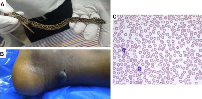
A multi-panel figure documenting a snakebite case. Panel A displays a clinical photograph of a juvenile Russell’s viper (Daboia russelii) held by gloved hands, identified by its characteristic dorsal brown blotches. Panel B is a clinical photograph of a patient's right lower leg and heel showing local envenomation effects, including a prominent dark, hemorrhagic, fluid-filled blister and two adjacent small puncture wounds consistent with fang marks. Panel C shows a diagnostic light microscopy image of a peripheral blood smear (Wright-Giemsa stain). The smear demonstrates normocytic, normochromic erythrocytes with regular morphology and no evidence of fragmentation, schistocytes, or dysmorphic red cells. Scattered multi-lobed leukocytes (neutrophils) are visible against the background of mature red blood cells. The educational focus is on the clinical presentation of viperine envenomation (local tissue destruction) and the exclusion of microangiopathic hemolytic anemia via blood film examination.
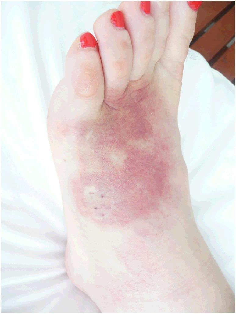
A clinical photograph of the dorsal aspect of a human foot demonstrating significant local envenomation effects following a death adder (Acanthophis species) bite. The image displays a prominent, irregularly shaped area of reddish-purple ecchymosis and bruising covering the midfoot and extending toward the base of the toes. Within this discolored region, several dark punctate lesions or fang marks are visible, consistent with a venomous snakebite. Associated features include localized edema (swelling) and a small, translucent bulla or blister located on the proximal aspect of the second toe. The surrounding skin shows faint, spreading erythema indicative of a local inflammatory response. This visual serves as a primary example of dermatological manifestations of elapid envenomation, highlighting tissue damage, hemorrhage, and localized inflammation which may precede complications such as cellulitis or secondary infection.
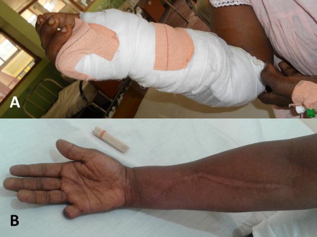
Clinical photograph comparison (Images A and B) demonstrating the acute management and long-term musculoskeletal outcomes of a hump-nosed viper bite. Image A shows a patient's right forearm and hand in the acute phase, heavily encased in white gauze and compression bandages extending from the mid-forearm to the digits, following an emergency decompression fasciotomy for compartment syndrome. Image B displays the same limb four years post-injury. Key visible features include a prominent, linear, longitudinal surgical scar along the volar aspect of the forearm, consistent with a healed fasciotomy. Additionally, the right fifth digit (small finger) shows partial amputation of the distal and middle phalanxes, a consequence of localized necrosis from the envenomation. This comparison highlights the potential for permanent musculoskeletal sequelae, such as tissue loss and scarring, following severe necrotic snakebite envenoming and its surgical management.
Snakebite Envenomation - Clinical Guidelines
Epidemiology & Global Burden
- ~5.4 million bites/year worldwide; 2.7 million envenomations
- 83,000-138,000 deaths/year; ~400,000 permanent disabilities (amputations, blindness)
- Most deaths in South Asia, sub-Saharan Africa, and Latin America
- USA: 7,000-8,000 venomous bites/year; <6 deaths/year (due to antivenom availability)
- 20-25% of pit viper bites and up to 75% of sea snake bites are "dry bites" (no venom injected)
Snake Families of Medical Importance
| Family | Examples | Primary Venom Effect |
|---|---|---|
| Viperidae (Viperinae) | Russell's viper, saw-scaled viper, Old World vipers | Cytotoxic, hemotoxic |
| Viperidae (Crotalinae / Pit vipers) | Rattlesnakes, copperheads, water moccasin, bushmaster | Cytotoxic, hemotoxic, occasionally neurotoxic |
| Elapidae | Cobras, kraits, mambas, coral snakes, taipans, sea snakes | Primarily neurotoxic; some also cytotoxic (cobras) |
| Colubridae | Boomslang, bird snake (Africa) | Hemotoxic (rare, severe coagulopathy) |
| Atractaspididae | Burrowing asps (Africa) | Cytotoxic, cardiotoxic |
Venom Syndromes
1. Cytotoxic / Hemotoxic (Vipers)
- Local: Pain, swelling, erythema, ecchymosis, hemorrhagic bullae, tissue necrosis
- Systemic: Coagulopathy (DIC pattern), thrombocytopenia, spontaneous bleeding (gums, IV sites), hematuria
- Renal: Myoglobinuria, acute kidney injury (Russell's viper especially)
- Shock: Hypovolemia from third spacing
2. Neurotoxic (Elapids)
- Pre-synaptic (kraits, taipans, some mambas): Irreversible - block acetylcholine release; antivenom less effective once bound
- Post-synaptic (cobras, most elapids): Reversible by antivenom + neostigmine
- Features: Ptosis (earliest sign), dysarthria, diplopia, bulbar palsy, progressive descending paralysis, respiratory failure (the killer)
- Minimal local effects (except cobras, which also cause necrosis)
- Onset may be delayed 12+ hours after bite
3. Compartment Syndrome
- Particularly with rattlesnake/viper bites on digits or confined spaces
- Distinguish from simple envenomation swelling - requires pressure measurement
- True compartment syndrome (compartment pressure >30 mmHg) requires fasciotomy
Warning Signs of Severe Envenomation
- Snake identified as highly dangerous species
- Rapid early spread of local swelling beyond the bitten segment
- Early tender lymphadenopathy (regional nodes)
- Early collapse, hypotension, shock
- Ptosis, ophthalmoplegia, inappropriate drowsiness
- Spontaneous systemic bleeding (gums, nose, GI, IV sites)
- Dark brown/black urine (hemoglobinuria/myoglobinuria)
First Aid (Field Management)
DO:
- Move patient away from the snake (stay beyond striking distance)
- Keep calm and still - movement accelerates venom absorption
- Immobilize the bitten limb in neutral position at approximately heart level (splint like a fracture)
- Remove rings, watches, bracelets, tight clothing near the bite
- Clean the wound with soap and water; apply a sterile dressing
- Rapid transport to a facility with antivenom - this is the single most important step
- Take a photo of the snake from a safe distance (for identification)
For Elapid bites ONLY (cobras, kraits, coral snakes, sea snakes):
- Pressure-immobilization bandage (PIB): Wrap the entire bitten limb with an elastic bandage (snug, not tight - finger should slip under), then splint and immobilize
- Bandage pressure: 40-70 mmHg upper limb; 55-70 mmHg lower limb
- The victim must be carried, not walk - muscle pumping disperses venom
- Removes once antivenom is running
DO NOT:
- Incise or suck the bite - damages tissue, no venom removed, increases infection risk
- Apply tourniquets - cause ischemia and worsen local necrosis
- Apply ice/cold water - worsens venom injury
- Give alcohol - vasodilator, increases venom absorption
- Use electric shock - ineffective, causes electrical burns
- Try to capture the snake - risk of second bite
- Give aspirin or NSAIDs - worsen coagulopathy/bleeding
Hospital Assessment
History
- Time of bite, snake description/photo
- Geographic location (narrows likely species)
- First aid applied (note if tourniquet placed - remove carefully after antivenom starts)
- Symptoms since bite (syncope, vomiting, numbness, weakness)
Physical Examination
- Vital signs (BP, HR, RR, SpO2, temperature)
- Fang marks, wound extent, swelling measurement (circumference q1h)
- Neurological: ptosis, eye movements, bulbar function, limb power
- Bleeding: IV sites, gums, urine, stool
Investigations
| Test | Purpose |
|---|---|
| 20-min Whole Blood Clotting Test (20WBCT) | Screening for coagulopathy (no clot = significant coagulopathy) |
| PT/INR, aPTT, fibrinogen | Severity of coagulopathy, DIC |
| FBC + differential | Thrombocytopenia |
| U&E, creatinine | Renal function |
| CK, LDH | Myotoxicity |
| Urinalysis | Hematuria, myoglobinuria |
| Blood group & crossmatch | If transfusion anticipated |
| ECG | Cardiotoxic species (Atractaspis) |
| CXR | Baseline before antivenom; respiratory failure monitoring |
Antivenom - The Cornerstone of Treatment
Indications for Antivenom
- Evidence of coagulopathy (20WBCT non-clotting, spontaneous bleeding)
- Neurotoxicity (ptosis, bulbar weakness, limb paralysis)
- Cardiovascular compromise (hypotension, arrhythmia)
- Renal failure (oliguria, rising creatinine, hematuria)
- Rhabdomyolysis/myotoxicity
- Thrombocytopenia (<100,000 with bleeding)
- Swelling extending beyond adjacent joint (beyond wrist for hand bite; beyond knee for foot bite)
- Rapid progressive swelling after bite
- Lymphangitis spreading proximally
- Any definite bite by a neurotoxic elapid - give antivenom prophylactically; do not wait for symptoms (symptoms may be irreversible)
- Ptosis = antivenom NOW (indicates imminent respiratory failure)
Antivenom - How to Give
| Parameter | Recommendation |
|---|---|
| Route | IV infusion ONLY (never IM or SC - unpredictable absorption) |
| Timing | Start within 4 hours ideally; effective up to 2+ hours after bite; late administration still beneficial for coagulopathy |
| Dilution | Dilute in 250-500 mL normal saline; infuse over 30-60 min |
| Initial infusion rate | Slow (first 10 min to watch for reactions), then increase |
| Premedication | Subcutaneous adrenaline 0.25 mg (adult) prophylactically before antivenom (WHO-endorsed practice; reduces early anaphylactoid reactions) |
Antivenom Types
- Polyvalent antivenom (India, Africa, Australia): Covers multiple species; does not require species identification
- Monovalent antivenom: Species-specific, used in some regions
- Fab fragment antivenoms (USA): CroFab (Crotalidae Polyvalent Immune Fab, Ovine); longer-acting F(ab')2 now available, reduces recurrent coagulopathy
Antivenom Dosing (General Principles)
- Children receive the same dose as adults (same volume of venom injected)
- Repeat dose in 6 hours if: coagulopathy persists, neurotoxicity worsens, swelling continues to progress, or vital signs do not stabilize
- USA rattlesnake (CroFab): Initial 4-6 vials IV; repeat 2 vials q6h x 3 doses to prevent recurrent coagulopathy
- Coral snake (USA): 3-5 vials prophylactically for any definite bite
- India polyvalent: 10 vials IV initially; repeat as needed based on 20WBCT at 6 hours
Antivenom Reactions
| Reaction | Management |
|---|---|
| Early anaphylactoid (within 30 min): urticaria, bronchospasm, hypotension | Stop infusion, give IM adrenaline 0.5 mg (adult), chlorphenamine IV, hydrocortisone IV; restart at slower rate once stable |
| Pyrogenic (1-2 h): chills, fever | Slow infusion, antipyretics; usually self-limiting |
| Serum sickness (7-14 days): fever, arthralgia, rash, lymphadenopathy | Oral prednisolone 5 mg/kg/day x 5-7 days; or antihistamines for mild cases |
Organ-Specific Management
Coagulopathy / DIC
- Antivenom is the treatment - FFP does not work until venom is neutralized
- After antivenom, FFP/cryoprecipitate/platelets for persistent bleeding
- Repeat 20WBCT at 6 hours; if still non-clotting, repeat antivenom dose
- Do NOT use heparin for viper-induced DIC
Neurotoxic Paralysis
- Airway and ventilation are paramount - intubate early if ptosis + swallowing difficulty
- Anticipate need for prolonged mechanical ventilation (days to weeks with krait bites)
- Anticholinesterase test: Give atropine 0.6 mg IV then neostigmine 1.5-2 mg IM; if improvement in 30 min, continue neostigmine 0.5 mg IM q30min (effective for post-synaptic block - cobras; ineffective for pre-synaptic block - kraits)
- Monitor spirometry and vital capacity serially (intubate if VC <15 mL/kg or <1 L)
- SpO2 monitoring unreliable alone - use ETCO2 or arterial blood gas
Acute Kidney Injury
- Common with Russell's viper, sea snakes, and some African vipers
- Maintain hydration; avoid nephrotoxins (aminoglycosides, NSAIDs, contrast)
- Furosemide for oliguria only if volume-replete
- Early dialysis (CRRT preferred) if AKI persists or severe electrolyte disturbance
- AKI from Russell's viper may recover fully with dialysis support
Hypotension / Shock
- IV fluid resuscitation (0.9% saline or Hartmann's 250-500 mL bolus)
- Vasopressors (norepinephrine) if fluid-refractory
- Watch for pulmonary edema (some vipers cause increased capillary permeability)
Local Wound Care
- Mark the margin of swelling with a pen every 30-60 min with time noted
- Elevate the limb after antivenom is given
- Fasciotomy ONLY for confirmed compartment syndrome (direct pressure measurement >30 mmHg with clinical features) - do not do prophylactically
- Debridement of necrotic tissue when demarcated (days to weeks later)
- Tetanus prophylaxis
- Antibiotics: Not routinely indicated; use only if wound infection signs develop (antibiotic stewardship - routine prophylaxis is not evidence-based per Brenes-Chacon et al., Toxins 2024)
Pain
- Paracetamol (acetaminophen) - preferred
- Codeine phosphate for moderate pain
- Avoid aspirin and NSAIDs (worsen bleeding)
Antivenom Is NOT Indicated When:
- Dry bite (no signs of envenomation after 6-12 hours observation)
- Minor, purely local swelling confined to the bite area without progression
- Non-venomous snake confirmed
- Psychological symptoms only (fright response)
Monitoring Protocol
| Time | Action |
|---|---|
| Admission | Baseline vitals, 20WBCT, bloods, mark swelling |
| q30-60 min | Mark swelling progression, neuro check, vitals |
| 6 hours post-antivenom | Repeat 20WBCT, coag screen, renal function |
| 12-24 hours | Clinical review for recurrent coagulopathy, wound assessment |
| Discharge | When swelling resolves, coagulopathy corrected, ambulatory |
| 7-14 days post-discharge | Serum sickness watch, wound review |
Disposition
- Observation minimum 12 hours for all suspected venomous bites (even if initially asymptomatic) - elapid envenomation may be delayed
- ICU for: respiratory compromise, shock, severe coagulopathy, renal failure, multi-organ involvement
- Discharge criteria: Swelling resolving, coagulopathy resolved, ambulatory and eating/drinking, no neurological deficits
- Instruct on serum sickness symptoms before discharge
Special Situations
Ophthalmia (Spitting Cobras)
- Venom spat directly into eyes causes intense pain, conjunctivitis, corneal ulceration
- Immediate copious irrigation with water (or any bland fluid)
- Do NOT rub the eye
- Antivenom is NOT instilled into the eye
- Ophthalmology review for corneal injury
Pregnancy
- Antivenom is NOT contraindicated - benefits outweigh risks
- Premature labor and fetal loss are recognized complications of envenomation
- Obstetric monitoring mandatory
Pediatric Considerations
- Same antivenom dose as adults
- Higher risk of severe envenomation relative to body mass (children receive same volume of venom)
- Rapid deterioration possible; lower threshold for ICU
WHO 2026 Update: Novel Therapeutics
Key Guideline Sources
| Source | Year | Focus |
|---|---|---|
| WHO Snakebite Strategy | 2019-2030 | Global burden, antivenom access, halving deaths by 2030 |
| JTS CPG ID-81 (US Military) | Apr 2026 | Regional treatment algorithms, field antivenom use |
| Tintinalli's Emergency Medicine Ch. 212 | 2024 ed. | Comprehensive ED management |
| Harrison's Principles 22e, Ch. 471 | 2025 | Global epidemiology, field and hospital management |
| Government of India National Protocol | 2007 | India-specific first aid and antivenom guidelines |
| WHO Guidelines for Antivenom Production | 2017 | Antivenom standards |