Now I have excellent diagrams. Let me now write the comprehensive, detailed notes with mnemonics for all 12 topics.
📚 SKULL BONES & SCALP - Week 1 Complete Notes
Gray's Anatomy for Students | Full Detail + Mnemonics
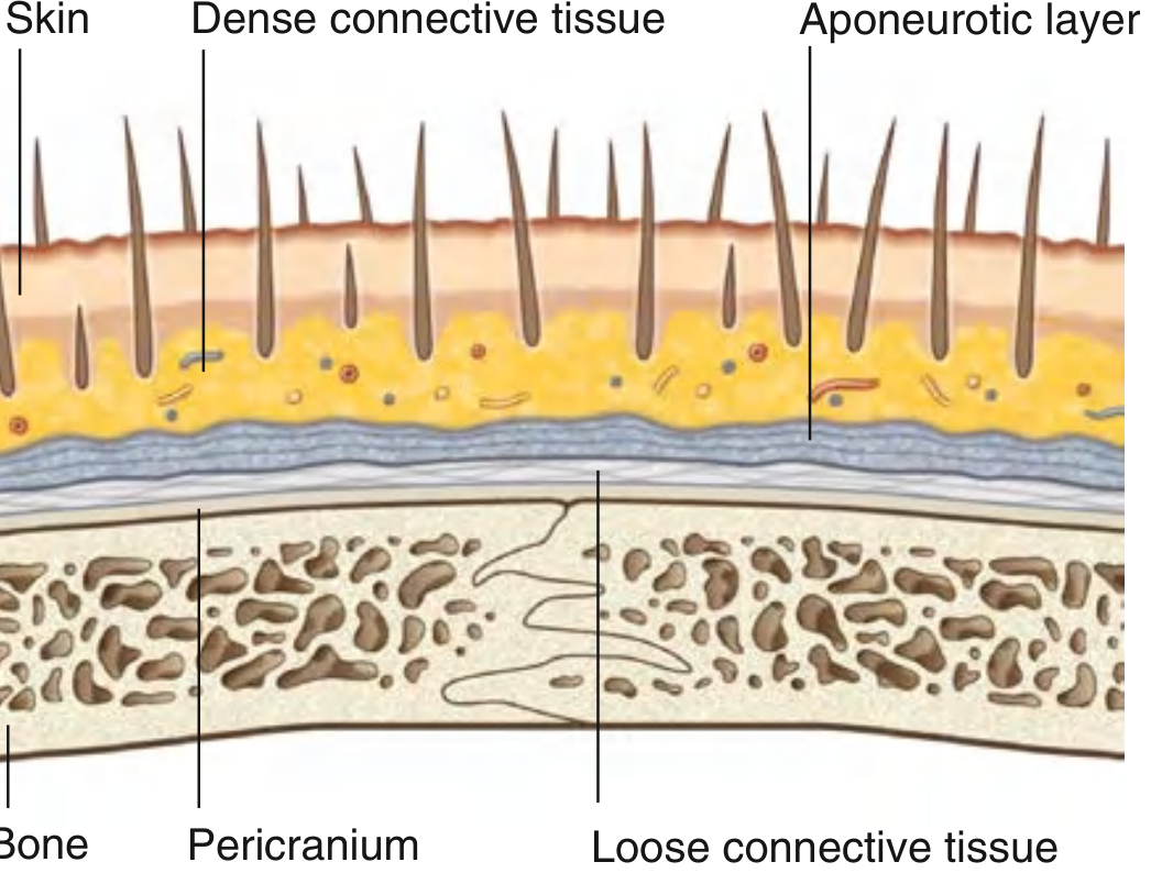
TOPIC 1 - LAYERS OF SCALP
Mnemonic: "SCALP"
The word SCALP itself is the mnemonic!
| Letter | Layer | Details |
|---|
| S | Skin | Thickest skin of body; contains hair follicles, sebaceous glands, sweat glands |
| C | Connective Tissue (Dense) | Contains arteries, veins, nerves; tightly anchors skin to layer 3 |
| A | Aponeurotic Layer (Galea Aponeurotica) | Epicranial aponeurosis; connects frontalis + occipitalis muscles |
| L | Loose Connective Tissue | "Dangerous zone" - allows spread of infection/blood |
| P | Pericranium | Periosteum of skull bones; firmly attached to bone |
(Gray's Anatomy for Students - Fig. 8.74)
Key Points:
- Layers S + C + A are FIRMLY attached to each other = "Scalp proper" (torn together in scalping injuries)
- Layer L (Loose CT) = "Danger Zone" - infection/pus/blood can spread freely here
- Layer P = tightly attached to bone, CANNOT be lifted easily
Extent of Scalp:
- Anteriorly: Superciliary arches
- Posteriorly: External occipital protuberance + Superior nuchal lines
- Laterally: Zygomatic arch
TOPIC 2 - VASCULAR SUPPLY OF SCALP
Mnemonic: "Super PASSion" (for arteries)
Supratrochlear | Posterior auricular | Anterior scalp (supraorbital) | Superficial temporal | Shield = Occipital
Arteries (5 pairs - all anastomose freely):
| Artery | Origin | Region Supplied |
|---|
| Supratrochlear | Ophthalmic a. (from ICA) | Anteromedial forehead |
| Supraorbital | Ophthalmic a. (from ICA) | Anterior scalp to vertex |
| Superficial temporal | External carotid a. | Temporal/lateral scalp |
| Posterior auricular | External carotid a. | Behind ear |
| Occipital | External carotid a. | Posterior scalp |
Mnemonic for blood supply origin: "2 from Inside, 3 from Outside"
- 2 from ICA (via ophthalmic): Supratrochlear + Supraorbital
- 3 from ECA: Superficial temporal + Posterior auricular + Occipital
Veins:
- Follow the same named arteries
- Important: Veins anastomose with diploic veins of skull and dural venous sinuses via emissary veins
- This connection = pathway for intracranial spread of infection
Lymphatics:
- Anterior to vertex → Parotid/preauricular nodes
- Posterior to vertex → Mastoid nodes (retro-auricular) → Upper deep cervical nodes
- Forehead → Submandibular nodes (follow facial artery)
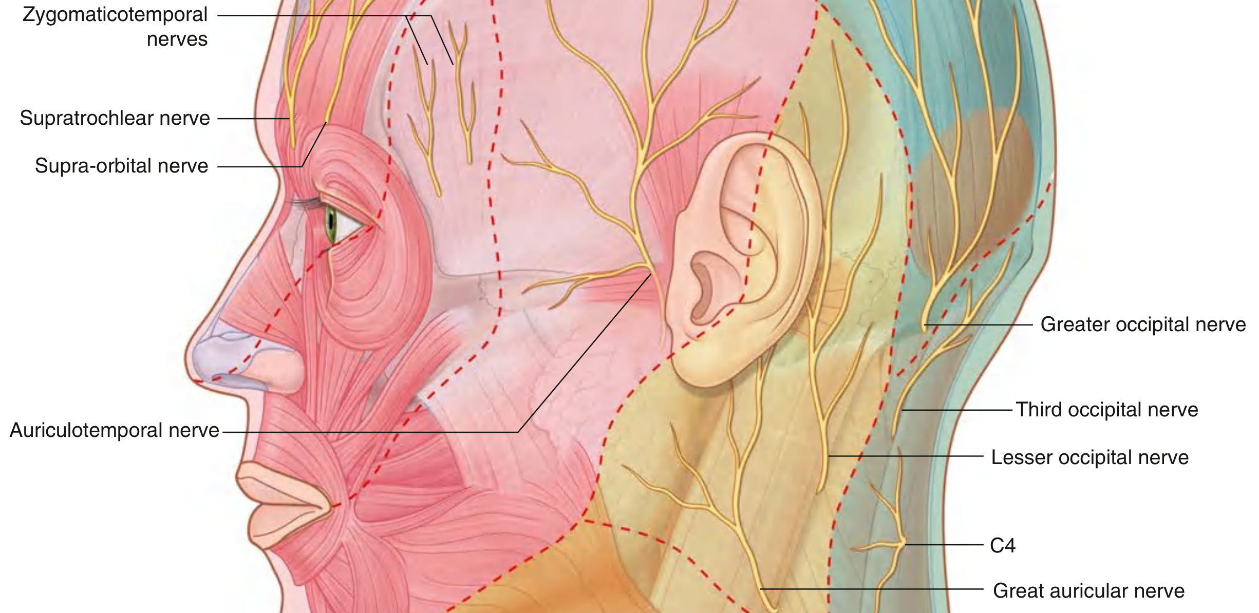
TOPIC 3 - NERVE SUPPLY OF SCALP
Mnemonic: "Scared Students Get Answers Quickly, Great Luck There"
(for all 8 nerves in order front to back)
(Gray's Anatomy for Students)
Anterior to ears + vertex - TRIGEMINAL (CN V):
| Nerve | Origin | Area |
|---|
| Supratrochlear | V1 (ophthalmic div.) | Anteromedial forehead near midline |
| Supraorbital | V1 (ophthalmic div.) | Forehead to vertex |
| Zygomaticotemporal | V2 (maxillary div.) | Small anterior temple area |
| Auriculotemporal | V3 (mandibular div.) | Temple + anterior to ear to near vertex |
Posterior to ears + vertex - CERVICAL NERVES (C2, C3):
| Nerve | Origin | Area |
|---|
| Great auricular | C2, C3 (anterior rami) | Small area posterior to ear |
| Lesser occipital | C2 (anterior ramus) | Posterior + superior to ear |
| Greater occipital | C2 (posterior ramus) | Large posterior scalp to vertex |
| Third occipital | C3 (posterior ramus) | Small lower posterior scalp |
Mnemonic: "V before C - Ventral (anterior) = V5, Back (posterior) = Cervical"
- Front half = Trigeminal (CN V)
- Back half = Cervical plexus (C2, C3)
Motor Innervation of Scalp Muscles:
- Frontalis (frontal belly) → Temporal branches of CN VII (facial nerve)
- Occipitalis (occipital belly) → Posterior auricular branch of CN VII
TOPIC 4 - APPLIED ASPECTS OF SCALP
Mnemonic: "SCALP problems = LABS"
Laceration (bleeding), Abscess (danger zone spread), Black eye (Layer C bleed tracking), Scalping (layers S+C+A)
1. Profuse Bleeding in Scalp Lacerations:
- Dense CT (Layer C) surrounds vessels and keeps them open (prevents vasoconstriction)
- Even small cuts bleed profusely
- Clinically: Even small scalp wounds may require suturing due to extensive bleeding
2. The "Dangerous Layer" (Layer L - Loose CT):
- Infection or hematoma in this layer can spread freely in all directions (no fascial barriers)
- Spread is limited: anteriorly by orbital septum, posteriorly by nuchal lines
- Infection here → can spread to intracranial sinuses via emissary veins = life-threatening
3. "Black Eye" after Scalp Injury:
- Blood from a forehead wound can track through loose connective tissue into periorbital tissue
- Produces bilateral periorbital bruising ("raccoon eyes" if bilateral)
4. Scalping Injuries:
- The first 3 layers (S+C+A) are firmly united = avulsed together
- Separation occurs at Layer L (loose CT) = the plane of cleavage
5. Subgaleal Hematoma (Cephalohematoma):
- Blood in Layer L (loose CT) - can spread across entire scalp
- Crosses suture lines (unlike cephalhematoma which is confined by periosteum)
6. Caput Succedaneum vs Cephalohematoma:
| Feature | Caput Succedaneum | Cephalohematoma |
|---|
| Layer | Loose CT (Layer L) | Subperiosteal (below Layer P) |
| Crosses sutures? | YES | NO |
| Onset | At birth | Hours after birth |
| Pitting edema | Yes | No |
TOPIC 5 - BASIC ANATOMY OF SKULL BONES
Mnemonic: "F.O.T.P.E.S.Z.N.M.V" - "Flip Over The Plate, Every Single Zygoma Needs More Vomers"
(Frontal, Occipital, Temporal x2, Parietal x2, Ethmoid, Sphenoid, Zygomatic x2, Nasal x2, Maxilla x2, Vomer x1, Lacrimal x2, Inferior nasal conchae x2, Mandible, Palatine x2)
The 8 Cranial Bones:
Mnemonic: "PEST OF 6" → Parietal (x2) + Ethmoid + Sphenoid + Temporal (x2) + Occipital + Frontal = 8 bones
| Bone | No. | Position |
|---|
| Frontal | 1 | Anterior cranium/forehead |
| Parietal | 2 | Sides and top |
| Temporal | 2 | Lateral sides |
| Occipital | 1 | Posterior/base |
| Sphenoid | 1 | Central base |
| Ethmoid | 1 | Anterior base/roof of nose |
The 14 Facial Bones:
Mnemonic: "Never Let Monkeys Intimidate Very Pale Zebras"
- Nasal (x2), Lacrimal (x2), Maxilla (x2), Inferior nasal conchae (x2), Vomer (x1), Palatine (x2), Zygomatic (x2), Mandible (x1) = 14
Skull Structure:
- Calvaria = Skull cap (frontal + parietals + occipital)
- Cranial Base = Floor (ethmoid + sphenoid + temporal + occipital + frontal)
- Diploe = Cancellous bone between outer and inner tables of calvaria
- Contains diploic veins that communicate with emissary veins and dural sinuses
TOPIC 6 - SUTURES
Mnemonic: "CLAMS" (or "Clever Lions Are Mostly Squamous")
Coronal | Lambdoid | Sagittal | Metopic | Squamosal
The 5 Main Sutures:
| Suture | Between | Shape/Note |
|---|
| Coronal | Frontal + Parietals | Coronal plane |
| Sagittal | 2 Parietal bones | Midline, antero-posterior |
| Lambdoid | Occipital + Parietals | Lambda (λ) shaped |
| Squamosal | Temporal + Parietal | Each side |
| Metopic | Frontal bone (between two halves) | Closes 1st-2nd year of life |
Key Junctions:
| Junction | Name | Sutures Meeting |
|---|
| Coronal + Sagittal | Bregma | Anterior fontanelle site |
| Lambdoid + Sagittal | Lambda | Posterior fontanelle site |
| Coronal + Squamosal + Sphenoid | Pterion | Weakest point of skull |
| Lambdoid + Squamosal + Mastoid | Asterion | |
Pterion - CLINICALLY IMPORTANT:
- Junction of frontal, parietal, temporal, and sphenoid bones
- Thinnest/weakest part of skull
- Located 2-3 cm posterior to zygomatic process of frontal bone
- Lies over middle meningeal artery (anterior division)
- Blow to pterion → Extradural (epidural) hematoma
- Mnemonic: "PTerion = Poor Temporal bone = breaks easily, bleeding from Middle Meningeal"
TOPIC 7 - EXTERIOR AND INTERIOR OF SKULL
NORMA (5 views of skull):
Mnemonic: "Very Lovely Beautiful Occipital Things"
- Verticalis (from above)
- Lateralis (from side)
- Basalis (from below)
- Occipitalis (from behind)
- Frontalis (from front)
Norma Verticalis (from above):
- Bregma (junction of coronal + sagittal sutures) - anteriorly
- Lambda (junction of lambdoid + sagittal sutures) - posteriorly
- Sagittal suture runs between them
Norma Lateralis (from side):
- Pterion (2.5-3 cm posterior to zygomatic arch)
- Temporal fossa
- Zygomatic arch (zygoma + temporal)
- Mastoid process
- External auditory meatus
- External occipital protuberance (inion)
Norma Basalis (base of skull - external):
- Hard palate (maxilla + palatine)
- Foramen magnum
- Occipital condyles
- Styloid process
- Mastoid process
- Carotid canal
- Jugular foramen
- Stylomastoid foramen
INTERIOR OF SKULL (3 cranial fossae):
Mnemonic: "All Bright Children" → Anterior, Middle, Posterior
Anterior Cranial Fossa:
- Bones: Frontal, Ethmoid (cribriform plate), Sphenoid (lesser wings)
- CN I (olfactory) passes through cribriform plate
- CN II exits via optic canal
Middle Cranial Fossa:
- Bones: Sphenoid (greater wings, body), Temporal (squamous + petrous part)
- Foramina: Mnemonic "ROSA"
| Foramen | Contents |
|---|
| Rotundum | CN V2 (maxillary) |
| Ovale | CN V3 (mandibular) + accessory meningeal a. |
| Spinosum | Middle meningeal a. + v. |
| Superior orbital fissure | CN III, IV, V1, VI + ophthalmic vein |
| Carotid canal (foramen lacerum) | Internal carotid artery |
Posterior Cranial Fossa:
- Bones: Occipital, Temporal (petrous), Sphenoid
- Foramen magnum: medulla, CN XI, vertebral arteries
- Internal acoustic meatus: CN VII + VIII
- Jugular foramen: CN IX, X, XI + IJV
Mnemonic for jugular foramen contents: "GVA" = Glossopharyngeal (IX), Vagus (X), Accessory (XI) + IJV
TOPIC 8 - FRONTAL AND OCCIPITAL BONES
FRONTAL BONE:
Parts:
- Squamous part - Vertical plate (forehead)
- Orbital parts (2) - Horizontal plates forming orbital roofs
- Nasal part - Forms nasal bridge
Key Landmarks - Mnemonic: "Super Glabella Fills Orbits Neatly"
| Landmark | Description |
|---|
| Glabella | Smooth midline elevation between eyebrows |
| Superciliary arch | Ridge above orbit (more prominent in males) |
| Supraorbital notch/foramen | Transmits supraorbital nerve + vessels |
| Frontal sinuses | Air spaces within squamous part |
| Metopic suture | Midline, closes 1st-2nd year of life |
| Zygomatic process | Articulates with zygomatic bone |
| Trochlear fossa | Small depression - attachment of trochlea (superior oblique) |
| Lacrimal fossa | Lateral orbital roof - lodges lacrimal gland |
OCCIPITAL BONE:
Parts - Mnemonic: "CLAB" = Condylar, Lateral, Basilar, Squamous
- Basilar part - Anterior to foramen magnum (clivus)
- Condylar parts (2) - Bear occipital condyles
- Squamous part - Posterior plate
Key Landmarks:
| Landmark | Description |
|---|
| Foramen magnum | Largest foramen; medulla, vertebral arteries, spinal accessory |
| External occipital protuberance (Inion) | Midline, palpable bump on posterior skull |
| Superior nuchal line | Extends from inion; muscle attachment |
| Inferior nuchal line | Below superior; deeper muscle attachment |
| Occipital condyles | Articulate with C1 (atlas) - atlantooccipital joint |
| Hypoglossal canal | Above condyle; transmits CN XII |
| Condylar canal | Behind condyle; emissary vein |
| Basilar part (Clivus) | Anteriorly fused with sphenoid |
| Internal occipital protuberance | Inside; cruciate eminence; attachment of falx + tentorium |
TOPIC 9 - MAXILLA
Two maxillae articulate at the intermaxillary suture to form the upper jaw.
Parts - Mnemonic: "Freddy Ants Zigged Past"
Frontal process | Alveolar process | Zygomatic process | Palatine process
| Process | Description |
|---|
| Frontal process | Projects upward to articulate with frontal bone; forms medial orbit wall |
| Alveolar process | Bears the upper teeth (8 teeth per side) |
| Zygomatic process | Laterally to zygomatic bone |
| Palatine process | Horizontal; forms anterior 3/4 of hard palate |
Body of Maxilla:
- Contains maxillary sinus (largest of paranasal sinuses)
- Opens into middle meatus via ostium (infundibulum)
- 4 surfaces: Anterior, Posterior (infratemporal), Superior (orbital), Medial (nasal)
Key Foramina:
| Foramen | Contents |
|---|
| Infraorbital foramen | Infraorbital nerve (V2) + vessels |
| Incisive foramen | Nasopalatine nerve + greater palatine vessels |
Applied:
- Cleft palate - failure of palatine processes to fuse
- Maxillary sinus infection - molar tooth roots project into floor of sinus
- Mnemonic: "Max Sinus DRANO: Drains Nasally (middle meatus), Roots Accessible from below (via teeth)"
TOPIC 10 - SPHENOID AND TEMPORAL BONES
SPHENOID BONE
Shape: Resembles a bat/butterfly (with wings spread)
Parts - Mnemonic: "Big Bat Has Greatness, Little Bat Stands Proud"
- Body (central) - contains sphenoid sinuses
- Greater wings (2) - form middle cranial fossa floor + orbital walls
- Lesser wings (2) - form posterior orbital roof; optic canals
- Pterygoid processes (2) - hang down; medial and lateral pterygoid plates
Key Foramina of Sphenoid - Mnemonic: "ROSS + Carotid"
| Foramen | Contents | Memory Tip |
|---|
| Foramen Rotundum | CN V2 (maxillary) | R = Round, goes to face |
| Foramen Ovale | CN V3 + accessory meningeal a. | O = Oval, bigger |
| Foramen Spinosum | Middle meningeal artery + vein | S = Spine nearby |
| Superior orbital fissure | CN III, IV, V1, VI; ophthalmic veins | S = Superior |
| Optic canal | CN II + ophthalmic artery | O = Optic |
| Carotid sulcus | ICA passes | C = Carotid |
Sella Turcica ("Turkish Saddle"):
- Depression in sphenoid body
- Contains pituitary gland (hypophysis)
- Anterior: Tuberculum sellae
- Posterior: Dorsum sellae
- Floor of sella = pituitary fossa
- Clinical: Pituitary tumors → compress optic chiasm → bitemporal hemianopia
Pterygoid Plates:
- Medial pterygoid plate - ends in hamulus (hooks for tensor palati tendon)
- Lateral pterygoid plate - attachment of pterygoid muscles
TEMPORAL BONE
Parts - Mnemonic: "PAST" = Petrous, (tympanic), Squamous, Styloid, (Mastoid)
| Part | Features |
|---|
| Squamous | Thin flat plate; zygomatic process; mandibular fossa |
| Tympanic | Surrounds external auditory meatus |
| Mastoid | Contains mastoid air cells; mastoid process; digastric notch |
| Petrous | Dense, pyramidal; contains inner ear; important canals |
| Styloid | Needle-like; origin of stylohyoid ligament + 3 muscles |
Key Features:
| Structure | Location/Note |
|---|
| External auditory meatus | Tympanic part |
| Zygomatic process | Forms zygomatic arch with zygomatic bone |
| Mandibular fossa | TMJ articulation with mandibular condyle |
| Mastoid process | Sternocleidomastoid attachment |
| Stylomastoid foramen | CN VII exits here |
| Internal acoustic meatus | CN VII + VIII enter petrous part |
| Carotid canal | ICA enters skull base |
| Jugular fossa | Jugular bulb/IJV |
| Petrotympanic fissure | Chorda tympani exits |
Mnemonic for Styloid Muscle attachments: "Stylo-3 muscles"
Stylohyoid, Stylopharyngeus, Styloglossus
Clinical Pearl - Mastoid:
- Mastoid air cells communicate with middle ear (via aditus)
- Infection (otitis media) → mastoiditis → potentially meningitis
- Digastric notch on medial mastoid = grooved for digastric posterior belly
TOPIC 11 - FOETAL SKULL
Why different from adult skull?
- Bones separated by membranous junctions (future sutures + fontanelles)
- Brain grows rapidly after birth
- Fontanelles allow skull deformation during birth (molding)
Fontanelles (6 total):
Mnemonic: "2 Big, 2 Small, 2 Tiny" → Anterior + Posterior + 2 Sphenoidal + 2 Mastoid
| Fontanelle | Location | Shape | Closes |
|---|
| Anterior (Bregma) | Coronal + Sagittal | Diamond/Rhomboid | 18-24 months |
| Posterior (Lambda) | Lambdoid + Sagittal | Triangular | 2-3 months |
| Sphenoidal (x2) | Anterolateral | Small | 6 months |
| Mastoid (x2) | Posterolateral | Small | 12-18 months |
Mnemonic: "Anterior Diamond-shape = 18 months; Posterior Triangle = 3 months"
(Bigger the fontanelle, longer to close)
Clinical Importance of Anterior Fontanelle:
- Bulging: ↑ ICP (meningitis, hydrocephalus, tumor)
- Sunken: Dehydration
- Closed early (craniosynostosis): premature fusion → abnormal skull shape
- Open too long: hypothyroidism, rickets, Down syndrome, hydrocephalus
Fontanelle Mnemonic: "MARCH" closes anterior
- Metabolic disease (rickets)
- Achondroplasia
- Rubella (congenital infection)
- Chromosomal (Down syndrome, trisomies)
- Hydrocephalus
→ These cause DELAYED closure of anterior fontanelle
Foetal Skull Proportions:
- Face:Cranium ratio = 1:8 (adult = 1:2.5)
- Frontal sinuses absent at birth (develop postnatally)
- Mastoid process absent at birth (develops after 2 years)
- Styloid process not ossified at birth
Moulding:
- Parietal bones can override frontal and occipital during labor
- Resolves within days of birth
TOPIC 12 - HYOID BONE
"The ONLY bone in the body with NO articulation with any other bone"
Mnemonic: "H-Y-O-I-D = Hangs by ligaments, yields to swallowing, only floating bone, it's unique, different"
Structure:
- Body (corpus) - central horizontal part
- Greater horns (2) - project posteriorly from body
- Lesser horns (2) - small, project superiorly from body-greater horn junction
Position:
- Level of C3 vertebra
- Located between mandible and thyroid cartilage
- In the anterior neck
Attachments - Mnemonic: "Supra = above muscles, Infra = below (strap) muscles"
ABOVE (Suprahyoid muscles):
| Muscle | Origin → Hyoid |
|---|
| Mylohyoid | Mylohyoid line of mandible → Body |
| Geniohyoid | Genial tubercle of mandible → Body |
| Digastric (posterior belly) | Mastoid notch → (via tendon loop) Body |
| Stylohyoid | Styloid process → Body |
BELOW (Infrahyoid/strap muscles):
| Muscle | Hyoid → Insertion |
|---|
| Sternohyoid | Body → Sternum |
| Omohyoid | Body → Scapula |
| Thyrohyoid | Body → Thyroid cartilage |
Ligaments:
- Stylohyoid ligament - From styloid process to lesser horn
- Thyrohyoid membrane - From body to thyroid cartilage
- Hyoepiglottic ligament - Body to epiglottis
Ossification:
- Body and greater horns: Ossify in fetal life (cartilage → bone by puberty)
- Lesser horns: Fibrocartilaginous throughout life
- Fusion of greater horns with body: 40s-50s
Clinical Importance:
- Fracture - Rare but significant; associated with strangulation/throttling (forensic importance)
- Thyrohyoid cyst - Thyroglossal duct cyst passes near hyoid body
- Eagle syndrome - Elongated styloid process/calcified stylohyoid ligament → pharyngeal pain
Applied Mnemonic: "SHELF" for hyoid facts
- Strangulation fracture
- Hangs freely (no joint)
- Epiglottis attached
- Level C3
- Floating bone
QUICK REVISION TABLE - ALL TOPICS
| Topic | Key Mnemonic | Must-Remember Fact |
|---|
| Scalp layers | SCALP | Layer L = Danger Zone |
| Scalp arteries | 2 from ICA, 3 from ECA | Anastomose freely |
| Nerve supply | V = Anterior, C = Posterior | Greater occipital = C2 posterior ramus |
| Applied scalp | Caput vs Cephalohematoma | Caput crosses sutures |
| Cranial bones | PEST OF 6 = 8 bones | Ethmoid + Sphenoid form base |
| Sutures | CLAMS | Pterion = weakest point |
| Skull views | VLBOF (5 Normas) | Pterion over MMA |
| Frontal bone | Glabella + supraorbital notch | Frontal sinus |
| Occipital | Foramen magnum | Inion = external protuberance |
| Maxilla | FAZP processes | Maxillary sinus → middle meatus |
| Sphenoid | Butterfly shaped | Sella turcica = pituitary |
| Temporal | PAST parts | Stylomastoid = CN VII exit |
| Foetal skull | Anterior fontanelle = 18 months | Bulging = ↑ICP; Sunken = dehydration |
| Hyoid bone | SHELF | Only floating bone; level C3 |
MCQ POINTS TO REMEMBER (3 per topic format):
Scalp Layers:
- Layer "L" (loose CT) = dangerous space - infection/hematoma spreads here
- Scalp bleeds profusely because dense CT holds cut vessels open
- Scalp proper = S + C + A (avulsed together)
Vascular Supply:
- Supratrochlear and supraorbital from ophthalmic artery (ICA branch)
- Emissary veins connect scalp veins to intracranial venous sinuses
- Forehead lymphatics drain to submandibular nodes
Nerve Supply:
- Greater occipital nerve = posterior ramus of C2 (most important posterior scalp nerve)
- Auriculotemporal nerve = branch of V3 (mandibular)
- Anterior scalp = trigeminal; posterior scalp = cervical plexus
Applied Scalp:
- Pterion blow → middle meningeal artery tear → extradural hematoma
- Caput succedaneum (loose CT) crosses sutures; cephalohematoma (subperiosteal) does NOT
- Scalp infections can spread to intracranial sinuses via emissary veins
Skull Bones:
- 8 cranial bones + 14 facial bones = 22 skull bones
- Diploe = spongy bone between inner + outer tables of skull
- Sphenoid = keystone bone of skull (articulates with all other cranial bones)
Sutures:
- Pterion = thinnest skull; overlies anterior branch of middle meningeal artery
- Bregma = junction of coronal + sagittal sutures (site of anterior fontanelle)
- Lambda = junction of lambdoid + sagittal sutures (site of posterior fontanelle)
Foetal Skull:
- Anterior fontanelle = diamond-shaped, closes 18-24 months
- Posterior fontanelle = triangular, closes 2-3 months
- Caput succedaneum resolves in days; cephalohematoma takes weeks
Hyoid:
- Only bone with no joint to any other bone
- Level C3; fracture suggests strangulation
- Stylohyoid ligament connects styloid process to lesser horn
Sources: Gray's Anatomy for Students (textbook library), Gray's Anatomy 42nd Ed. principles