
Mucopolysachharides
Reading File
Mucopolysaccharides (MPS) are long-chain complex carbohydrates (glycosaminoglycans) found in connective tissue. Deficiencies in lysosomal enzymes that degrade them lead to storage disorders. Let me search the medical library for detailed coverage.
Searching sources...
Finding Sources
Finding Sources
Reading File
Reading File
Reading File
Reading File
Reading File
Finding Sources
Reading File
Reading File
Reading File
Finding Sources
I now have comprehensive coverage from multiple authoritative sources. Let me compile the full answer.
Mucopolysaccharides (Glycosaminoglycans) and the Mucopolysaccharidoses (MPS)
What Are Mucopolysaccharides?
Mucopolysaccharides - now more correctly called glycosaminoglycans (GAGs) - are long-chain, sulfated polysaccharides that form the carbohydrate side-chains of proteoglycans found in connective tissues, bone, cartilage, and extracellular matrix. The major GAGs include:
- Dermatan sulfate
- Heparan sulfate
- Keratan sulfate
- Chondroitin sulfate (4- and 6-sulfate)
- Hyaluronan (not sulfated)
They are normally degraded within lysosomes by a series of specific hydrolases. When these enzymes are deficient, GAGs accumulate within lysosomes throughout the body - in the brain, spinal cord, heart, viscera, bone, and connective tissue - producing a group of diseases called the Mucopolysaccharidoses (MPS).
The overall prevalence of MPS is approximately 1 in 8,000 births.
General Features of All MPS Disorders
- Storage of GAGs within lysosomes (lysosomal storage diseases)
- Progressive accumulation due to defective degradation
- Multisystem involvement: skeletal, cardiac, respiratory, neurologic, ocular, hepatosplenic
- All are autosomal recessive except MPS II (Hunter), which is X-linked recessive
- Diagnosis: urinary GAG excretion pattern + specific enzyme assay + gene analysis
- Characteristic "gargoyle" or coarse facial features in several types
Classification of the Mucopolysaccharidoses
| Type | Eponym | Enzyme Deficiency | GAGs Excreted | Key Features |
|---|---|---|---|---|
| MPS I H | Hurler syndrome | α-L-Iduronidase | Dermatan sulfate, Heparan sulfate | Most severe; corneal clouding, coarse facies, intellectual disability, organomegaly, kyphosis, cardiac/respiratory death by mid-adolescence |
| MPS I S | Scheie syndrome | α-L-Iduronidase (partial) | Dermatan sulfate, Heparan sulfate | Milder allele; normal intelligence, normal life span |
| MPS I H/S | Hurler-Scheie | α-L-Iduronidase (intermediate) | Dermatan sulfate, Heparan sulfate | Severe somatic disease, usually without major neurologic degeneration |
| MPS II | Hunter syndrome | Iduronate sulfate sulfatase | Dermatan sulfate, Heparan sulfate | X-linked; similar to Hurler but milder; no corneal clouding |
| MPS III A-D | Sanfilippo syndrome | 4 different enzymes (see below) | Heparan sulfate | Most common MPS; predominant CNS involvement (behavioral, intellectual decline, seizures); mild somatic features |
| MPS IV A/B | Morquio syndrome | Galactosamine-6-sulfatase (A) / β-Galactosidase (B) | Keratan sulfate, Chondroitin 6-sulfate | Normal intelligence; prominent skeletal dysplasia, kyphoscoliosis, odontoid hypoplasia, cervical instability risk |
| MPS V | (No longer used) | - | - | Reclassified as Scheie (now MPS I S) |
| MPS VI | Maroteaux-Lamy | N-acetylgalactosamine-4-sulfatase (Arylsulfatase B) | Dermatan sulfate | Hurler-like somatic features; normal intelligence; corneal clouding; cardiac valve disease |
| MPS VII | Sly syndrome | β-Glucuronidase | Dermatan sulfate, Heparan sulfate, Chondroitin 4-sulfate | Wide severity range; can present as fetal hydrops |
Sources: Adams and Victor's Principles of Neurology 12th Ed; Harrison's Principles of Internal Medicine 22E (2025); Emery's Elements of Medical Genetics and Genomics
Individual Syndromes - Clinical Detail
MPS I - Hurler Syndrome
- Onset: end of the first year of life
- Clinical triad: coarse "gargoyle" facies, corneal clouding, intellectual disability
- Skeletal: dwarfism, kyphosis, broad hands with stubby fingers, flexion contractures at knees and elbows
- Hearing loss (conductive), hepatosplenomegaly, valvular heart disease, recurrent respiratory infections, hernias
- Biochemistry: absence of α-L-iduronidase → accumulation of dermatan + heparan sulfate in tissues and urine
- Also increased ganglioside content in brain neurons
- Death: mid-adolescence from cardiac failure and respiratory infections
- Diagnosis: metachromatic granules in leukocytes; increased urinary GAGs; confirmed by enzyme assay (α-L-iduronidase) and IDUA gene analysis
MPS II - Hunter Syndrome
- X-linked recessive (only X-linked MPS)
- Onset: 2-5 years; males affected
- Hurler-like but milder: coarse facies, joint stiffness, deafness, organomegaly, developmental delay
- No corneal clouding (key distinguishing feature from Hurler)
- Two forms: severe (death in mid-teens) and mild (relatively normal intelligence, survival to middle age)
- Enzyme deficiency: iduronate sulfate sulfatase (IDS gene)
- Urine: excess dermatan + heparan sulfate
MPS III - Sanfilippo Syndrome
- Most common MPS
- Predominant neuropsychiatric presentation: behavioral problems, intellectual regression, hyperactivity, seizures
- Mild somatic features (unlike other MPS types)
- Four subtypes (A-D) from four different enzymes, all degrading heparan sulfate:
- IIIA: N-sulphoglucosamine sulphohydrolase (SGSH gene)
- IIIB: α-N-acetylglucosaminidase (NAGLU gene)
- IIIC: Heparan-α-glucosaminide N-acetyltransferase (HGSNAT gene)
- IIID: N-acetylglucosamine-6-sulfatase (GNS gene)
- Types A and B account for 90% of cases
- Death: early adult life
MPS IV - Morquio Syndrome
- Onset: 2-3 years
- Skeletal dysplasia without intellectual impairment (intelligence normal)
- Short stature, thoracic deformity, kyphoscoliosis
- Risk: spinal cord compression from odontoid hypoplasia and cervical instability
- Slight corneal clouding; cardiac and respiratory complications
- Urine: keratan sulfate
- Enzyme: galactosamine-6-sulfatase (MPS-IVA, GALNS gene) or β-galactosidase (MPS-IVB, GLB1 gene)
- Prevalence: ~1 in 200,000-300,000
MPS VI - Maroteaux-Lamy Syndrome
- Hurler-like somatic features: coarse facies, short stature, kyphosis, joint restriction, corneal clouding, cardiac valve abnormalities
- Intelligence normal
- Enzyme deficiency: N-acetylgalactosamine-4-sulfatase / arylsulfatase B (ARSB)
- Urine: dermatan sulfate
- Spinal cord compression can occur
- Two forms: severe (survival to early adulthood) and mild
MPS VII - Sly Syndrome
- Enzyme deficiency: β-Glucuronidase (GUSB gene)
- Wide range of severity
- Severe form: fetal hydrops - can cause stillbirth or perinatal death
- Later-onset form: short stature, coarse facies, hepatosplenomegaly
Diagnosis
- Urine GAG quantification and fractionation - screening test (increased dermatan, heparan, keratan, or chondroitin sulfate excretion patterns are type-specific)
- Enzyme activity assay - in serum, leukocytes, or cultured fibroblasts - confirmatory
- Molecular gene analysis - IDUA, IDS, NAGLU, GALNS, GUSB, etc.
- Newborn screening (NBS): MPS I added to recommended NBS panels in several US states; Pompe disease similarly screened
Treatment
Enzyme Replacement Therapy (ERT)
Available FDA-approved ERTs:
- MPS I: Laronidase (recombinant α-L-iduronidase) - effective for visceral/cardiac/respiratory manifestations; does not cross blood-brain barrier (no direct CNS benefit)
- MPS II: Idursulfase - weekly IV; addresses visceral manifestations; no CNS penetration
- MPS IVA: Elosulfase alfa - approved; improves ambulatory mobility and decreases pain; no treatment for MPS IVB
- MPS VI: Galsulfase (arylsulfatase B) - FDA approved
- MPS VII: Vestronidase alfa-vjbk - approved
Hematopoietic Stem Cell Transplantation (HSCT)
- Standard treatment for MPS I presenting at <2 years of age with neurologic involvement
- Stabilizes CNS disease, reverses hepatosplenomegaly, improves cardiac/respiratory disease
- Does not improve corneal disease or progressive skeletal disease
- HSCT has not been successful in treating CNS disease in MPS II
- Not helpful in Hunter or Sanfilippo diseases
- Combination ERT + HSCT is being tried (ERT before transplant to reduce disease burden)
Supportive Management
- For MPS (airway): adenotonsillectomy, CPAP, or tracheotomy (tonsil/adenoid/tongue hypertrophy and tracheobronchial deposits cause obstructive sleep apnea and chronic pulmonary disease - a major cause of death)
- Regular lifelong cardiac and respiratory follow-up even after successful HSCT
Wound Healing Role
Mucopolysaccharides (particularly hyaluronic acid and chondroitin sulfate) also play important roles in wound healing - they facilitate fibroblast migration, cell proliferation, and tissue remodeling during the repair process.
Key Memory Aids:
- "Hunter = Heparan + dermatan + Hemizygous (X-linked)" - the only X-linked MPS
- "Sanfilippo = Smart (normal IQ early) then degenerates + Sulfate of heparan only"
- "Morquio = Musculoskeletal (skeletal dysplasia) + Mind intact (normal intelligence)"
- "Maroteaux-Lamy = like Hurler but Lucid (normal intelligence)"
Sources: Harrison's Principles of Internal Medicine 22E (2025, McGraw Hill); Adams and Victor's Principles of Neurology 12th Ed; Emery's Elements of Medical Genetics and Genomics; Cummings Otolaryngology; Goldman-Cecil Medicine
Explain glucose transporter glut in intestine.
Glucose transport in the intestine involves SGLT1 (sodium-glucose co-transporter) on the apical/brush border membrane and GLUT2 on the basolateral membrane, working together to absorb dietary glucose into the bloodstream. Let me search the medical library for detailed coverage.
Searching sources...
Finding Sources
Finding Sources
Reading File
Reading File
Reading File
Reading File
Now let me fetch the key diagram from Guyton and the one from Sleisenger for visual clarity.
Loading Image
Loading Image
Excellent - both diagrams retrieved. I now have everything needed for a thorough explanation.
Glucose Transporters (GLUT/SGLT) in the Intestine
Overview
Dietary carbohydrates are digested to monosaccharides (glucose, galactose, fructose) at the brush border by enzymes like lactase, sucrase-isomaltase, and maltase before absorption. Absorption occurs predominantly in the jejunum via three distinct membrane carrier systems:
- SGLT1 (apical/brush-border membrane) - for glucose and galactose
- GLUT5 (apical/brush-border membrane) - for fructose
- GLUT2 (basolateral membrane) - exit route for all three monosaccharides into portal blood
The Classic Diagram (Normal/Low Glucose Load)
(Guyton and Hall Textbook of Medical Physiology)
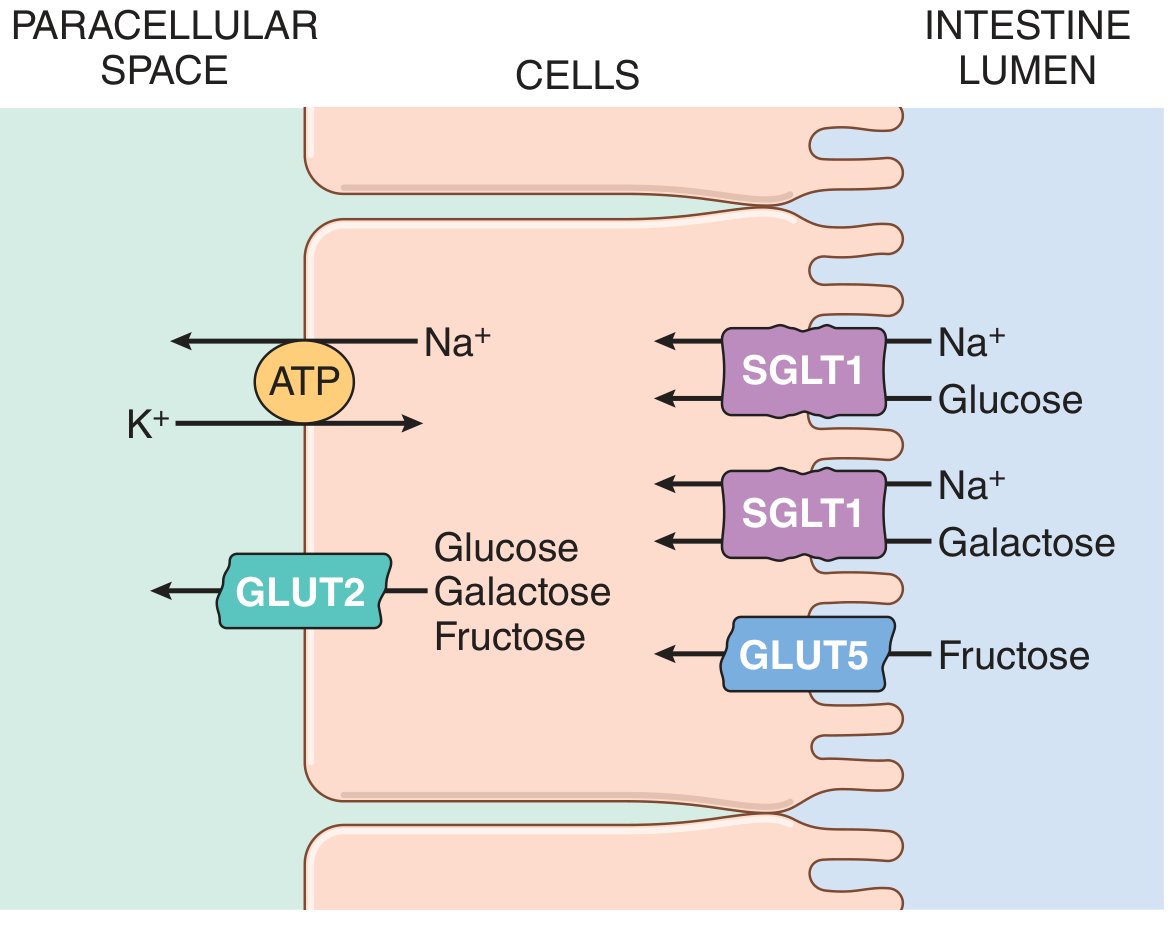
Step-by-Step Mechanism
Step 1: Na⁺/K⁺-ATPase Creates the Driving Force (Basolateral Membrane)
- The Na⁺/K⁺-ATPase pump on the basolateral membrane (BLM) continuously pumps Na⁺ out of the enterocyte and K⁺ in, using ATP
- This keeps intracellular Na⁺ low and maintains an inside-negative membrane potential
- The resulting electrochemical Na⁺ gradient is the ultimate energy source for glucose absorption - this is secondary active transport
Step 2: SGLT1 - Apical/Brush-Border Membrane Entry (Glucose & Galactose)
| Property | Detail |
|---|---|
| Full name | Sodium-Glucose Cotransporter 1 |
| Gene | SLC5A1 |
| Location | Apical/brush-border membrane (BBM) of enterocytes |
| Substrates | Glucose AND galactose (not both simultaneously) |
| Coupling ratio | 2 Na⁺ : 1 monosaccharide per transport cycle |
| Transport type | Secondary active transport (electrogenic) |
| Affinity | High affinity (efficient at low luminal sugar concentrations) |
Mechanism: Na⁺ flows down its electrochemical gradient into the cell through SGLT1, dragging glucose (or galactose) along with it. Since 2 positive charges enter per cycle, the process is electrogenic - it depolarizes the BBM. SGLT1 is a high-affinity transporter, meaning it works well at normal/low luminal glucose concentrations.
Step 3: GLUT5 - Apical Membrane Entry (Fructose Only)
| Property | Detail |
|---|---|
| Full name | Glucose Transporter 5 |
| Gene | SLC2A5 |
| Location | Apical/brush-border membrane |
| Substrate | Fructose only (not glucose or galactose) |
| Transport type | Facilitated diffusion (passive, energy-independent) |
| Na⁺ dependence | None |
- Fructose enters the enterocyte purely by concentration gradient via GLUT5
- No Na⁺, no ATP required
- Rate of fructose absorption is ~half that of glucose/galactose as a result
Step 4: GLUT2 - Basolateral Membrane Exit (All Three Sugars)
| Property | Detail |
|---|---|
| Full name | Glucose Transporter 2 |
| Gene | SLC2A2 |
| Location | Basolateral membrane (under normal conditions) |
| Substrates | Glucose, galactose, and fructose |
| Transport type | Facilitated diffusion (passive) |
| Affinity | Low affinity |
- All three monosaccharides that enter the enterocyte exit across the BLM via GLUT2 into the paracellular space and then into portal blood
- The low affinity is physiologically important - it ensures sugars only exit when intracellular concentrations exceed portal blood concentrations (i.e., net export only when the cell is loaded)
- GLUT2 also functions in the pancreas (β-cells) as a glucose sensor to trigger insulin secretion proportional to blood glucose
The Dynamic Model: GLUT2 Trafficking Under High-Glucose Load
(Sleisenger and Fordtran's Gastrointestinal and Liver Disease, Fig. 102.5)
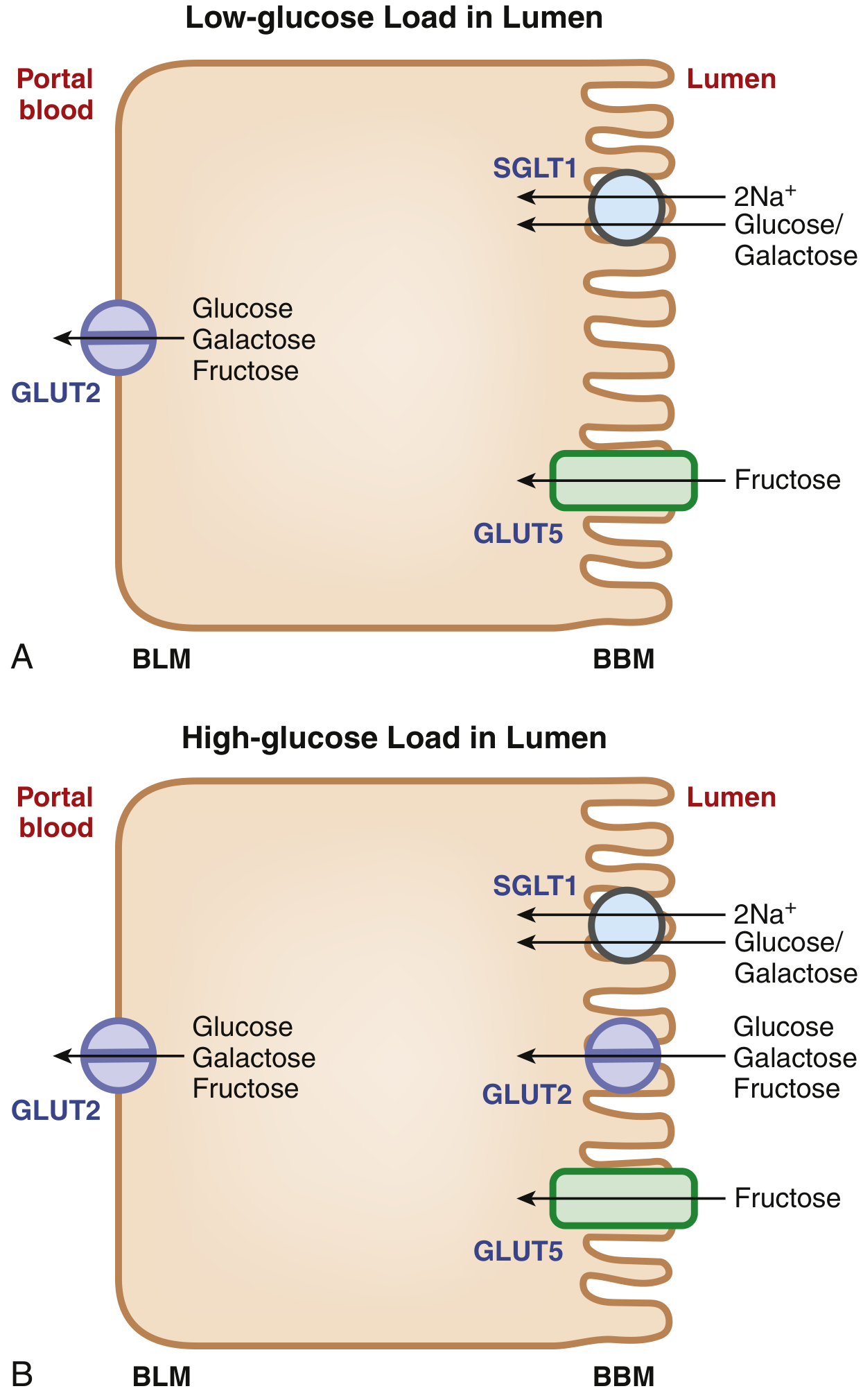
The classical model shows GLUT2 only on the BLM. However, when the intestinal lumen faces a high sugar load:
- SGLT1-mediated glucose entry acts as the signal that triggers trafficking of GLUT2 from the BLM to the BBM (apical membrane)
- GLUT2 then appears on both membranes simultaneously
- Because GLUT2 is a low-affinity, high-capacity transporter, its presence on the BBM under high-load conditions allows rapid, high-volume absorption that SGLT1 alone (high affinity but lower capacity) cannot achieve
- This GLUT2 recruitment to the BBM also facilitates fructose absorption - since SGLT1 cannot transport fructose, the apical GLUT2 provides an extra route for fructose entry beyond GLUT5
- Evidence from SGLT1 knockout mice confirms this: Sglt1-null mice cannot recruit Glut2 to the BBM, confirming SGLT1-mediated entry as the trigger
Summary Table: The Three Intestinal Sugar Transporters
| Transporter | Gene | Membrane | Substrates | Mechanism | Na⁺ Dependent? | Affinity |
|---|---|---|---|---|---|---|
| SGLT1 | SLC5A1 | Apical (BBM) | Glucose, Galactose | Secondary active (2Na⁺:1 sugar) | Yes | High |
| GLUT5 | SLC2A5 | Apical (BBM) | Fructose only | Facilitated diffusion | No | - |
| GLUT2 | SLC2A2 | Basolateral (BLM); also BBM at high load | Glucose, Galactose, Fructose | Facilitated diffusion | No | Low |
Knockout Mouse Evidence
| Gene Deleted | Effect |
|---|---|
| Sglt1 | Glucose/galactose malabsorption; no fructose effect; no GLUT2 trafficking to BBM |
| Glut5 | Fructose malabsorption; glucose/galactose unaffected |
| Glut2 | Surprisingly, no defect in intestinal glucose absorption (suggesting alternative exit mechanisms like exocytosis); however lethal systemically due to loss of pancreatic β-cell glucose sensing → hypoinsulinemia and hyperglycemia |
Some Fructose Becomes Glucose Inside the Enterocyte
A portion of fructose entering the cell is:
- Phosphorylated
- Converted to glucose intracellularly
- Transported as glucose into the portal circulation
This is why fructose contributes to hepatic glucose/triglyceride load.
Clinical Relevance
| Condition | Mechanism |
|---|---|
| Glucose-Galactose Malabsorption | Rare autosomal recessive mutation in SGLT1 (SLC5A1); severe osmotic diarrhea in neonates on glucose/galactose feeds; responds to fructose-only diet |
| Fructose malabsorption | Reduced GLUT5 expression or capacity; bloating, diarrhea after fructose intake |
| SGLT2 inhibitors (gliflozins) | Act on SGLT2 in the kidney (not intestine) to block renal glucose reabsorption; diabetes treatment |
| Lactose intolerance | Deficient brush-border lactase; undigested lactose reaches colon → osmotic diarrhea, hydrogen gas (basis of breath hydrogen test) |
Sources: Guyton and Hall Textbook of Medical Physiology; Sleisenger and Fordtran's Gastrointestinal and Liver Disease; Sabiston Textbook of Surgery; Yamada's Textbook of Gastroenterology 7th ed.
This is a shared conversation. Sign in to Orris to start your own chat.