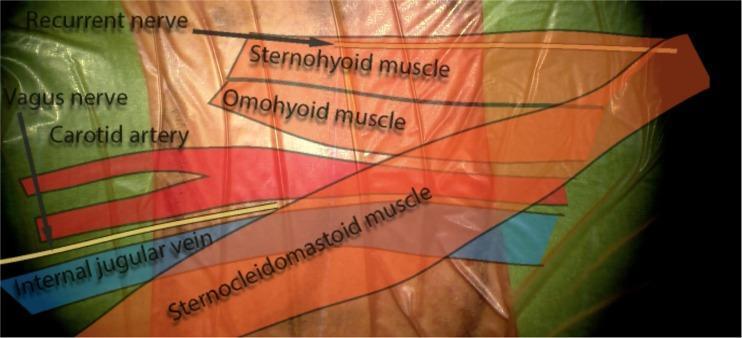
Uncus short answer questions
Uncus - Short Answer Questions
- Ipsilateral pupillary dilation (blown pupil) from compression of the oculomotor nerve (CN III) as it runs along the edge of the tentorium
- Progressive oculomotor palsy (ptosis, eye deviates down and out)
- Contralateral hemiparesis from compression of the ipsilateral cerebral peduncle/corticospinal tract
- Decreasing level of consciousness as the herniation progresses and compresses the midbrain reticular formation


Explain these topics according to professional exam 1st year mbbs 5 mark answer
Topic Map (from your notes)
How I'll format each answer
Which section should I start writing full 5-mark answers for first?
Uncus - Short Answer Questions
- Ipsilateral pupillary dilation (blown pupil) from compression of the oculomotor nerve (CN III) as it runs along the edge of the tentorium
- Progressive oculomotor palsy (ptosis, eye deviates down and out)
- Contralateral hemiparesis from compression of the ipsilateral cerebral peduncle/corticospinal tract
- Decreasing level of consciousness as the herniation progresses and compresses the midbrain reticular formation
Topic Map (from your notes)
How I'll format each answer
1. Breast (Mammary Gland)
2. Cubital Fossa
- Base (proximal): imaginary line joining medial and lateral epicondyles
- Lateral border: medial margin of brachioradialis
- Medial border: lateral margin of pronator teres
- Roof: deep fascia reinforced by bicipital aponeurosis, superficial fascia, and skin (crossed by median cubital vein)
- Floor: brachialis and supinator muscles Contents (lateral to medial - "TAN," or better remembered as Tendon-Artery-Nerve): Biceps tendon, Brachial artery (dividing into radial and ulnar arteries at its apex), Median nerve. Radial nerve lies deep, between brachioradialis and brachialis. Clinical correlation: Site for venepuncture (median cubital vein), BP measurement (brachial artery auscultation), and IV cannulation. Supracondylar fracture of humerus risks injury to brachial artery and median nerve here.
3. Axillary Artery
- 1st part (proximal to pectoralis minor): 1 branch - Superior thoracic artery
- 2nd part (behind pectoralis minor): 2 branches - Thoracoacromial and Lateral thoracic
- 3rd part (distal to pectoralis minor): 3 branches - Subscapular, Anterior circumflex humeral, Posterior circumflex humeral (Mnemonic: "1-2-3, S-TL-SAP") Relations: Related to cords of brachial plexus which surround the 2nd part (medial, lateral, posterior cords named accordingly). Clinical correlation: Site of surgical exposure for axillary block anesthesia; aneurysm can compress brachial plexus; important in axillary lymph node dissection for breast cancer.
4. Intermuscular Septa/Spaces (of Arm)
5. Rotator Cuff
6. Axilla (Contents)
- Axillary artery and its 3 parts/branches
- Axillary vein (medial to artery)
- Brachial plexus (cords and terminal branches)
- Axillary lymph nodes (5 groups - anterior/pectoral, posterior/subscapular, lateral, central, apical)
- Axillary tail of breast
- Fat and loose connective tissue Clinical correlation: Site of axillary lymph node clearance in breast cancer; long thoracic nerve (to serratus anterior) and thoracodorsal nerve (to latissimus dorsi) are at risk during axillary surgery - injury causes winging of scapula or weak arm adduction respectively.
7. Muscles Producing Abduction of Shoulder Joint
- 0-15 deg: Supraspinatus (initiator)
- 15-90 deg: Deltoid (middle fibers) - prime mover
- 90-180 deg (full overhead): Trapezius and Serratus anterior producing scapular rotation (scapulohumeral rhythm, ratio 2:1 glenohumeral to scapulothoracic movement) Clinical correlation: Axillary nerve injury paralyzes deltoid, causing loss of abduction beyond 15 deg and flattened "regimental badge" sensory loss over lower deltoid. Painful arc (60-120 deg) indicates rotator cuff impingement/tear.
8. Carpal Tunnel
- 4 tendons of flexor digitorum superficialis
- 4 tendons of flexor digitorum profundus
- 1 tendon of flexor pollicis longus (each profundus/FPL tendon has its own synovial sheath)
- Median nerve (most superficial, lies just deep to flexor retinaculum) (Flexor carpi radialis lies in a separate compartment split off the retinaculum; palmaris longus and ulnar nerve/artery pass superficial to the retinaculum, NOT through the tunnel.) Clinical correlation - Carpal Tunnel Syndrome: Compression of median nerve within the tunnel causes pain/paraesthesia in radial 3.5 fingers, thenar wasting, weak thumb opposition/abduction (positive Tinel's and Phalen's tests). Common in pregnancy, hypothyroidism, rheumatoid arthritis, and repetitive strain. Treated by surgical division of flexor retinaculum.
9. Brachial Plexus
- Erb's palsy (upper trunk, C5-C6): "waiter's tip" position - arm adducted, medially rotated, forearm pronated; from birth trauma or excessive traction.
- Klumpke's palsy (lower trunk, C8-T1): claw hand with possible Horner's syndrome, from breech delivery or sudden upward traction.
10. Palmar Spaces
- Thenar space: deep to flexor tendons of index finger, communicates with the space around the adductor pollicis.
- Midpalmar space: deep to flexor tendons of middle, ring, little fingers. Clinical correlation: Pyogenic infection (from penetrating injury/spread of tendon sheath infection) collects in these spaces causing a tense, painful swelling with loss of the normal palmar concavity; may require surgical drainage. Infection can also spread proximally into the forearm via the "space of Parona."
11. Clavipectoral Fascia
12. Pulp Space of Thumb (Terminal Pulp Space of Digits)
13. Median Nerve and Anterior Interosseous Nerve (Muscles Supplied)
- Forearm (via its main trunk in the front of forearm): Pronator teres, Flexor carpi radialis, Palmaris longus, Flexor digitorum superficialis
- Hand (via recurrent/motor branch, "LOAF" muscles): Lateral 2 Lumbricals, Opponens pollicis, Abductor pollicis brevis, Flexor pollicis brevis (superficial head) Anterior interosseous nerve (branch of median nerve given off just below elbow):
- Flexor pollicis longus
- Lateral half of Flexor digitorum profundus (to index and middle fingers)
- Pronator quadratus Clinical correlation: Anterior interosseous nerve syndrome causes weakness of pinch grip (inability to make an "OK" sign - the Kiloh-Nevin sign) with no sensory loss (pure motor nerve). Complete median nerve injury at the wrist gives "Ape thumb deformity" with sensory loss over radial 3.5 digits.
14. Flexor Retinaculum and Extensor Retinaculum
- A strong fibrous band on the front of the wrist, converting the concave carpal arch into the carpal tunnel.
- Attachments: laterally to tubercles of scaphoid and trapezium; medially to pisiform and hook of hamate.
- Structures deep to it: 9 flexor tendons + median nerve (see Carpal Tunnel above); FCR passes through a split in it.
- Structures superficial to it: ulnar nerve/artery, palmaris longus tendon, palmar cutaneous branch of median nerve. Extensor retinaculum:
- Located on the dorsum of the wrist, a thickened band of deep fascia holding extensor tendons in place, preventing "bowstringing" during wrist extension.
- Attachments: laterally to distal radius; medially to pisiform and triquetrum.
- Forms 6 osseofibrous compartments for the extensor tendons (deep to it), each with its own synovial sheath. Clinical correlation: Flexor retinaculum division relieves carpal tunnel syndrome; extensor retinaculum thickening/inflammation contributes to De Quervain's tenosynovitis (1st compartment - APL/EPB tendons).
15. Claw Hand
- Ulnar nerve palsy (main cause): paralysis of medial 2 lumbricals and all interossei -> claw deformity of ring and little fingers ("ulnar claw"). Paradoxically, a high (proximal) lesion at the elbow produces a less obvious claw ("ulnar paradox") because the flexor digitorum profundus to ring/little fingers is also paralyzed, removing the flexing force at the IP joints.
- Combined median + ulnar nerve palsy: claw affects all four fingers ("main-en-griffe").
- Also seen in leprosy (ulnar nerve most commonly affected peripheral nerve) and Klumpke's palsy. Clinical correlation: Tested by asking the patient to make a fist or by Froment's sign / card test for adductor pollicis weakness (ulnar). Treated by tendon transfer surgery (e.g., lasso procedures) if nerve recovery fails.
16. Wrist Drop
17. Dorsum of Hand - Cutaneous Innervation Diagram
- Radial side (thumb, index, middle, half of ring finger, dorsally up to distal IP joints only): Superficial branch of radial nerve
- Ulnar side (little finger, half of ring finger dorsally): Dorsal branch of ulnar nerve
- Tips of index, middle, ring, little fingers on the dorsum (beyond the DIP joints): supplied by palmar digital nerves (median/ulnar) which wrap around to supply the dorsal tips, since these areas are used for fine touch.
- Central dorsum of hand/wrist: Posterior cutaneous nerve of forearm (from radial nerve) proximally. Clinical correlation: In radial nerve injury, sensory loss is limited to a small area over the first dorsal web space because the median and ulnar nerves compensate for the rest of the dorsum via their digital branches wrapping around the fingertips.

Exam oriented 5 mark answer according to bdc with diagram
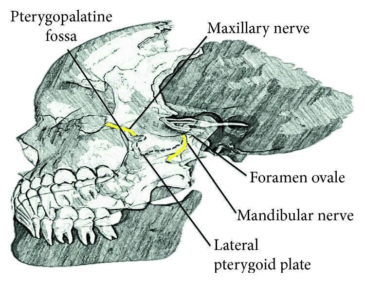
You are a 1st year MBBS anatomy tutor writing EXAM-ORIENTED 5-mark answers in the style of B.D. Chaurasia's "Human Anatomy" textbook (concise, structured, exam-pattern), for the following Head & Neck topics: 1. Dural venous sinuses (with focus on Cavernous sinus) 2. Anatomy of the orbit (margins of the orbit / bony orbit) 3. Paranasal sinuses 4. Nasal cavity 5. Pterygopalatine fossa 6. Extraocular muscles (muscles of the eye) 7. Lateral wall of the nose 8. Pharyngeal arches 9. Tongue 10. Middle ear For EACH topic, produce a complete 5-mark exam answer following this structure (matching how BD Chaurasia presents topics): - Definition/Introduction - Location/Extent/Boundaries (as applicable) - Parts/Divisions/Relations/Contents (as applicable, use tables or bullet points for nerve/muscle/artery lists as BDC does) - Applied Anatomy / Clinical correlation (BDC always has a clinical note - e.g. cavernous sinus thrombosis, orbital fractures, sinusitis, DNS, epistaxis, ptosis/squint, pharyngeal arch derivative anomalies, tongue-tie/carcinoma tongue, otitis media) - A note describing what diagram should be drawn (labeled parts) for full marks You have access to query_library_db (medical textbook library with FTS search), search_image and query_images_db (medical image caption database), and fetch_image. For EACH of the 10 topics, use search_image or query_images_db to try to find ONE relevant existing labeled anatomical diagram/image (e.g. search for 'cavernous sinus diagram', 'bony orbit margins', 'paranasal sinuses diagram', 'pterygopalatine fossa', 'extraocular muscles diagram', 'lateral wall of nose', 'pharyngeal arches diagram', 'tongue papillae dorsum diagram', 'middle ear anatomy diagram'). If you find a good match, include the image_url in your answer using markdown image syntax so it can be embedded. If no good image is found for a topic, just note "diagram: [describe what to draw and label]" instead - do not fabricate a URL. Also use query_library_db to verify facts against standard anatomy/clinical textbooks in the library (e.g. Gray's Anatomy for Students, THIEME Atlas of Anatomy) where useful, but keep answers concise and exam-focused (not textbook-length) - this is for 5-mark short answer questions, so each answer should be roughly 150-300 words plus the diagram note/image. Return your FINAL answer as a single well-organized markdown document with a heading for each of the 10 topics, in the order listed above, each containing the full 5-mark answer and diagram/image. Do not include your intermediate search process, only the final compiled answers.
You are a 1st year MBBS anatomy tutor writing EXAM-ORIENTED 5-mark answers in the style of B.D. Chaurasia's "Human Anatomy" textbook (concise, structured, exam-pattern), for the following topics: 1. Cervical fascia (superficial and deep cervical fascia - layers of deep cervical fascia) 2. Parotid gland 3. Ligament of Berry (of the thyroid gland) 4. Tracheal ring / T4 vertebral level landmark (transpyloric-type landmark question - "T4 landmark" refers to the vertebral level of the sternal angle / bifurcation of trachea / arch of aorta - cover the key structures found at the T4 (sternal angle) vertebral level as a short-answer landmark topic) 5. Contents of the suprasternal space (space of Burns) 6. Spaces related to the deep cervical fascia (e.g. pretracheal space, retropharyngeal space, carotid sheath, space of Burns - give a structured overview of fascial spaces of the neck) 7. Thyroid gland 8. Epistaxis and Little's area (Kiesselbach's plexus) 9. Shoulder joint and Rotator cuff 10. Lymphatic drainage of the breast 11. Clavipectoral fascia 12. Otic ganglion 13. Histology (brief microscopic structure, 1 short paragraph each, exam short-note style) of: (a) Cerebellum, (b) Cerebral cortex/cerebrum, (c) Spinal cord (grey and white matter arrangement) For topics 1-12, produce a complete 5-mark exam answer following BD Chaurasia's structure: - Definition/Introduction - Location/Extent/Boundaries/Attachments (as applicable) - Parts/Divisions/Relations/Contents (use bullet points or tables for nerve/muscle/artery lists as BDC does) - Applied Anatomy / Clinical correlation (BDC always includes this - e.g. cavernous sinus/Ludwig's angina for cervical fascia, parotid tumors/Frey's syndrome for parotid, recurrent laryngeal nerve injury near ligament of Berry during thyroidectomy, retropharyngeal abscess for neck spaces, thyroid swellings/thyroglossal cyst, epistaxis management, rotator cuff tear/frozen shoulder, breast cancer axillary spread, brachial plexus/lateral pectoral nerve injury near clavipectoral fascia, otic ganglion in parotid secretomotor pathway) - A note describing what diagram should be drawn (labeled parts) for full marks For topic 13 (histology), give 3 short exam-style micro-anatomy notes (~80-120 words each) covering key layers/cell types (e.g. cerebellum: molecular, Purkinje, granular layers; cerebrum: 6 layers of neocortex; spinal cord: grey matter H-shape with horns, white matter tracts) with a note on what histology diagram to draw. You have access to query_library_db (medical textbook library with FTS search), search_image and query_images_db (medical image caption database), and fetch_image. For as many topics as possible, use search_image or query_images_db to find ONE relevant existing labeled diagram or histology micrograph (e.g. search for 'deep cervical fascia layers', 'parotid gland diagram', 'thyroid gland diagram relations', "Kiesselbach's plexus little's area", 'rotator cuff shoulder diagram', 'axillary lymph nodes breast drainage', 'clavipectoral fascia diagram', 'otic ganglion diagram', 'cerebellum histology micrograph Purkinje cells', 'cerebral cortex histology layers', 'spinal cord cross section histology grey matter'). If you find a good match, include the image_url in your answer using markdown image syntax. If no good image is found, note "diagram: [describe what to draw and label]" instead - do not fabricate a URL. Also use query_library_db to verify facts against standard anatomy/clinical textbooks in the library where useful, but keep answers concise and exam-focused (5-mark short answers, not textbook-length essays, roughly 150-300 words each plus diagram/image; histology notes shorter as specified). Return your FINAL answer as a single well-organized markdown document with a heading for each topic in the order listed above, each containing the full answer and diagram/image. Do not include your intermediate search process, only the final compiled answers.
Uncus - Short Answer Questions
- Ipsilateral pupillary dilation (blown pupil) from compression of the oculomotor nerve (CN III) as it runs along the edge of the tentorium
- Progressive oculomotor palsy (ptosis, eye deviates down and out)
- Contralateral hemiparesis from compression of the ipsilateral cerebral peduncle/corticospinal tract
- Decreasing level of consciousness as the herniation progresses and compresses the midbrain reticular formation
Topic Map (from your notes)
How I'll format each answer
1. Breast (Mammary Gland)
2. Cubital Fossa
- Base (proximal): imaginary line joining medial and lateral epicondyles
- Lateral border: medial margin of brachioradialis
- Medial border: lateral margin of pronator teres
- Roof: deep fascia reinforced by bicipital aponeurosis, superficial fascia, and skin (crossed by median cubital vein)
- Floor: brachialis and supinator muscles Contents (lateral to medial - "TAN," or better remembered as Tendon-Artery-Nerve): Biceps tendon, Brachial artery (dividing into radial and ulnar arteries at its apex), Median nerve. Radial nerve lies deep, between brachioradialis and brachialis. Clinical correlation: Site for venepuncture (median cubital vein), BP measurement (brachial artery auscultation), and IV cannulation. Supracondylar fracture of humerus risks injury to brachial artery and median nerve here.
3. Axillary Artery
- 1st part (proximal to pectoralis minor): 1 branch - Superior thoracic artery
- 2nd part (behind pectoralis minor): 2 branches - Thoracoacromial and Lateral thoracic
- 3rd part (distal to pectoralis minor): 3 branches - Subscapular, Anterior circumflex humeral, Posterior circumflex humeral (Mnemonic: "1-2-3, S-TL-SAP") Relations: Related to cords of brachial plexus which surround the 2nd part (medial, lateral, posterior cords named accordingly). Clinical correlation: Site of surgical exposure for axillary block anesthesia; aneurysm can compress brachial plexus; important in axillary lymph node dissection for breast cancer.
4. Intermuscular Septa/Spaces (of Arm)
5. Rotator Cuff
6. Axilla (Contents)
- Axillary artery and its 3 parts/branches
- Axillary vein (medial to artery)
- Brachial plexus (cords and terminal branches)
- Axillary lymph nodes (5 groups - anterior/pectoral, posterior/subscapular, lateral, central, apical)
- Axillary tail of breast
- Fat and loose connective tissue Clinical correlation: Site of axillary lymph node clearance in breast cancer; long thoracic nerve (to serratus anterior) and thoracodorsal nerve (to latissimus dorsi) are at risk during axillary surgery - injury causes winging of scapula or weak arm adduction respectively.
7. Muscles Producing Abduction of Shoulder Joint
- 0-15 deg: Supraspinatus (initiator)
- 15-90 deg: Deltoid (middle fibers) - prime mover
- 90-180 deg (full overhead): Trapezius and Serratus anterior producing scapular rotation (scapulohumeral rhythm, ratio 2:1 glenohumeral to scapulothoracic movement) Clinical correlation: Axillary nerve injury paralyzes deltoid, causing loss of abduction beyond 15 deg and flattened "regimental badge" sensory loss over lower deltoid. Painful arc (60-120 deg) indicates rotator cuff impingement/tear.
8. Carpal Tunnel
- 4 tendons of flexor digitorum superficialis
- 4 tendons of flexor digitorum profundus
- 1 tendon of flexor pollicis longus (each profundus/FPL tendon has its own synovial sheath)
- Median nerve (most superficial, lies just deep to flexor retinaculum) (Flexor carpi radialis lies in a separate compartment split off the retinaculum; palmaris longus and ulnar nerve/artery pass superficial to the retinaculum, NOT through the tunnel.) Clinical correlation - Carpal Tunnel Syndrome: Compression of median nerve within the tunnel causes pain/paraesthesia in radial 3.5 fingers, thenar wasting, weak thumb opposition/abduction (positive Tinel's and Phalen's tests). Common in pregnancy, hypothyroidism, rheumatoid arthritis, and repetitive strain. Treated by surgical division of flexor retinaculum.
9. Brachial Plexus
- Erb's palsy (upper trunk, C5-C6): "waiter's tip" position - arm adducted, medially rotated, forearm pronated; from birth trauma or excessive traction.
- Klumpke's palsy (lower trunk, C8-T1): claw hand with possible Horner's syndrome, from breech delivery or sudden upward traction.
10. Palmar Spaces
- Thenar space: deep to flexor tendons of index finger, communicates with the space around the adductor pollicis.
- Midpalmar space: deep to flexor tendons of middle, ring, little fingers. Clinical correlation: Pyogenic infection (from penetrating injury/spread of tendon sheath infection) collects in these spaces causing a tense, painful swelling with loss of the normal palmar concavity; may require surgical drainage. Infection can also spread proximally into the forearm via the "space of Parona."
11. Clavipectoral Fascia
12. Pulp Space of Thumb (Terminal Pulp Space of Digits)
13. Median Nerve and Anterior Interosseous Nerve (Muscles Supplied)
- Forearm (via its main trunk in the front of forearm): Pronator teres, Flexor carpi radialis, Palmaris longus, Flexor digitorum superficialis
- Hand (via recurrent/motor branch, "LOAF" muscles): Lateral 2 Lumbricals, Opponens pollicis, Abductor pollicis brevis, Flexor pollicis brevis (superficial head) Anterior interosseous nerve (branch of median nerve given off just below elbow):
- Flexor pollicis longus
- Lateral half of Flexor digitorum profundus (to index and middle fingers)
- Pronator quadratus Clinical correlation: Anterior interosseous nerve syndrome causes weakness of pinch grip (inability to make an "OK" sign - the Kiloh-Nevin sign) with no sensory loss (pure motor nerve). Complete median nerve injury at the wrist gives "Ape thumb deformity" with sensory loss over radial 3.5 digits.
14. Flexor Retinaculum and Extensor Retinaculum
- A strong fibrous band on the front of the wrist, converting the concave carpal arch into the carpal tunnel.
- Attachments: laterally to tubercles of scaphoid and trapezium; medially to pisiform and hook of hamate.
- Structures deep to it: 9 flexor tendons + median nerve (see Carpal Tunnel above); FCR passes through a split in it.
- Structures superficial to it: ulnar nerve/artery, palmaris longus tendon, palmar cutaneous branch of median nerve. Extensor retinaculum:
- Located on the dorsum of the wrist, a thickened band of deep fascia holding extensor tendons in place, preventing "bowstringing" during wrist extension.
- Attachments: laterally to distal radius; medially to pisiform and triquetrum.
- Forms 6 osseofibrous compartments for the extensor tendons (deep to it), each with its own synovial sheath. Clinical correlation: Flexor retinaculum division relieves carpal tunnel syndrome; extensor retinaculum thickening/inflammation contributes to De Quervain's tenosynovitis (1st compartment - APL/EPB tendons).
15. Claw Hand
- Ulnar nerve palsy (main cause): paralysis of medial 2 lumbricals and all interossei -> claw deformity of ring and little fingers ("ulnar claw"). Paradoxically, a high (proximal) lesion at the elbow produces a less obvious claw ("ulnar paradox") because the flexor digitorum profundus to ring/little fingers is also paralyzed, removing the flexing force at the IP joints.
- Combined median + ulnar nerve palsy: claw affects all four fingers ("main-en-griffe").
- Also seen in leprosy (ulnar nerve most commonly affected peripheral nerve) and Klumpke's palsy. Clinical correlation: Tested by asking the patient to make a fist or by Froment's sign / card test for adductor pollicis weakness (ulnar). Treated by tendon transfer surgery (e.g., lasso procedures) if nerve recovery fails.
16. Wrist Drop
17. Dorsum of Hand - Cutaneous Innervation Diagram
- Radial side (thumb, index, middle, half of ring finger, dorsally up to distal IP joints only): Superficial branch of radial nerve
- Ulnar side (little finger, half of ring finger dorsally): Dorsal branch of ulnar nerve
- Tips of index, middle, ring, little fingers on the dorsum (beyond the DIP joints): supplied by palmar digital nerves (median/ulnar) which wrap around to supply the dorsal tips, since these areas are used for fine touch.
- Central dorsum of hand/wrist: Posterior cutaneous nerve of forearm (from radial nerve) proximally. Clinical correlation: In radial nerve injury, sensory loss is limited to a small area over the first dorsal web space because the median and ulnar nerves compensate for the rest of the dorsum via their digital branches wrapping around the fingertips.
Which topic should I write the full 5-mark exam answer for first?
Which topic should I write up first as a full 5-mark exam answer (with diagram/image)?
Explain all
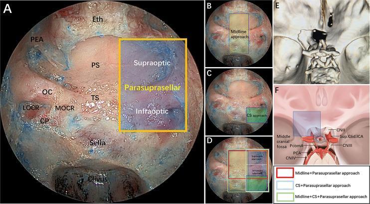
bony orbit margins anatomy diagram
paranasal sinuses diagram anatomy
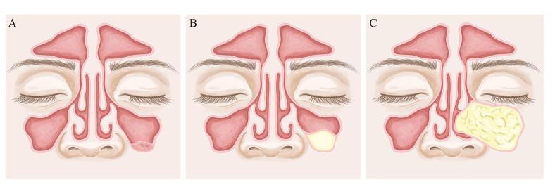
Anatomical diagram in a frontal (coronal) view of the midface, illustrating three distinct types of benign paranasal sinus lesions within the maxillary sinuses. All three panels (A, B, and C) display consistent anatomy, including the frontal sinuses, nasal cavity with turbinates and septum, and the maxillary sinuses. Panel A depicts a Pseudocyst, characterized by a small, dome-shaped, pinkish lesion originating from the floor of the left maxillary sinus. Panel B illustrates a Mucous Retention Cyst, shown as a smooth, pale-yellow, well-circumscribed mass located in the inferior aspect of the right maxillary sinus. Panel C demonstrates a Mucocele, represented as a significantly larger, yellowish, lobulated, and complex mass that occupies most of the right maxillary sinus, potentially exerting pressure on the sinus walls. This comparison focuses on the differential diagnosis of paranasal sinus opacifications, highlighting variations in morphology, size, and internal texture. The illustration is intended for educational use in otolaryngology and radiology to distinguish between fluid-filled cysts and more expansive sinus pathologies.
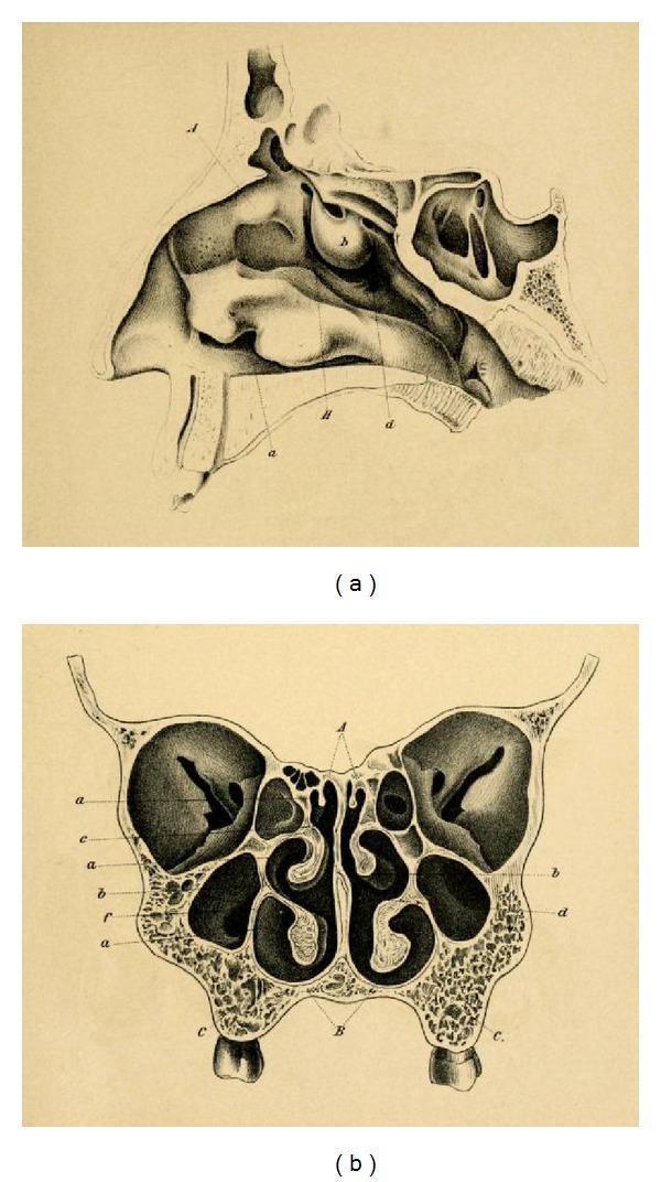
This historical anatomical diagram by Emil Zuckerkandl (1882) illustrates the paranasal sinuses and nasal anatomy in two planes. Image (a) is a sagittal section of the nasal cavity, detailing the complex arrangement of the superior, middle, and inferior turbinates (conchae) along the lateral wall. It visualizes the frontal sinus superiorly, the sphenoid sinus posteriorly, and the relationship between the nasal cavity and the hard palate. Image (b) presents a frontal (coronal) section, showcasing the bilateral symmetry of the mid-facial anatomy. Key features include the central nasal septum, the convoluted turbinates projecting into the nasal passages, and the expansive maxillary sinuses situated laterally within the maxilla bones. Small ethmoid air cells are visible medially to the orbits and superior to the maxillary sinuses. This anatomical illustration serves as a foundational reference for otorhinolaryngology and neurosurgical approaches, particularly regarding the development of transsphenoidal surgery for pituitary lesions.
extraocular muscles of eye diagram
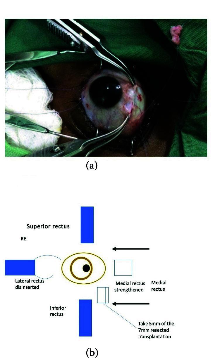
A composite image consisting of a clinical photograph and a schematic diagram illustrating a strabismus surgical procedure. Image (a) is a close-up clinical photograph of an intraoperative ophthalmological field showing the right eye. Two surgical forceps are retracting tissue and muscle, while 6-0 double-ended Vicryl sutures are visible within the moist, erythematous surgical site. A small portion of resected extraocular muscle tissue is seen on the green sterile drape in the top right. Image (b) is an anatomical schematic of the right eye (RE) detailing the surgical plan. It labels the superior, inferior, and medial rectus muscles. The diagram indicates a 'disinserted' lateral rectus and a 'strengthened' medial rectus, with specific instructions for a 5mm graft from a 7mm resected muscle transplantation. The content demonstrates complex extraocular muscle repositioning or transpositioning techniques used in corrective ophthalmic surgery for ocular motility disorders.
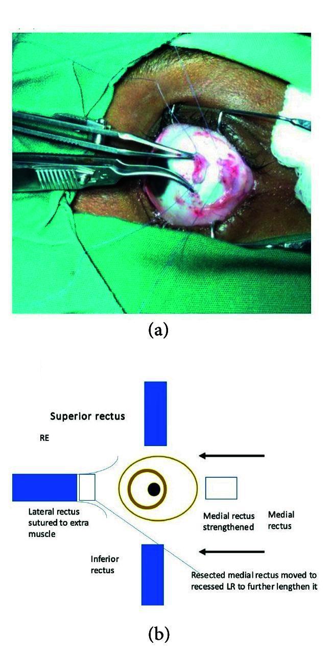
This composite educational resource consists of a clinical photograph and a corresponding anatomical diagram illustrating a complex strabismus surgical procedure. The photograph (a) depicts an intraoperative view of a human eye draped for surgery. The sclera is exposed, showing localized vascular congestion and surgical trauma. Fine blue monofilament sutures are visible, and ophthalmic forceps are seen manipulating a resected extraocular muscle tendon. The diagram (b) provides a schematic representation of the 'transplantation of resected medial rectus to the lateral rectus.' It labels the four rectus muscles—Superior, Inferior, Lateral (LR), and Medial (MR)—and uses arrows and annotations to explain the surgical logic: the resected MR is being moved to the recessed LR to further lengthen it, a technique typically used for ocular alignment in conditions like severe nerve palsy. This visual illustrates key concepts in ophthalmic reconstructive surgery and extraocular muscle transposition techniques for advanced strabismus management.
lateral wall of nose diagram anatomy
pharyngeal arches diagram embryology
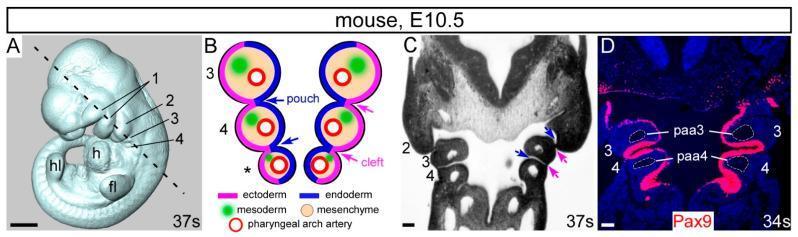
This composite educational image illustrates the developmental anatomy of the pharyngeal arches in a mouse embryo at embryonic day 10.5 (E10.5). (A) A 3D whole-mount embryo reconstruction showing anatomical landmarks including the forelimb bud (fl), hindlimb bud (hl), heart (h), and the sequential pharyngeal arches (1-4). (B) A schematic diagram of a coronal section detailing the embryological layers: outer ectoderm (pink), internal endoderm (blue) forming pouches, and a mesenchymal core containing mesoderm (green) and pharyngeal arch arteries (red circles). (C) A corresponding histological coronal section showing the physical arrangement of arches 2, 3, and 4. (D) An immunofluorescence-stained coronal section showing Pax9 expression (red signal) localized within the pharyngeal endoderm. The image highlights the specific positioning of the 3rd and 4th pharyngeal arch arteries (paa3, paa4) within their respective arches. This material demonstrates key concepts in embryology, specifically the rostral-caudal development of the pharyngeal apparatus and the molecular markers of the pharyngeal endoderm essential for cardiovascular and craniofacial morphogenesis.
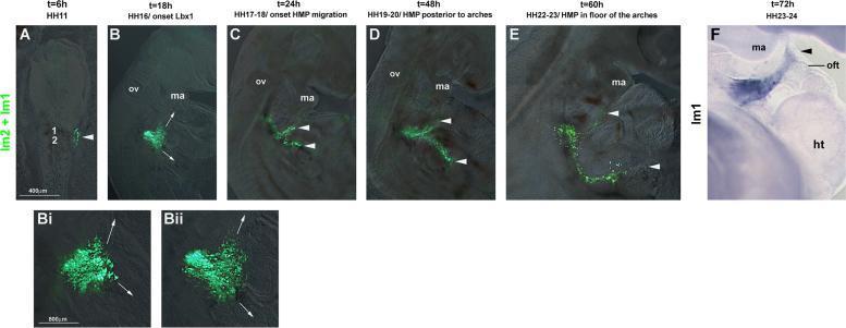
This composite diagnostic and pathophysiology diagram illustrates the migration of occipital lateral mesoderm in a developing avian embryo using GFP electroporation and in situ hybridization. Panels A through E display a temporal sequence from HH11 (t=6h) to HH22-23 (t=60h). Fluorescent GFP-labeled cells, initially localized near somites 1 and 2 (lm1+lm2), are shown organizing into two distinct migratory streams: a rostral stream projecting toward the pharyngeal arches and a lateral-caudal stream. Higher magnification panels (Bi, Bii) at HH16 highlight the onset of this bifurcation. Panel F shows GFP mRNA expression (blue staining) at HH23-24, demonstrating the accumulation of these lateral mesoderm-derived cells near the mandibular arch (ma) and the heart's outflow tract (oft). Anatomical landmarks include the otic vesicle (ov), heart (ht), and mandibular arch. This content serves as an educational resource for embryology and developmental biology, specifically regarding circumpharyngeal cell movements and the origins of cranial and cardiac mesodermal structures.
tongue dorsum papillae diagram
middle ear anatomy diagram
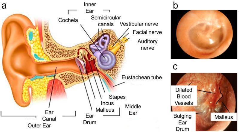
This medical educational graphic comprises an anatomical diagram and clinical otoscopic photographs illustrating the human ear in healthy and pathological states. Panel (a) is a cross-sectional anatomical diagram of the human ear, labeling key structures including the outer ear (pinna, ear canal), middle ear (eardrum/tympanic membrane, malleus, incus, stapes), and inner ear (cochlea, semicircular canals, vestibular and auditory nerves, Eustachian tube). Panel (b) shows a healthy tympanic membrane via white light otoscopy, characterized by a translucent, pinkish-gray appearance, a normal light reflex, and a concave shape. Panel (c) provides a comparative otoscopic view of acute otitis media. Pathological markers labeled include a bulging eardrum due to middle ear pressure, intense erythema (redness), and dilated blood vessels (vascular injection) across the membrane surface. The malleus remains visible but appears distorted by the inflammatory swelling. The image serves to teach the visual diagnostic criteria for middle ear infections compared to normal anatomy.
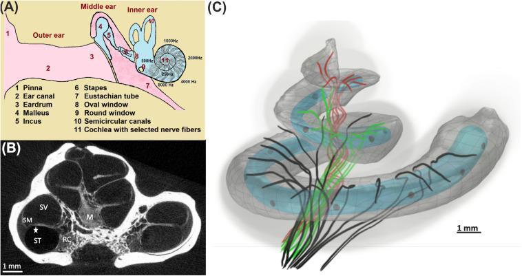
Educational composite detailing human ear anatomy and cochlear mechanics. Panel (A) is an anatomical diagram of the outer, middle, and inner ear, highlighting the conduction pathway from the pinna (1) and ear canal (2) to the ossicles (4-6) and the tonotopic frequency mapping of the cochlea (11). Panel (B) is a high-resolution cross-sectional diagnostic image of the cochlea, identifying the scala vestibuli (SV), scala media (SM), and scala tympani (ST), alongside the central modiolus (M) and Rosenthal’s canal (RC). Panel (C) presents a 3D pathophysiology diagram illustrating the tonotopic organization of auditory nerve fibers: basal (black), middle (green), and apical (red) turns. A light blue 12-channel cochlear implant electrode array is shown inserted into the translucent scala tympani. This visual content illustrates the relationship between cochlear microanatomy and medical device placement for treating hearing loss. Scale bars of 1 mm are provided for anatomical reference.