I have all the content needed. Let me now compile the comprehensive oral exam prep.All sourced directly from Schwartz's Principles of Surgery, 11th Edition - Chapter 29: Colon, Rectum, and Anus.
ORAL EXAM PREP: ANAL ABSCESS
Schwartz's Principles of Surgery, 11th Ed.
PART 1 - ANATOMY (Brief)
Anal Canal Structure
- The anatomic anal canal extends from the dentate (pectinate) line to the anal verge
- The dentate line marks the transition between columnar rectal mucosa (above) and squamous anoderm (below)
- The dentate line is surrounded by longitudinal mucosal folds called anal columns (columns of Morgagni) - between these are the anal crypts
- Anal glands open into these crypts at the dentate line level - their ducts traverse the internal sphincter into the intersphincteric plane (this is KEY for abscess pathogenesis)
Sphincter Complex
- Internal anal sphincter (IAS): smooth muscle, involuntary, distal extension of the inner circular muscle of the rectum - innervated by sympathetic + parasympathetic fibers
- External anal sphincter (EAS): skeletal muscle, voluntary - innervated by the inferior rectal branch of the pudendal nerve
- Puborectalis: part of the levator ani, forms the anorectal ring, innervated by pudendal nerve and direct branches of S3-S5
- The intersphincteric space separates the IAS and EAS, continuous with the perianal space distally, extends cephalad into the rectal wall
Perianal/Perirectal Spaces (Memorize These - They Determine Abscess Type)
| Space | Location | Boundaries |
|---|
| Perianal | Surrounds anus | Laterally continuous with fat of buttocks |
| Intersphincteric | Between IAS and EAS | Continuous with perianal space distally |
| Ischiorectal (ischiorectal fossa) | Lateral + posterior to anus | Medially: EAS; Laterally: ischium; Superiorly: levator ani; Inferiorly: transverse septum |
| Deep postanal | Posterior, above anococcygeal ligament, below levator ani | Connects both ischiorectal spaces posteriorly |
| Supralevator | Above levator ani, each side of rectum | Communicate posteriorly |
Vascular Supply (Brief)
- Superior rectal artery - from inferior mesenteric artery - upper rectum
- Middle rectal artery - from internal iliac
- Inferior rectal artery - from internal pudendal artery (branch of internal iliac)
Lymphatic Drainage
- Above dentate line: drains to inferior mesenteric AND internal iliac nodes
- Below dentate line: primarily to inguinal lymph nodes (also to inferior mesenteric and internal iliac)
This is clinically important - anal canal cancer below dentate line can spread to inguinal nodes
Nerve Supply
- Sympathetic: T6-T12, L1-L3 (inhibitory to colon motility)
- Parasympathetic: nervi erigentes from S2-S4 (stimulatory)
- Rectum is relatively insensate; anal canal below dentate line has somatic innervation (painful!)
PART 2 - PHYSIOLOGY (Brief)
Fluid & Electrolyte Exchange
- Colon absorbs ~1000-2000 mL/day of water (up to 5000 mL capacity)
- Na+ absorbed actively via Na+/K+ ATPase (up to 400 mEq/day)
- Water follows Na+ passively along osmotic gradient
- K+ is actively secreted and also passively absorbed
- Cl- absorbed via chloride-bicarbonate exchange
Motility, Defecation & Continence
- Continence depends on the anorectal angle (maintained by puborectalis) and the sphincter complex
- The rectum is a reservoir; when distended, the rectoanal inhibitory reflex relaxes the IAS
- Voluntary contraction of EAS maintains continence until defecation is appropriate
Colonic Flora
- Anaerobes predominate - Bacteroides species most common (10^11-10^12/mL)
- E. coli most numerous aerobe
- Flora important for vitamin K production and colonization resistance
PART 3 - ANAL ABSCESS (Anorectal Sepsis & Cryptoglandular Abscess)
Pathogenesis (Cryptoglandular Theory)
- Anal glands sit in the intersphincteric plane; their ducts traverse the IAS and open into anal crypts at the dentate line
- Infection of an anal gland → abscess in the intersphincteric plane
- The abscess enlarges and spreads along perianal/perirectal spaces → determines the TYPE of abscess
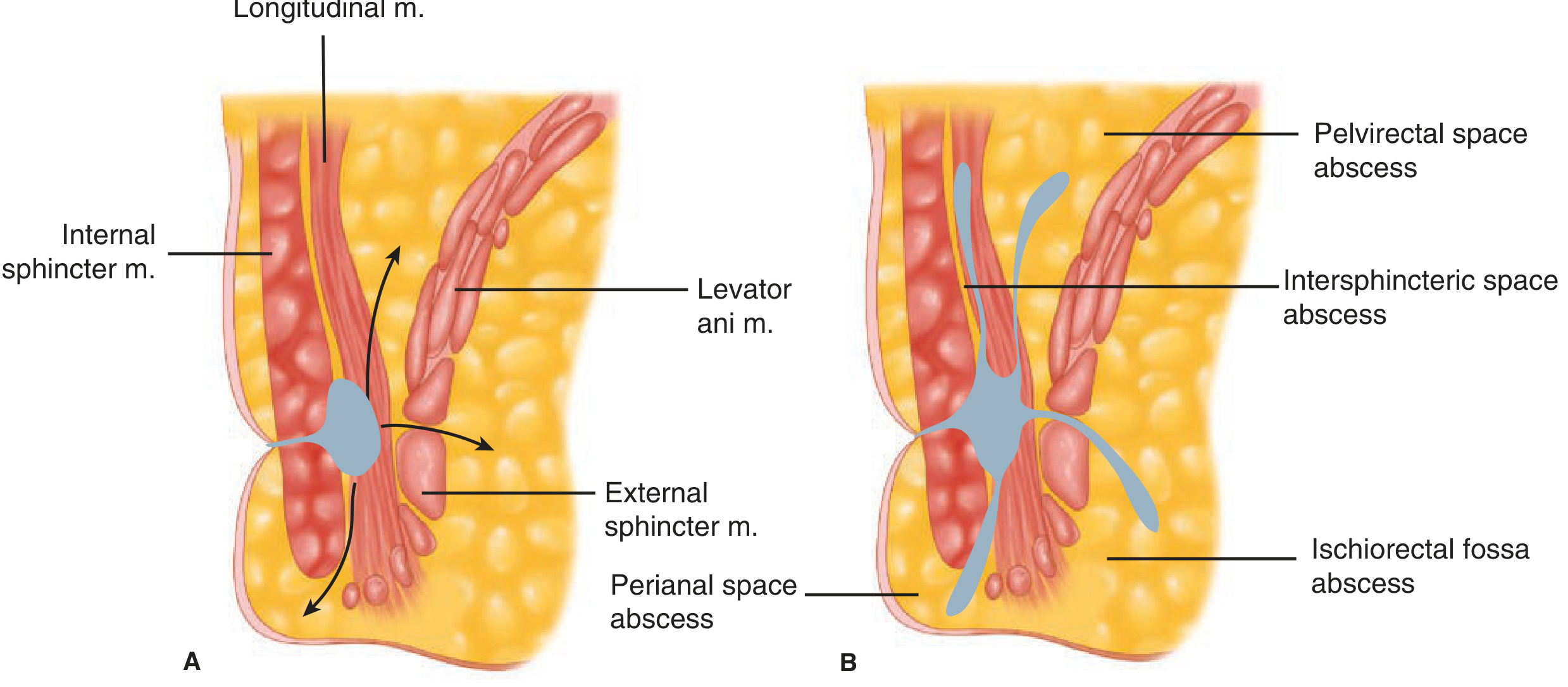
Types of Abscess (Based on Spread)
| Type | Spread/Location | Clinical Feature |
|---|
| Perianal | Most common; spreads to perianal space | Painful swelling at anal verge, visible |
| Ischiorectal | Spreads through EAS below puborectalis into ischiorectal fossa | Diffuse swelling in ischiorectal fossa, may be very large, may not be visible in perianal region; DRE reveals painful swelling laterally |
| Intersphincteric | Remains in intersphincteric space | Little external swelling, few perianal signs; pain is deep, "up inside," worsened by coughing/sneezing; so intense it precludes DRE; requires EUA |
| Supralevator | Uncommon; upward extension of intersphincteric or ischiorectal abscess, OR downward extension of intra-abdominal abscess | DRE: indurated, bulging mass above anorectal ring; can mimic intra-abdominal conditions |
| Horseshoe abscess | Ischiorectal abscess involving both sides via deep postanal space | Bilateral swelling, requires drainage of deep postanal space + counter-incisions |
Fig 29-36 - Pathways of anorectal infection (Schwartz's, p.1343)
Diagnosis
Symptoms:
- Severe anal pain - most common presenting complaint
- Palpable mass on inspection or DRE
- Fever
- Urinary retention
- Life-threatening sepsis (in severe/delayed cases)
Workup:
- Perianal and ischiorectal: usually diagnosed by physical exam alone
- Intersphincteric: requires Examination Under Anesthesia (EUA) - high index of suspicion
- Complex/atypical: CT or MRI to delineate anatomy
PART 4 - MANAGEMENT
General Principle
Anorectal abscesses should be drained as soon as the diagnosis is established. - Schwartz's, p.1341
Antibiotics: Only indicated if:
- Extensive overlying cellulitis
- Immunocompromised patient
- Diabetes mellitus
- Valvular heart disease
Antibiotics alone are INEFFECTIVE at treating perianal/perirectal infection. Drainage is the definitive treatment.
By Type:
Perianal Abscess:
- Most can be drained under local anesthesia in office/clinic/ER
- Larger/complicated → OR under general/regional anesthesia
- Skin incision + disk of skin excised to prevent premature closure
- No packing needed; sitz baths start next day
Ischiorectal Abscess:
- Incision in overlying skin
- Horseshoe abscess: drainage of deep postanal space (incising anococcygeal ligament) + counter-incisions over one or both ischiorectal spaces
Intersphincteric Abscess:
- Drained through a limited, usually posterior, internal sphincterotomy
- If diagnosis in doubt: EUA is both diagnostic and therapeutic
Supralevator Abscess (Critical - Know the Drainage Route):
| Origin | Correct Drainage Route | Wrong Route Results In |
|---|
| Upward extension of intersphincteric abscess | Through the rectum | Draining through ischiorectal fossa → suprasphincteric fistula |
| Upward extension of ischiorectal abscess | Through the ischiorectal fossa | Draining through rectum → extrasphincteric fistula |
| Intra-abdominal disease origin | Treat primary process; drain most direct route (transabdominal, rectal, or ischiorectal) | - |
Immunocompromised Patient:
- Leukopenia = may have no cardinal signs of inflammation despite serious infection
- Broad-spectrum antibiotics may help, but EUA should not be delayed due to neutropenia
- Any indurated area: incise + drain + biopsy (to exclude leukemic infiltrate) + culture
- Increased pain/fever/deterioration = mandatory EUA
PART 5 - COMPLICATIONS
1. Fistula in Ano (Most Common Complication)
- Drainage of anorectal abscess results in cure in only ~50% of patients
- The other 50% develop a persistent fistula in ano
- Fistula usually originates from the infected crypt (internal opening) and tracks to the external opening (usually site of prior drainage)
- Goodsell's Rule predicts internal opening location:
- External opening anterior to anus → short, radial tract to internal opening
- External opening posterior to anus → curvilinear tract to posterior midline
- Exception: anterior opening >3 cm from anal margin usually tracks to posterior midline
Fistula Classification (Park's):
- Intersphincteric: tracks through distal IAS and intersphincteric space to near anal verge
- Transsphincteric: often from ischiorectal abscess; extends through both IAS and EAS
- Suprasphincteric: originates intersphincteric plane, loops over puborectalis
- Extrasphincteric: entirely outside sphincter complex
2. Necrotizing Soft Tissue Infection (Fournier's Gangrene)
- Rare but lethal complication
- Usually polymicrobial and synergistic
- Source: undrained/inadequately drained cryptoglandular abscess or urogenital infection
- Also seen post-hemorrhoidectomy
- Risk factors: immunocompromised, diabetes
- Signs: necrotic skin, bullae, crepitus; systemic toxicity, hemodynamic instability
- Treatment: urgent surgical debridement of all nonviable tissue (multiple operations may be needed) + broad-spectrum antibiotics + possible colostomy
- Mortality: ~50% despite early treatment
3. Recurrence
- Occurs when source (infected crypt/gland) is not addressed at time of drainage
4. Septicemia
- Delayed or inadequate treatment → extensive life-threatening suppuration, massive tissue necrosis, septicemia
5. Complex Fistula Formation (from Incorrect Drainage of Supralevator Abscess)
- Wrong drainage route → suprasphincteric or extrasphincteric fistula (very difficult to manage)
6. Incontinence
- Risk with extensive sphincter involvement in treatment
PART 6 - MOCK ORAL EXAM CASE (Anal Abscess)
Case:
A 35/M presents to ER with 3-day history of progressively worsening perianal pain. He reports difficulty sitting and notes a painful swelling near his anus. He has low-grade fever. No history of diabetes. On examination: erythematous, tender, fluctuant swelling at the anal verge, 2 o'clock position. Vital signs: BP 118/76, HR 96, T 38.2, RR 18.
Q1: What is your assessment?
Perianal abscess - most likely cryptoglandular in origin
Q2: What is your initial workup?
CBC (WBC for infection), blood culture if systemic sepsis suspected, glucose (rule out DM), physical exam is usually sufficient - CT/MRI only if complex presentation
Workup returns:
- WBC 18, Hemoglobin 130, Plt 220
- Blood glucose: normal
- No valvular disease, not immunocompromised
Q3: What is your management?
Incision and drainage under local anesthesia; excise a disk of skin to prevent premature closure; no packing; sitz baths next day. Antibiotics NOT indicated (no extensive cellulitis, not immunocompromised, no DM, no valvular disease).
Q4: What complication will you counsel the patient about?
50% chance of developing a fistula in ano; if persistent drainage from the wound after healing, he must return for evaluation.
Source: Schwartz's Principles of Surgery, 11th Edition - Chapter 29: Colon, Rectum, and Anus, pp. 1259-1344
All the best for your oral exam today! You've got this 💪 The key examiner favorites here are: cryptoglandular theory, space anatomy, the 50% fistula rule, and the supralevator drainage route rule - make sure those are sharp.