
Battered baby syndrome
Reading File
Now let me search the medical library for Battered Baby Syndrome in parallel with a PubMed search.
Finding Sources
Finding Sources
Searching PubMed
"battered child syndrome" OR "non-accidental trauma" AND child
Good results. Let me now read the key sections from multiple forensic medicine textbooks in parallel.
Reading File
Reading File
Reading File
Excellent content. Let me now read more from the Essentials textbook (which has the fullest content) and Parikh's for the medico-legal aspects.
Reading File
Reading File
I now have comprehensive information. Let me also read the surface injuries and internal injuries section from the Essentials textbook, and the management/legal aspects.
Reading File
Reading File
I now have comprehensive information from multiple authoritative forensic medicine textbooks. Let me compile the full answer.
Battered Baby Syndrome (Caffey's Syndrome)
Definition and Nomenclature
Battered Baby Syndrome (BBS) is a clinical condition in young children - usually under 3 years of age - who have received non-accidental violence or injury, on one or more occasions, at the hands of an adult responsible for the child's welfare. The term was coined by Henry Kempe in 1962.
Synonyms:
- Caffey's syndrome
- Child abuse syndrome
- Maltreatment syndrome
- Non-accidental injury (NAI) of childhood
The Brandeis University (Massachusetts) definition: "Non-accidental physical attack or physical injury, including minimal as well as fatal injury, inflicted upon children by persons caring for them."
Types of Child Abuse (Six Patterns)
- Physical abuse
- Nutritional deprivation
- Sexual abuse
- Intentional drugging/chemical abuse (intentional poisoning)
- Neglect of medical care or safety
- Emotional/psychological abuse
Classical Features (5 Key Features)
- Obscure illness or unexplained injury in infants from 6-8 weeks up to 4-5 years of age
- Repeated abuse - physical hurt over a period of weeks or months
- Inflicted by either/both parents, guardian, or babysitter
- Failure to report or delay in reporting the incidents
- Deliberate deception of medical personnel about the cause of injury
The hallmark is an obvious discrepancy between the nature of injuries and the explanation offered by parents, combined with an unexplained delay between injury and seeking medical attention.
Epidemiology / Profile
| Factor | Details |
|---|---|
| Age | Usually <3 years (most <1 year); can occur at any age |
| Sex | Slightly more common in males (55-63%) |
| Birth order | Often the eldest or youngest; commonly an unwanted child (illegitimate, failed contraception, doubted paternity) |
| Socioeconomic | Parents typically 20-30 years old, lower social class and education, financially stressed, socially isolated |
| Parents | Father may have criminal record or be unemployed; mother may have psychiatric problems, low IQ, or multiple social problems. Many battering parents were themselves battered children |
| Precipitating factor | Child's crying, refusal to be quiet, persistent soiling of nappies - causing sudden loss of temper |
In Eastern/Indian culture, classical BBS is rare; however, child labour and domestic servant abuse cases are documented.
Injuries
Surface Injuries
- Head, face, neck: Bruises, abrasions, and lacerations of different ages (pathognomonic)
- Frenulum tear: Laceration/tear of the frenulum of the upper lip - the most characteristic lesion - from blows to the mouth to silence a crying child
- Multiple bruises of various ages from beating, kicking, rough handling
- Grip marks: Bruises on chest, axillae, elbows, knees from gripping and shaking
- Slap marks: Clear lines of petechial hemorrhages
- Knuckle punches: Rows of 3-4 roughly round bruises
- Pinch marks: Butterfly-shaped bruises
- Belt/strap marks: Bruising on buttocks and thighs
- Traumatic alopecia: Bald patches from hair pulling (very characteristic)
- Subgaleal hematoma: From vigorous scalp pulling
- Bite marks: On cheeks, shoulders, chest, abdomen
Eye Injuries
- Retinal hemorrhages and separation
- Subconjunctival hemorrhages
- Vitreous hemorrhages
- Subhyaloid hemorrhages
- Lens displacement
- Black eye ("raccoon eyes")
Burns
- Cigarette burns: Small circular pitted burns - pink/red when fresh, silvery center with red rim when healing
- Burns from hot stoves or immersion in hot fluids
Visceral Injuries
- Subdural hemorrhage in ~40% of fatal cases
- Ruptured liver, mesenteric hemorrhages
- Perforation of hollow viscera (stomach, intestine, urinary bladder)
- Duodenal/jejunal transection from deceleration/whipping forces
- Extensive internal injuries may coexist with minimal external signs
Skeletal Injuries (X-ray is essential)
- Skull fractures: Multiple, depressed, wide - especially occipito-parietal region
- Periosteal hematomas - large (periosteum easily stripped in infants); calcification appears as an extra line of opacity on X-ray
- Epiphyseal separation and periosteal shearing from pulling/twisting
- Transverse and spiral fractures of long bones
- Rib fractures: Anteroposterior compression → midaxillary line fractures; side-to-side squeezing → costochondral junction fractures; posterior angle fractures
- "String of beads" appearance (nobbing fractures) on X-ray of paravertebral gutter after 1-2 weeks (callus formation)
- Metaphyseal chip fractures: Avulsion of metaphyses - specific to BBS ("corner fractures," "bucket-handle fractures")
Caffey's "Whiplash-Shaking" mechanism (1974): Shaking causes subdural hematoma and intraocular bleeding ("infantile whiplash syndrome"). Note: Recent research has cast doubt on pure shaking without head impact as the sole mechanism.
Diagnosis
Suspect BBS in any child:
- Where degree/type of injury is at variance with the history given
- When injuries of different ages and stages of healing are found
- When there is purposeful delay in seeking medical attention
- Who exhibits fractures, subdural hematoma, failure to thrive, soft tissue swelling, or skin bruising
- Who dies suddenly (unexplained)
Differential Diagnosis (must exclude)
- Scurvy
- Congenital syphilis
- Osteomyelitis
- Leukemia
- Rickets
- Juvenile osteoporosis with stress fractures
- Osteogenesis imperfecta (brittle bone disease)
- Infantile cortical hyperostosis (Caffey's disease)
- Paralytic disease with fractures
Radiological metaphyseal lesions are specific to BBS and help differentiate it from the above conditions.
Autopsy in BBS
The history is often completely misleading. A thorough autopsy should include:
- Photographs: Entire body distribution of injuries + close-ups (color photos show age differences between bruises)
- Sketches of injury distribution
- Whole-body X-ray (radiological skeletal survey) - done before autopsy to detect old fractures
- Microscopic sections of pertinent lesions
- Toxicological analysis
- External: note weight, height, head circumference, nutrition, hygiene, diaper rash, insect bites
- Special attention: frenulum tear, burn scars, joint swelling, eye findings (retinal hemorrhages, subconjunctival hemorrhages), genitalia (sexual abuse)
- Internal: brain, subdural space, liver, mesentery
Head injury is the most common cause of death, followed by rupture of an abdominal organ.
Related Condition: Munchausen Syndrome by Proxy (MSBP)
A related but distinct form of child abuse where a parent (usually the mother, >90% of cases) fabricates or induces illness in a child to gain medical attention and sympathy.
Rosenberg's (1989) diagnostic criteria:
- Illness produced, alleged, or both - by a parent
- Repeated requests for medical care leading to multiple procedures
- Parental denial of knowledge of the cause of symptoms
- Regression of symptoms when child is separated from parent (most important clue)
Methods used include: adding blood to urine samples, airway obstruction, smothering, insulin injection, etc. Most common in first 2 years of life.
Medico-legal Importance
- BBS is a form of homicide by omission or commission
- Mandatory reporting laws exist in many countries (though India lags behind)
- Physician's duty: report suspicion to child welfare authorities
- Evidence collection at autopsy is critical for prosecution
- The syndrome must be considered when a child presents with unexplained injuries, failure to thrive, or sudden death
Sources: The Essentials of Forensic Medicine and Toxicology, 36th ed. (2026), pp. 431-432 | Parikh's Textbook of Medical Jurisprudence, Forensic Medicine and Toxicology, pp. 397-398 | P.C. Dikshit Textbook of Forensic Medicine and Toxicology, pp. 335-336
Recent review literature (2024-2025) continues to focus on abusive head trauma as a key presentation of BBS - see Sailer et al., 2025 (PMID: 40335171) in Pediatric Clinics of North America and Hartman & Greene, 2024 (PMID: 38964810) for updated guidance on acute presentation and ophthalmologic findings in abusive head trauma.
Sids
Searching sources...
Finding Sources
Searching PubMed
"sudden infant death syndrome"[MeSH Terms]
Finding Sources
Searching PubMed
"sudden infant death syndrome" AND pathogenesis
Excellent sources identified. Now reading the key textbook sections in parallel.
Reading File
Reading File
Reading File
The Robbins Pathology (large edition) has very rich content - let me read more of it, and also the forensic autopsy and Dikshit sections.
Reading File
Reading File
Reading File
Good. Let me now read the Robbins large pathology section for the pathogenesis/theories, and the Dikshit section on typical history, autopsy, and theories.
Reading File
Reading File
I now have comprehensive, high-quality content from multiple authoritative sources. Let me compile the full answer.
Sudden Infant Death Syndrome (SIDS)
Definition
SIDS is defined by the National Institute of Child Health and Human Development (NICHD) as:
"The sudden death of an infant under 1 year of age which remains unexplained after a thorough case investigation, including performance of a complete autopsy, examination of the death scene, and review of the clinical history."
The International Conference at Seattle (1990) defined it as: "The sudden death of any infant/young child which is unexpected by history and in whom a thorough necropsy fails to demonstrate an adequate cause of death."
Synonyms: Cot death (UK), Crib death (USA)
Important distinction: Cases where autopsy reveals an anatomic or biochemical cause should be labeled Sudden Unexpected Infant Death (SUID), not SIDS. SIDS accounts for approximately half of all SUID cases. SIDS is therefore a diagnosis of exclusion.
Epidemiology
| Parameter | Data |
|---|---|
| Incidence | ~0.6/1000 live births (India); UK: 2/1000; USA: 2.3/1000 historically; now ~35/100,000 after "Safe to Sleep" campaigns |
| Age | Official range: 2 weeks to 2 years; peak at 2-4 months (lowest point of infant immunity as maternal antibodies decline); 90% of cases under 6 months |
| Sex | Males more affected - M:F ratio = 1.3:1 |
| Twins | 3-5x increased risk; sometimes both twins die on the same day |
| Season | More common in colder months (October to April in Northern Hemisphere; July to August in Australasia) |
| Time of death | Most before mid-morning; usually found dead after night sleep or morning feed |
| Race | Non-Hispanic Black and American Indian/Alaskan Native infants: 70-77/100,000 - more than double the rate of White infants (racial disparities linked to sleep positioning practices) |
India: SIDS is not a recognized common cause of death in India because high infant mortality from infections and malnutrition overshadows cot deaths. No reliable incidence studies available.
The introduction of the "Back to Sleep" (now "Safe to Sleep") campaign in 1992 reduced SIDS mortality dramatically - from ~120/100,000 to ~35/100,000 live births in the US by 2017.
Risk Factors
Parental Factors
- Young maternal age (<20 years)
- Maternal smoking during pregnancy (>2x risk - impairs brainstem arousal centers via nicotine)
- Drug abuse in either parent (paternal marijuana; maternal opiate, cocaine)
- Short inter-gestational intervals
- Late or no prenatal care
- Low socioeconomic group
Infant Factors
- Brainstem abnormalities - delayed development of arousal and cardiorespiratory control
- Prematurity and/or low birth weight (risk increases with decreasing gestational age)
- Male sex
- Product of a multiple birth (twins)
- SIDS in a prior sibling (5-fold relative risk of recurrence - note: child abuse must be excluded)
- Antecedent respiratory infections
- Germline polymorphisms in autonomic nervous system genes (serotonergic signaling genes)
Environmental Factors
- Prone or side sleeping position (single most modifiable risk factor)
- Sleeping on a soft surface
- Hyperthermia (overheating)
- Co-sleeping in the first 3 months of life
Pathogenesis: "Triple-Risk" Model
The most widely accepted framework is the Triple-Risk Model, which proposes that SIDS results from the convergence of three factors:
1. A VULNERABLE INFANT
        +
2. A CRITICAL DEVELOPMENTAL PERIOD (first 6 months)
        +
3. AN EXOGENOUS STRESSOR (prone position, infection, overheating)
        ↓
      SIDS
Central Hypothesis - Brainstem Serotonergic Dysfunction
The most compelling hypothesis is that SIDS reflects delayed development of arousal and cardiorespiratory control centered in the medulla oblongata.
- The serotonergic (5-HT) system of the medulla regulates arousal responses to noxious stimuli (hypercapnia, hypoxia, thermal stress during sleep), respiratory drive, blood pressure, and upper airway reflexes
- Abnormalities in serotonin-dependent signaling in the brainstem are the underlying basis in many SIDS cases
- Genetic polymorphisms in serotonergic and autonomic innervation genes further increase vulnerability
Laryngeal Chemoreceptor "Missing Link"
Upper respiratory tract infections → increased secretions → stimulation of laryngeal chemoreceptors → inhibitory cardiorespiratory reflex → in a vulnerable infant with impaired arousal = fatal apnea. The prone position worsens this by impairing swallowing and airway clearance.
Other Proposed Theories
- Cow's milk protein allergy
- House mite allergy
- Deliberate suffocation / overlaying
- Infant botulism
- Nutritional deficiencies (selenium, vitamins E, C, D, thiamine, calcium, magnesium)
- Staphylococcus aureus anaphylaxis from unabsorbed milk
- Hypoglycemia / hypothyroidism
- CO/CO₂ poisoning
- Prolonged sleep apnea (hypoxia → apnea → bradycardia → cardiac arrest cycle; popular in 1970s-80s but deflated by prospective studies)
- Cardiac conduction anomalies
- Deficient/abnormal pulmonary surfactant
- Immunodeficiency / hypogammaglobulinemia
Summary: SIDS is a final common pathway - death resulting from multiple deleterious factors summating to cause cardiorespiratory failure in a vulnerable infant at a critical developmental window.
Typical History
The child was found:
- Either quite well the previous evening, or with only a minor upper respiratory tract infection (cold/sneezes) or minor GI disturbance
- Dead in the morning in their sleeping place
- Parents had no warning; death entirely unexpected
Autopsy Findings
SIDS is a diagnosis of exclusion - no single pathognomonic finding.
In ~85% of cases: No significant post-mortem findings
In ~15% of cases: Coincidental findings (do NOT explain death)
- Frank pneumonia
- Congenital heart disease
- Down syndrome
- Tracheobronchitis
Consistent (but non-specific) Autopsy Findings:
| Finding | Details |
|---|---|
| Petechial hemorrhages | On visceral surfaces of heart, lungs, and thymus - in 70-75% of cases; agonal, from terminal respiratory efforts against a closed glottis |
| Blood-stained fluid at mouth and nose | ~50% of cases |
| Milky vomit in trachea and main bronchi | Common; with shedding of tracheobronchial epithelial cells |
| Froth in air passages | Common |
| Facial pallor | Present |
| Clenched hands around bed-clothing fibers | Characteristic |
| Petechiae on face/eyes | May be present |
| Lungs | Patchy/uniform purplish discoloration; firm, congested, edematous; patchy alveolar collapse; increased weight; thickened alveolar walls with lymphocytic ± neutrophil infiltration; peribronchiolar cell infiltration is the main histological finding |
| Respiratory tract changes | Laryngitis, tracheitis, bronchitis, bronchiolitis, pneumonitis, pleuritis - individually or in combination |
The extent of pathology found is rarely sufficient to explain death. Chemical examination of viscera and laboratory investigations must always be carried out.
Differential Diagnosis (SUID Causes - Must Exclude)
| Category | Examples |
|---|---|
| Infections | Viral myocarditis, meningitis, septicemia |
| Metabolic | Medium-chain acyl-CoA dehydrogenase (MCAD) deficiency, other fatty acid oxidation disorders |
| Cardiac | Long QT syndrome, congenital heart defects |
| Structural/CNS | Brain malformations |
| Non-accidental injury | Battered baby syndrome, smothering (especially if prior sibling SIDS) |
Prevention
- Supine sleeping position ("Back to Sleep") - single most effective intervention
- Firm, flat sleep surface
- Avoid soft bedding, pillows, bumpers
- Avoid overheating the baby
- Avoid smoking near the baby (prenatal and postnatal)
- Avoid co-sleeping (especially in first 3 months)
- Breastfeeding (associated with reduced SIDS risk)
- Pacifier use at sleep (evidence-based protective effect)
Medico-legal Importance
- SIDS must be distinguished from infanticide by smothering/overlaying - this is the critical medico-legal challenge
- Petechiae in the lungs/thymus may suggest smothering but are also found in natural SIDS
- If a prior sibling also died of "SIDS," battered baby syndrome / non-accidental injury must be carefully excluded
- A complete scene investigation, clinical history review, and autopsy are mandatory before a SIDS diagnosis is assigned
Sources: Robbins, Cotran & Kumar Pathologic Basis of Disease (10th ed.), pp. 447-449 | Robbins & Kumar Basic Pathology, p. 139 | The Essentials of Forensic Medicine and Toxicology, 36th ed. (2026), pp. 431-433 | P.C. Dikshit Textbook of Forensic Medicine and Toxicology, pp. 337-338
Recent evidence: A 2024 umbrella review of meta-analyses (PMID: 38684567) in World Journal of Pediatrics comprehensively catalogued prenatal and postnatal SIDS risk factors, confirming smoking and sleep position as the most modifiable risks. A 2025 review in Pediatric Clinics of North America (PMID: 41193137) provides updated guidance on sleep-related infant death, reflecting the current "Safe to Sleep" evidence base.
Postmortem caloricity
Searching sources...
Finding Sources
Finding Sources
Reading File
Reading File
Reading File
Now let me get the Parikh's section that covers the full context - the initial rise, causes, and what happens in heat deaths.
Reading File
Reading File
I now have all the content needed for a thorough answer. Let me compile it.
Postmortem Caloricity
Etymology and Definition
The term comes from Latin: post = after, mortem = death, calor = heat.
Postmortem caloricity is a phenomenon in which the body temperature remains elevated, or actually rises further, in the first 1-2 hours after death, instead of immediately beginning to fall as expected. After this initial period, the body then begins to cool as usual (algor mortis).
It is a notable exception to the normal pattern of postmortem cooling (algor mortis).
Context: Normal Algor Mortis
Under normal circumstances, after death, the body temperature falls progressively toward the ambient environmental temperature. The average rate of fall is approximately 0.6°C per hour (rectal temperature). The classic formula used to estimate time since death is:
Time since death (hours) = (37.2°C − Rectal temperature) ÷ 0.6°C
However, this formula applies mainly to cold or temperate climates. In tropical conditions (e.g., parts of India in summer), the room temperature may exceed body temperature, meaning the body does not cool at all - making temperature-based time-of-death estimation unreliable.
Causes of Postmortem Caloricity
Three main mechanisms produce postmortem caloricity:
1. Severe Disturbance of Heat-Regulation Centers (Before Death)
Conditions that profoundly disrupt the thermoregulatory mechanism:
- Sunstroke (Heat stroke) - the most classic cause; temperature remains high after death or may rise further
- Pontine hemorrhage - brainstem damage disrupts central temperature regulation
2. Greatly Increased Muscular Heat Production (Before Death)
Conditions associated with violent, sustained convulsions leading to excess metabolic heat in muscles:
- Tetanus
- Strychnine poisoning
- Other convulsive states
3. Excessive Bacterial/Viral Activity (Continuing After Death)
Ongoing microbial metabolic activity continues generating heat in tissues for a period after death:
- Septicemia / septic conditions
- Cholera
- Lobar pneumonia
- Typhoid fever
- Encephalitis and encephalomyelitis
- Other acute bacterial or viral infections with pronounced microbial activity
Summary Table
| Mechanism | Examples |
|---|---|
| Disturbed heat regulation (CNS) | Sunstroke, pontine hemorrhage |
| Increased muscular heat production (convulsions) | Tetanus, strychnine poisoning |
| Excessive bacterial/viral activity | Septicemia, cholera, lobar pneumonia, typhoid, encephalitis |
Key Exam Point (MCQ)
"Postmortem caloricity is found in all of the following EXCEPT: A. Sunstroke, B. Septicemia, C. Barbiturates, D. Pontine hemorrhage"Answer: C. Barbiturates - Barbiturate poisoning causes CNS/respiratory depression and hypothermia; it does not produce convulsions, hyperpyrexia, or excessive bacterial activity, and therefore does NOT cause postmortem caloricity.
Medicolegal Importance
- Interferes with time-of-death estimation - if postmortem caloricity is present, the body temperature at the time of examination will be higher than expected, leading to an underestimate of the time since death if the standard formula is applied without recognizing this phenomenon.
- Suggests the manner/cause of death - the presence of an elevated or rising postmortem temperature should prompt consideration of heatstroke, sepsis, or a convulsive cause.
- In heat stroke deaths: postmortem caloricity is associated with early, rapid rigor mortis (that also disappears early), rapid putrefaction, marked lividity, and petechial hemorrhages in the skin and viscera.
- Tropical climate caution - in hot climates, routine algor mortis calculations are already unreliable; postmortem caloricity makes them even more so.
Sources: The Essentials of Forensic Medicine and Toxicology, 36th ed. (2026), p. 431 | Parikh's Textbook of Medical Jurisprudence, Forensic Medicine and Toxicology, pp. 176, 237
Cadaveric spasm
Searching sources...
Finding Sources
Finding Sources
Reading File
Reading File
Excellent content. Let me now read the comparison table in Essentials and the medicolegal importance sections.
Reading File
Reading File
I have comprehensive content from multiple authoritative sources. Let me also fetch the textbook image of cadaveric spasm.
Loading Image
I now have everything needed for a thorough answer.
Cadaveric Spasm
Definition and Synonyms
Cadaveric spasm is a rare postmortem phenomenon in which muscles that were actively contracted at the exact moment of death remain in a state of stiffness and rigidity instantaneously after death, without passing through the normal stage of primary (flaccid) relaxation that precedes rigor mortis.
Synonyms:
- Instantaneous rigor
- Cataleptic rigidity
- Instantaneous rigor mortis
Mechanism
The exact mechanism is not fully understood (unlike true rigor mortis, whose mechanism is well established). Two main hypotheses exist:
-
Neurogenic mechanism - the spasm is primarily a vital phenomenon, originating from normal nervous stimulation of the muscles at the moment of death. It persists because the neural/chemical processes required for active muscular relaxation fail to occur during the transition to molecular death.
-
ATP depletion - adrenocortical exhaustion (from intense preceding physical/emotional stress) impairs resynthesis of ATP, preventing the dissociation of actin-myosin cross-bridges needed for relaxation.
The muscles involved do NOT undergo molecular death (unlike in true rigor mortis), which is why they still respond to electrical stimuli and retain an alkaline reaction.
Characteristics
- Onset: Instantaneous - at the very moment of death, with no intervening relaxation phase
- Muscles involved: Usually a single group of voluntary muscles, most commonly the hands and forearms; occasionally the whole body (e.g., soldiers shot in battle)
- Intensity: The contraction is more pronounced than in rigor mortis - very great force is required to break it
- Duration: Passes off only when putrefactive changes dissolve the muscle groups involved; it passes without interruption into normal rigor mortis
- Body temperature: Still warm at the time (as death is recent and instantaneous)
- Electrical response: Muscles still respond to electrical stimuli
- Muscular reaction: Alkaline (compared to acidic in rigor mortis)
Predisposing Factors
Cadaveric spasm occurs especially when death is sudden and is preceded by:
- Intense emotional tension - fear, excitement, terror
- Intense physical exertion / exhaustion
- Severe pain
- Cerebral hemorrhage
- Injury to the nervous system
- Firearm wound of the head
- Drowning
- Convulsant poisons - strychnine, etc.
The three essential preconditions are: (1) extremely rapid death, (2) great emotional tension, and (3) muscles in active physical activity at the moment of death.
Classic Examples
| Scenario | Object Grasped |
|---|---|
| Suicide (cut-throat) | Knife or razor firmly clenched in the hand |
| Suicide (firearm) | Pistol tightly gripped |
| Drowning | Grass, weeds, water plants, or leaves from the river/pond bed |
| Homicide (assault) | Clothing, button, or hair of the assailant |
| Soldiers killed in battle | Weapon; whole body frozen in fighting posture |
| Mountain/climbing accidents | Branches or shrubs gripped in the hands |
| Electrocution | The live electrical wire/aerial firmly grasped |
The image below shows two classic examples of cadaveric spasm:
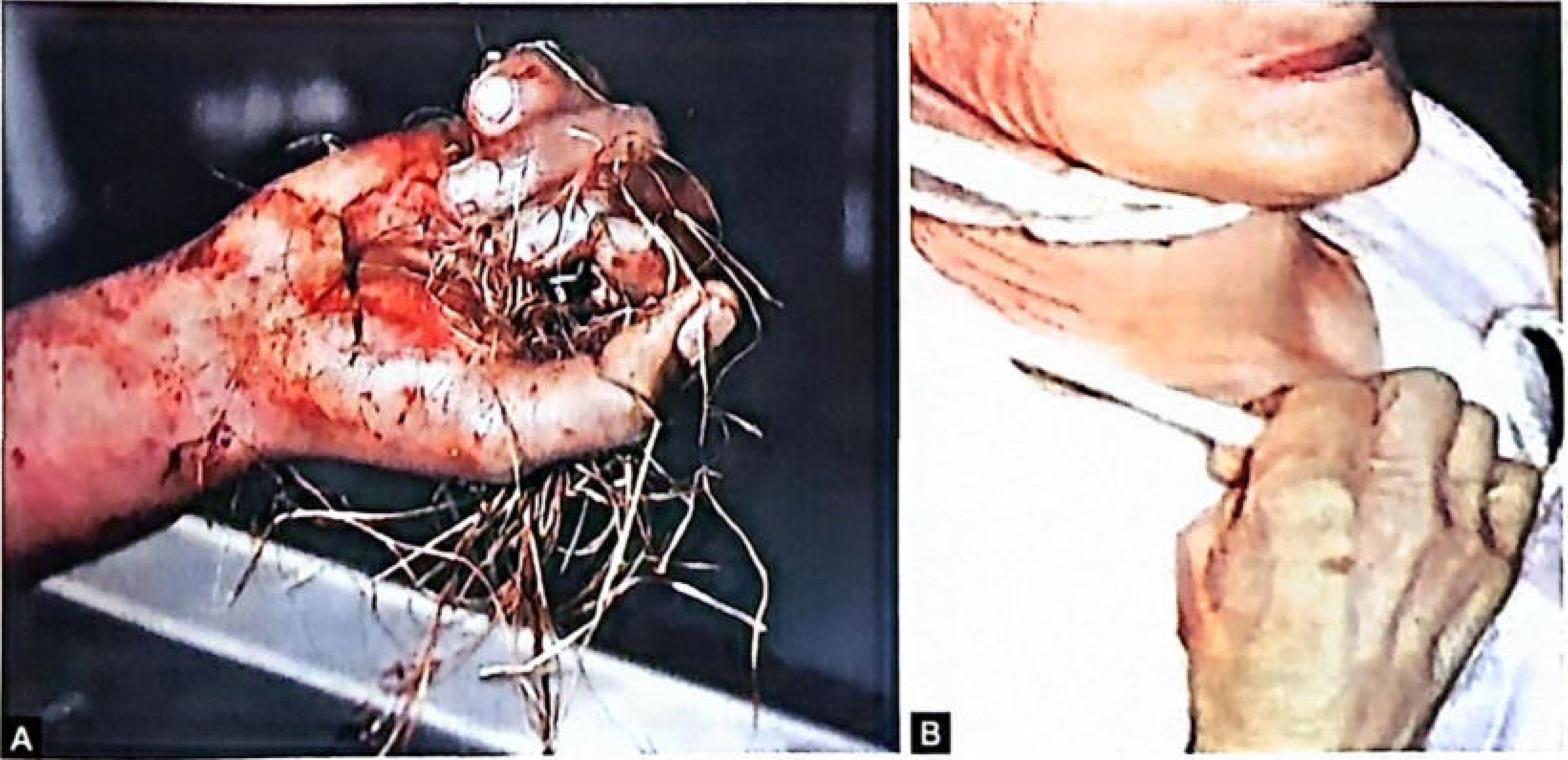
(A) Drowning victim with weeds firmly grasped in the hand; (B) Suicidal cut-throat with blade still gripped - both classic cadaveric spasm - The Essentials of Forensic Medicine and Toxicology, 36th ed.
Comparison: Cadaveric Spasm vs. Rigor Mortis
| Trait | Rigor Mortis | Cadaveric Spasm |
|---|---|---|
| Mechanism | Known - reduction/depletion of ATP | Not clearly known; possibly neurogenic |
| Onset | 1-2 hours after death (preceded by primary relaxation) | Instantaneous at moment of death |
| Preconditions | None - occurs in all deaths | Required: sudden death, emotional tension, physical activity |
| Muscles involved | All muscles (voluntary AND involuntary), in a fixed sequence | Usually one group of voluntary muscles only |
| Intensity | Moderate; broken by moderate force | Marked; very great force required to break |
| Molecular death | Occurs | Does NOT occur |
| Body temperature | Cold (delayed from time of death) | Warm |
| Electrical stimulation | Muscles do NOT respond | Muscles respond |
| Muscular reaction | Acidic | Alkaline |
| Can be produced after death? | Yes (freezing, heating >65°C) | Cannot be reproduced by any method after death |
| Medicolegal importance | Indicates time of death | Indicates mode/manner of death |
| Resolution | Disappears with putrefaction | Passes into normal rigor mortis, then resolves with putrefaction |
Key Point: Cannot Be Simulated
"It is impossible to simulate cadaveric spasm."
An object cannot be placed in the hand of a dead person to simulate cadaveric spasm - ordinary rigor mortis does not produce the same firm, natural grip. The position of gripping in cadaveric spasm would be anatomically consistent with how the person actually held the object, whereas a weapon placed post-mortem would rest unnaturally.
This makes cadaveric spasm conclusive proof that an object was gripped at or about the moment of death - it cannot be fabricated by a murderer attempting to simulate suicide.
Medicolegal Importance
-
Indicates sudden death associated with great emotional tension at the time of dying.
-
Indicates muscles in physical activity at the moment of death - provides a "snapshot" of the last act.
-
Helps determine the manner of death (suicide vs. homicide vs. accident):
- Suicide: Weapon (knife, razor, pistol) found firmly clenched in the victim's own hand - strong presumptive evidence of suicide. Murderers may attempt to place a weapon in the deceased's hand to simulate suicide, but this is distinguishable because ordinary rigor does not produce the same natural firm grip.
- Homicide/assault: Clothing, buttons, or hair of the assailant firmly grasped in the deceased's hands - can help identify the murderer.
- Drowning: Grass, weeds, or aquatic vegetation in the hands - proves the victim was alive on entering the water (distinguishes drowning from post-mortem submersion).
- Accidents: Mountain climbers may be found grasping branches/shrubs.
-
Evidence is conclusive - the nature of the grip and what is held cannot be artificially reproduced, making it invaluable forensic evidence.
Sources: The Essentials of Forensic Medicine and Toxicology, 36th ed. (2026), pp. 167-168 | Parikh's Textbook of Medical Jurisprudence, Forensic Medicine and Toxicology, pp. 183-184 | DiMaio's Forensic Pathology, 3rd ed.
Embalming
Searching sources...
Finding Sources
Finding Sources
Reading File
Reading File
Reading File
Reading File
Now let me read the cavity embalming, medicolegal importance, and artifact sections.
Reading File
Reading File
I now have comprehensive information from multiple authoritative textbooks. Let me compile the complete answer.
Embalming
Definition
Embalming is the treatment of a dead body with antiseptics and preservatives to prevent or delay putrefaction (decomposition). It causes coagulation and fixation of tissue proteins, rendering them resistant to bacterial digestion.
It should ideally be done as early as possible after death - for satisfactory results, within 6 hours of death.
Purposes / Aims
- Preservation - to delay putrefaction and allow safe handling, transport, or viewing of the body
- Hygiene and safety - to prevent spread of infection from the corpse
- Cosmetic - to restore a natural, peaceful appearance for funeral viewing
- Transport - required for transporting bodies over long distances or international repatriation
- Teaching and research - to preserve bodies for anatomical dissection and study
Embalming Fluid - Composition
The main arterial embalming fluid contains:
| Component | Role |
|---|---|
| Formalin (formaldehyde) | Primary fixative and preservative; coagulates proteins |
| Sodium borate | Buffer/antiseptic |
| Sodium citrate | Anticoagulant |
| Sodium chloride | Osmotic balance |
| Glycerin | Humectant (prevents dehydration and hardening of tissues) |
| Water | Solvent/vehicle |
Recommended Cavity Fluid Composition
| Component | Concentration |
|---|---|
| Formalin | 60% |
| Methanol (preservative) | 25% |
| Liquefied phenol (germicide) | 10% |
| Glycerin | 25% |
| Sodium lauryl sulphate | 1% |
| Mercuric chloride | 1% |
| Eucalyptus oil | 1% |
Methods of Embalming
Phase 1: Arterial Embalming
The embalming fluid is forced into the arterial system so it diffuses through arterioles and capillaries into cells and tissues.
Preparation of the Body
- Embalmer wears impervious apron, cap, mask, and gloves (infection control)
- Body placed supine; clothing and surgical dressings removed; washed with antiseptic soap and warm water
- Rigor mortis broken by bending, massaging, and rotating limbs/head
- Nostrils cleaned and plugged with cotton soaked in arterial solution
- Cheeks filled with cotton; mouth and eyelids closed; sunken eyeballs injected with arterial solution
- Head elevated 8-10 cm on a head rest; feet raised to facilitate drainage
- Anal orifice and vagina plugged with cotton soaked in cavity fluid
Choice of Injection Site
- The nearer the vessel to the heart, the better the result (ensures distribution and drainage)
- Single-point injection often leaves unperfused patches; multiple sites required for traumatic deaths, autopsied cases, and postmortem mutilations
The "Six-Point" Injection System (Standard Method)
| Artery | Region Served |
|---|---|
| Right and left common carotid arteries | Head and neck |
| Right and left axillary arteries | Upper limbs |
| Right and left femoral arteries | Lower limbs |
The trunk is perfused by directing fluid through these arteries back toward the heart. After injection, cannulae are removed and vessels ligated to prevent leakage. Each side of the face must be injected separately through the ipsilateral carotid to prevent facial distortion.
Injection Devices
- Hand/foot pump - manual
- Stirrup pump - manual
- Bulb syringe (Higginson-type) - rubber bulb with one-way valves; suction on one stroke, ejection on the other
- Gravity injector - simplest and safest; bottle raised by pulley; 1 metre height = 0.6 kg/cm² pressure; 2 metres = ~1 kg/cm²; slow and uneven distribution
- Motorized injector - best method; fluid from a compression tank forced under controlled pressure (~2 kg/cm²); ~10 litres injected in 30 minutes
Methods of Injection and Drainage
| Method | Description |
|---|---|
| Continuous injection + continuous drainage | Vein tube kept open throughout; quickest but gives poorest tissue saturation and drainage - least satisfactory |
| Continuous injection + interrupted drainage | Vein tube periodically closed; blood builds resistance in veins; better tissue saturation |
| Discontinuous (interrupted) injection + interrupted drainage | Best method - small quantities (~1-2 litres) injected alternately with drainage; best tissue saturation |
Phase 2: Cavity Embalming
Done 0.5-1 hour after arterial embalming (allowing visceral hardening for easier trocar puncture).
Technique
- A 30 cm trocar inserted through a small incision 5-6 cm above the umbilicus in the midline
- Sequential aspiration:
- Trocar directed upward, backward, and to the left → pierces and aspirates the stomach
- Slightly withdrawn, pushed up-right → pierces and aspirates the right side of the heart
- Trocar directed through diaphragm → aspirates right and left pleural sacs
- Multiple punctures in small intestine, caecum, and colon → aspirated
- Urinary bladder, sigmoid colon, and rectum aspirated
- After aspiration: 1 litre of cavity fluid into the abdominal cavity + 1 litre into the thoracic cavity, distributed evenly
Post-Embalming Drainage
- Undrained blood in large trunk vessels removed by aspiration during cavity treatment
- For transport: body wrapped in impermeable protective cover; covered with sawdust to absorb leakage
If left untended, an embalmed body will gradually dehydrate and ultimately mummify (shriveled to skin and bones).
Medicolegal Importance of Embalming
1. Preservation for Exhumation and Re-examination
- Embalming allows preservation of a body for later forensic examination, particularly useful in exhumation cases where the original autopsy findings are disputed
2. Interference with Toxicological Analysis (Critical Importance)
Embalming seriously interferes with toxicological investigation - hence, autopsy should always be performed BEFORE embalming in suspected poisoning cases.
Specific interferences:
| Poison / Substance | Interference |
|---|---|
| Cyanide | Reacts chemically with formalin → no longer identifiable |
| Volatile poisons | Embalming fluid contains methyl/ethyl alcohol → analyses for alcohol/volatiles rendered meaningless |
| Non-volatile organic drugs | Formalin-fixed tissues become resistant to organic solvents used for extraction → zero or very low drug recovery |
| Alcohol | Embalming fluid contains methanol/ethanol → blood and tissue alcohol levels rendered unreliable |
Practical rules for embalmed exhumed bodies:
- Provide the toxicologist with a sample of the embalming fluid used (as a control)
- Collect tissues least penetrated by embalming fluid for analysis: buttock muscle, centre of liver, vitreous humor (most protected)
3. Embalming as a Source of Postmortem Artifacts
Embalming can produce misleading postmortem findings:
- Trocar wounds (30 cm long) in the trunk → may be mistaken for stab wounds or bullet entry/exit wounds
- Blood forced from disrupted vessels may accumulate in tissues → simulates antemortem hemorrhage
- Incisions in the axillary or supraclavicular area → may be confused with antemortem injuries
- Skin bruises become markedly accentuated - embalming fluid increases skin transparency, making underlying bruises appear more prominent
- Air forced into blood vessels by the embalming process → may simulate air embolism
4. Effect on Bruise Visibility
In an embalmed body, bruises become more prominent - the embalming fluid makes the skin lighter/more transparent, and more blood is forced into damaged tissues under pressure, enhancing the bruise appearance.
5. India-Specific Context
- Embalming is not widely practiced in India due to religious customs (most Hindu and Muslim deaths are disposed of quickly)
- Bodies are sometimes embalmed for overseas repatriation
- Toxicological problems from embalming are therefore less commonly encountered in India compared to Western countries
Summary Table
| Aspect | Detail |
|---|---|
| Definition | Treatment with antiseptics/preservatives to prevent putrefaction |
| Timing | Within 6 hours of death for best results |
| Main fixative | Formalin (coagulates proteins) |
| Standard injection sites | Six-point: bilateral carotid, axillary, femoral arteries |
| Best injection method | Discontinuous injection with interrupted drainage |
| Cavity access | Trocar, 5-6 cm above umbilicus |
| Key toxicological concern | Autopsy BEFORE embalming in suspected poisoning |
| Key artifacts | Trocar wounds mimicking stab/bullet wounds; false hemorrhages; accentuated bruising |
| Long-term fate | Mummification if left untreated |
Sources: The Essentials of Forensic Medicine and Toxicology, 36th ed. (2026), pp. 178-179 | P.C. Dikshit Textbook of Forensic Medicine and Toxicology, p. 139 | Parikh's Textbook of Medical Jurisprudence, Forensic Medicine and Toxicology, pp. 162, 569
Hanging
Searching sources...
Finding Sources
Finding Sources
Good - the best sources are Parikh's and The Essentials (forensic). Let me read both in parallel.
Reading File
Reading File
Reading File
Reading File
Reading File
Reading File
Reading File
Reading File
Reading File
Reading File
I now have comprehensive material from multiple authoritative sources. Let me also fetch the key images.
Loading Image
Loading Image
I have all the content needed for a comprehensive answer. Let me compile it now.
Hanging
Definition
Hanging is a form of violent asphyxia resulting from suspension of the body by a ligature around the neck, where the constricting force is the weight of the body itself. The ligature constricts the neurovascular bundles in the neck and/or the upper airways.
Types
| Type | Description |
|---|---|
| Complete hanging | Feet do not touch the ground; weight of the whole body acts as the constricting force |
| Partial hanging | Body is partially supported (kneeling, sitting, reclining); weight of the head alone acts as the constricting force. Still potentially fatal - the head weighs enough (~3.5 kg) to compress carotid arteries |
A person can die from hanging even with knees on the floor, head resting against a bed-post, or in a sitting position. It is NOT necessary for the feet to be above the ground.
Pressure Required to Compress Neck Structures
| Structure | Pressure Required |
|---|---|
| Jugular veins | 2 kg |
| Carotid arteries | 3.5 kg |
| Trachea | 15 kg |
| Vertebral arteries | 16.6 kg |
This explains why vascular occlusion (not airway obstruction) is the primary mechanism of death in most hangings.
Cause of Death
Death in hanging may result from one or more of the following mechanisms:
- Cerebral ischemia and anoxia - compression of carotid arteries, blocking arterial blood supply to the brain (most common mechanism)
- Cerebral congestion - compression of jugular veins preventing venous drainage from the brain
- Blockage of air passages - direct compression of the trachea, or the root of the tongue pulled upward by the ligature
- Vagal inhibition - pressure on the vagus nerve or carotid sinus causing sudden cardiac arrest (reflex cardiac death)
- Injury to the spinal column/cord - particularly in judicial hanging with a significant drop; may cause instantaneous death
- Combination of any of the above
Important: Contrary to popular belief, airway obstruction is NOT the most common cause of death in hanging. Compression of the neurovascular bundles (carotids + jugulars) is the primary mechanism. Evidence: (a) persons with a tracheostomy below the level of the noose have died of hanging, and (b) vomitus is often found in the bronchi below the level of neck constriction.
Symptoms (In a Survivor or Witness Account)
Symptoms follow extremely rapidly:
- Flashes of light before the eyes
- Ringing in the ears
- Sudden loss of consciousness
- Death
Fatal Period
| Situation | Duration |
|---|---|
| Judicial hanging (with significant drop) | Death may be instantaneous from cervical fracture/spinal cord injury; heart may continue beating 15-20 minutes |
| Incomplete airway blockage | 5-8 minutes is the usual fatal period |
Postmortem Appearances
External Appearances
| Feature | Description |
|---|---|
| Neck | Stretched and elongated due to upward pull of the ligature |
| Head position | Always inclined to the side opposite the knot (gravitational force) |
| Face | Usually pale; congested and swollen if there is significant venous obstruction |
| Petechiae | On the face, head, neck, and conjunctivae if venous obstruction predominant |
| Eyes | Prominent and congested; pupils usually dilated; conjunctivae congested |
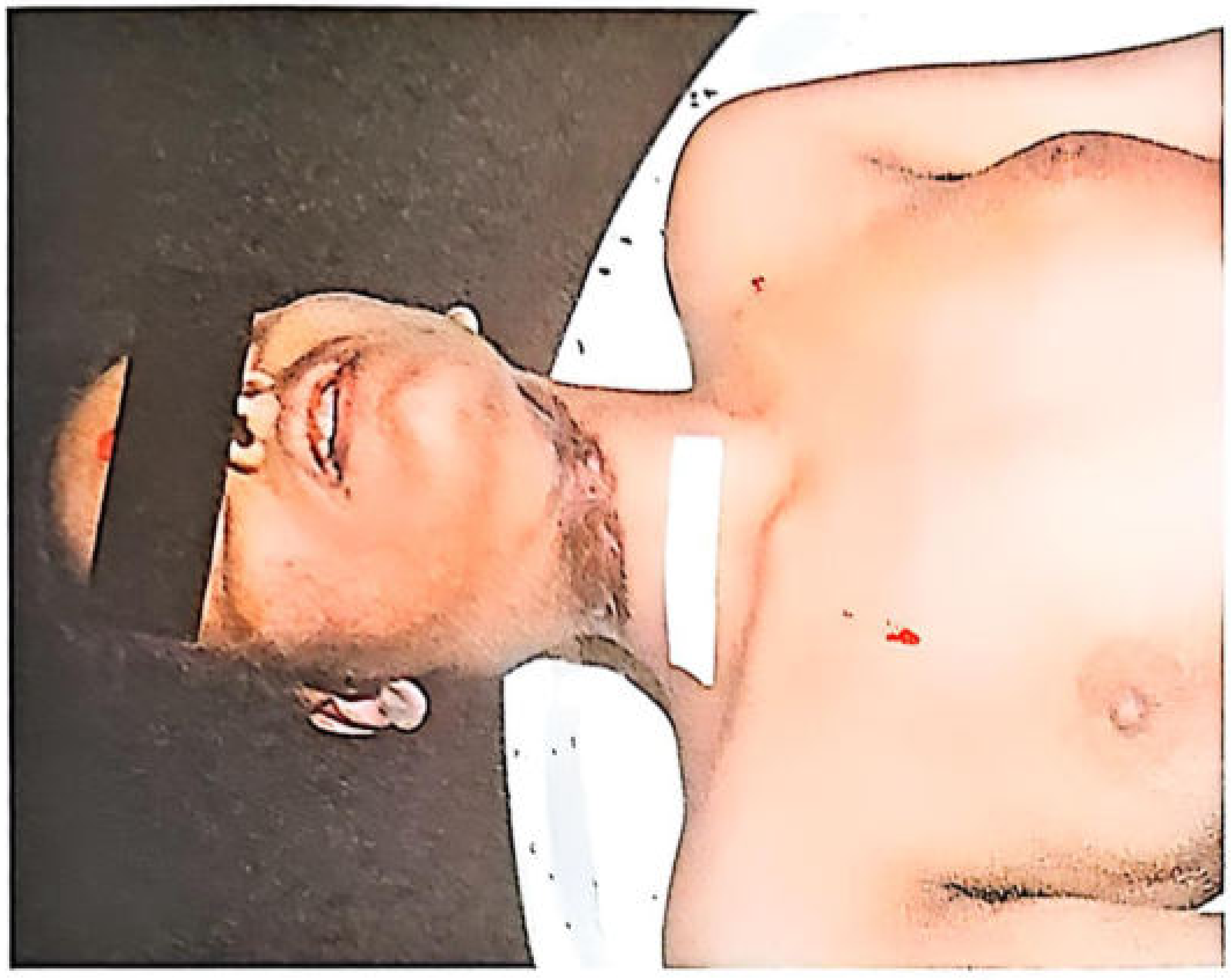
| Le facie sympathique | If the knot presses on the cervical sympathetic chain, the eye on that side remains open with a dilated pupil - indicates antemortem hanging |
| Tongue | Swollen, turgid, blue especially at base; may protrude between teeth; protruding part turns dark brown or black from drying |
| Saliva | Dribbles from the angle of mouth opposite the knot (due to pressure on salivary glands/pterygopalatine ganglion stimulation) |
| Hands | Clenched, especially in violent hanging; nail beds cyanosed |
| Genitalia | Penis engorged (hypostasis); may be semi-erect; semen may be found at the tip |
| Sphincters | Urine and feces may escape due to sphincter relaxation |
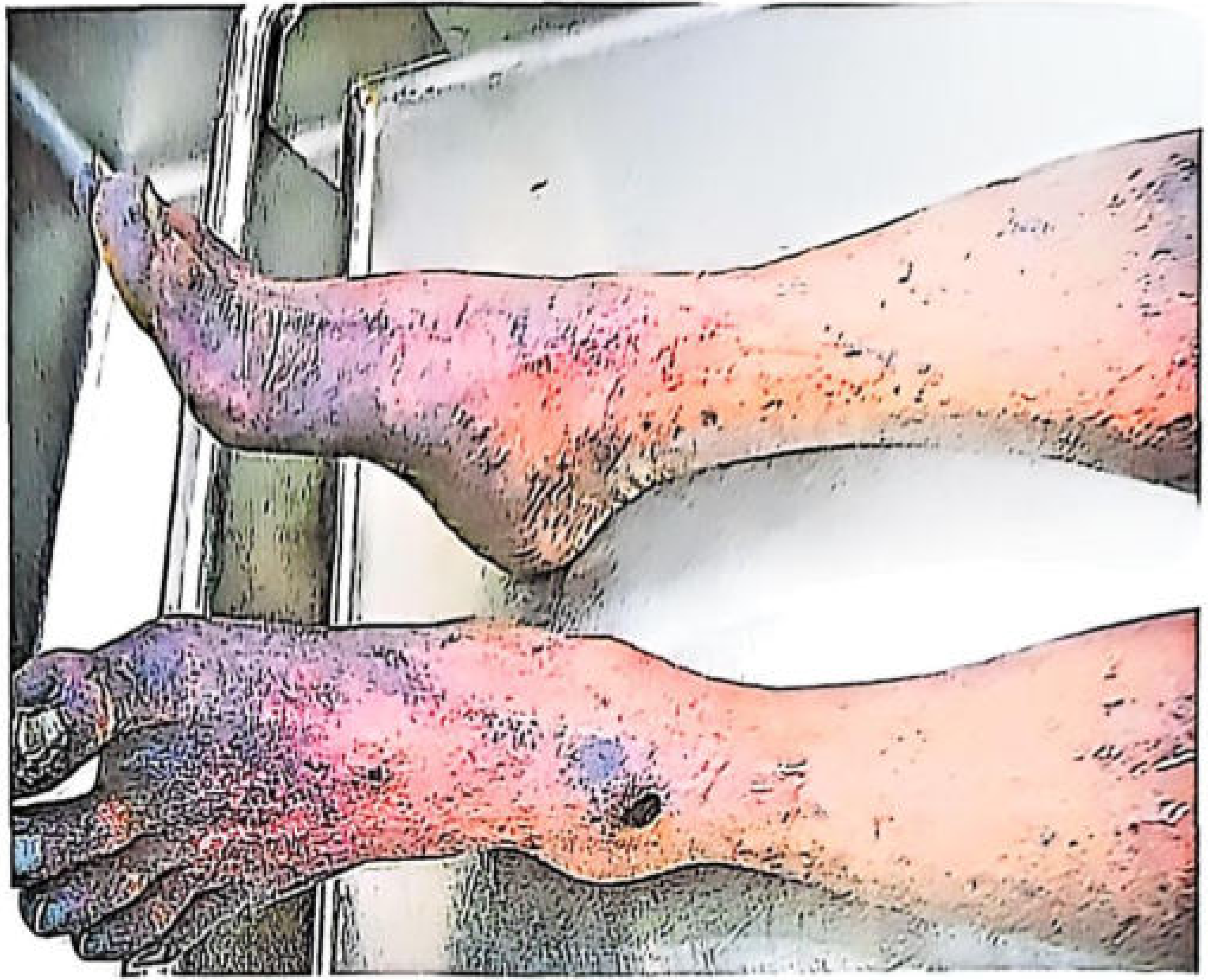
| Postmortem lividity | Circumferential on the skin of dependent arms and legs, face and neck above the ligature (glove-and-stocking pattern); if body removed within 4 hours and placed supine, limb lividity will fade |
Ligature mark of hanging with characteristic oblique groove:
Petechial hemorrhages on legs and feet in complete hanging (dependent hypostasis):
The Ligature Mark
The ligature mark is the most important external sign of hanging. It is a pressure mark (groove) on the neck at the site of the ligature.
Characteristics
| Feature | Description |
|---|---|
| Shape | Oblique (inverted V / upward slant toward the knot) - the classic distinguishing feature from strangulation which is horizontal |
| Position | Above the thyroid cartilage; usually in upper part of neck |
| Completeness | Incomplete - gap (interruption) at the knot site, usually at the nape or side of neck |
| Depth | Deepest at the points farthest from the knot; shallowest / absent near the knot |
| Appearance | Pale early; later becomes yellowish-brown, hard, dry, and parchment-like (from drying) |
| Pattern | Mirror image of the ligature texture may be imprinted (pressure abrasion) |
| Force required | Very great force required to break - resists putrefaction |
Factors Affecting the Ligature Mark
- Thin rope - deep, narrow mark; pattern imprinted on skin
- Broad soft cloth (sari, towel, scarf) - wide, shallow, ill-defined mark; may even be absent
- Soft material (nylon, silk, terylene) - may leave a mark only 2-3 mm wide; the knot may leave an abrasion due to its firmness
- Heavier body + longer suspension - more prominent, well-defined mark
- Multiple turns of ligature - corresponding number of marks, one above the other, with possible skin pinching between turns
- Beard / clothing between ligature and skin - no mark may be found in that area
- Ligature removed immediately - mark may disappear after several hours
A ligature mark can be reproduced if a body is suspended within 2 hours of death - this is a critical limitation in distinguishing antemortem from postmortem hanging.
Microscopy of Ligature Mark
Skin and deeper tissue at the ligature mark should be examined histologically for evidence of vital reaction (inflammatory changes). If present → confirms antemortem hanging. However, absence of tissue reaction does NOT exclude antemortem hanging.
Internal Appearances
Without Significant Drop (Suicidal/Accidental Hanging)
- Fibres of platysma and sternomastoid sometimes torn
- Posterior horns of thyroid cartilage may be fractured (from pressure on the thyrohyoid ligament)
- Hyoid bone fracture is rare, except in persons over 40 years (due to increased calcification and brittleness); involves the greater cornua at the junction of inner 2/3 and outer 1/3
- Carotid artery intima may show tears (Paltauf's hemorrhages)
- Hemorrhage into cervical muscles
- General asphyxia findings: congested and edematous brain, petechial hemorrhages on serous surfaces (Tardieu's spots), congested lungs
With Significant Drop (Judicial Hanging)
- Fracture-dislocation of cervical vertebrae (classically C2-C3)
- Transection of spinal cord at the level of fracture
- Massive hemorrhage in soft tissues of the neck
- Head may be nearly separated from the body in a violent drop
Diagnosis of Hanging
- Presence of oblique ligature mark around the neck
- Abrasions, ecchymoses, and redness about the ligature mark
- Dribbling of saliva from the mouth
- Rupture of the intima of the carotid arteries (Paltauf's hemorrhages)
- Congestion and hemorrhage in lymph nodes above and below the ligature mark
- Absence of any other cause of death
- Postmortem signs of asphyxia
- Le facie sympathique (sign of antemortem hanging)
Medicolegal Questions in Hanging
1. Was Death Due to Hanging?
In India, a common practice is to kill a victim by other means and then suspend the body (postmortem hanging) to simulate suicide or conceal murder. A ligature mark alone does NOT prove that death was due to hanging, since:
- A ligature mark can be reproduced by suspending a body within 2 hours of death
- Petechiae and lividity in dependent parts also occur in any suspended body
- The critical distinguishing investigations are: histology of ligature mark (vital reaction), general autopsy for other causes of death, and scene examination
2. Suicide, Homicide, or Accident?
Suicidal Hanging (Most Common)
- Most common method of suicide in many countries (especially among men)
- Ligature attached to a fixed high point (beam, fan, tree branch, window casing)
- Body in a position compatible with self-suspension
- No signs of struggle
- Farewell/suicide note in ~25% of cases
- History of depression, despondency, or psychiatric disorder
- A suicide may tie his own hands/feet to "make more certain of the result"
- Sometimes adopted as a last resort after other suicide methods have failed
Homicidal Hanging (Rare)
Practically impossible by a single assailant on a healthy adult. Suspect homicidal hanging when:
- (a) Knot is tied on the back of the neck
- (b) Mouth is gagged
- (c) Limbs are tied
- (d) Injuries present on the body that could not have been self-inflicted
- (e) Victim was intoxicated, elderly, frail, or physically helpless
- (f) Position of body or ligature is incompatible with self-suspension
Accidental Hanging
- Falls with neck caught in a noose or rope unexpectedly
- Children's necks caught in cribs, clothing, or playground equipment
- Sexual asphyxia (autoerotic hanging) - deliberate partial asphyxia for sexual gratification; accidental death common
Judicial Hanging
A legal method of execution involving a calculated long drop to cause instant death by cervical fracture and spinal cord transection rather than slow asphyxia. The drop length is calculated based on body weight to produce fracture at the atlantoaxial (C1-C2) or C2-C3 level.
Pressure Abrasion vs. Ligature Mark
The ligature mark in hanging is a pressure abrasion - not a bruise. It is produced by the crushing of superficial skin layers. Narrow ligatures leave a deeper mark with a pattern imprint ("mirror image phenomenon").
Sources: Parikh's Textbook of Medical Jurisprudence, Forensic Medicine and Toxicology, pp. 200-210 | The Essentials of Forensic Medicine and Toxicology, 36th ed. (2026), pp. 336-341 | P.C. Dikshit Textbook of Forensic Medicine and Toxicology
This is a shared conversation. Sign in to Orris to start your own chat.