
from Guyton and Hall textbook of medical physiology, 4th South Asia edition write a 5 mark short note on Heart Block-enlist types ,explain AV Nodal Block
Reading File
Finding Sources
Finding Sources
Finding Sources
Finding Sources
Reading File
Reading File
Reading File
Loading Image
Loading Image
Loading Image
Loading Image
I now have all the textbook content and ECG images. Here is the complete short note:
Short Note: Heart Block
(Source: Guyton and Hall Textbook of Medical Physiology, 4th South Asia Edition)
Definition
Heart block refers to a condition in which the conduction of the cardiac impulse through the intracardiac conduction pathways is delayed or completely blocked. The block can occur at the sinoatrial (SA) node, the AV node, the AV bundle (bundle of His), or within the bundle branches (intraventricular).
Types of Heart Block
Heart block is classified based on the site of block:
A. Sinoatrial (SA) Block
The impulse from the sinus node is blocked before it enters the atrial muscle. On ECG, there is sudden cessation of P waves with standstill of the atria. The ventricles pick up a new rhythm - usually originating in the AV node - so the QRS-T complex is preserved but slowed.
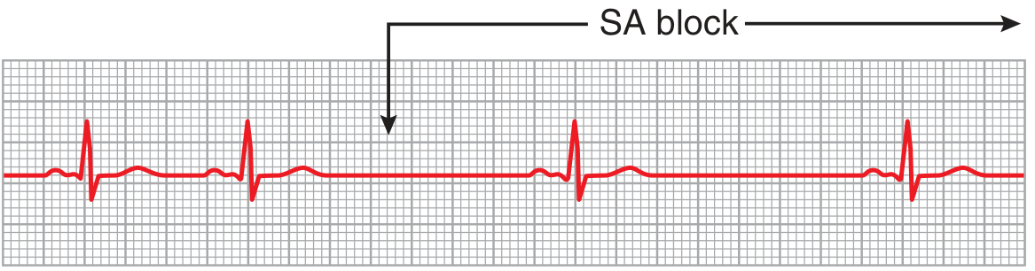
Figure 13.4: SA nodal block with AV nodal escape rhythm (Lead III). Note the absent P waves and the escape beats.
Causes: Myocardial ischemia affecting the sinus node, inflammation/infection, drug side effects, or seen in well-trained athletes.
B. Atrioventricular (AV) Block
Delayed or blocked conduction through the AV bundle. Further classified as:
| Degree | ECG Feature | Significance |
|---|---|---|
| 1st degree | P-R interval > 0.20 s (prolonged) | Delay only, no dropped beat |
| 2nd degree | Some P waves not followed by QRS (dropped beats) | Mobitz I or II |
| 3rd degree | Complete dissociation of P and QRS | Ventricular escape rhythm |
C. Intraventricular (Bundle Branch) Block
Blockage of impulse conduction in the peripheral ventricular Purkinje system. Results in altered, widened QRS complexes. Electrical alternans - a partial intraventricular block every other heartbeat - is one recognized pattern.
AV Nodal Block - Detailed Explanation
Causes of AV Block
The AV bundle (bundle of His) is the only pathway through which impulses can ordinarily pass from the atria to the ventricles. Conditions that cause AV block include:
- Ischemia of the AV node or AV bundle fibers - due to coronary insufficiency
- Compression of the AV bundle by scar tissue or calcified cardiac portions
- Inflammation of the AV node/bundle (from diphtheria, rheumatic fever, endocarditis, myocarditis)
- Vagal overstimulation - extreme vagal excitation (e.g., carotid sinus syndrome) can occasionally block AV conduction
- Degeneration of the AV conduction system (in older patients)
- Medications - digitalis, beta-adrenergic antagonists
First-Degree AV Block
- Normal P-R interval: ~0.16 s
- When P-R interval exceeds 0.20 s, it is prolonged - classified as 1st-degree block
- This is a delay of conduction, not actual blockage - impulses still reach the ventricles
- P-R interval rarely exceeds 0.35-0.45 s; beyond that, conduction stops entirely
- Useful as a marker of disease severity (e.g., acute rheumatic fever)
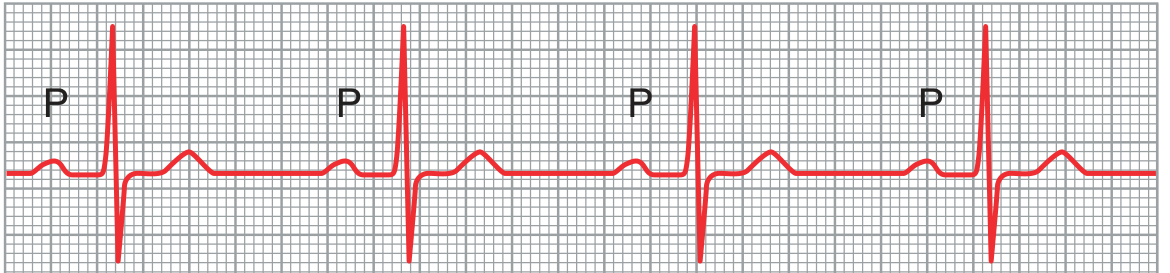
Figure 13.5: First-degree AV block showing prolonged P-R interval (~0.30 s instead of normal ≤0.20 s), Lead II.
Second-Degree AV Block
- P-R interval prolonged to 0.25-0.45 s
- The action potential is sometimes strong enough to conduct, sometimes not
- Results in "dropped beats" - a P wave with no QRS/T following it
Two subtypes:
Mobitz Type I (Wenckebach periodicity):
- Progressive lengthening of the P-R interval with each successive beat until a QRS is dropped
- After the dropped beat, the P-R interval resets and the cycle repeats
- Almost always due to AV node abnormality
- Usually benign; no specific treatment typically needed
Mobitz Type II:
- Fixed ratio of non-conducted P waves to QRS complexes (e.g., 2:1, 3:2, 3:1 block)
- P-R interval does not change before the dropped beat - it remains fixed
- Caused by abnormality of the bundle of His-Purkinje system
- More serious - may require pacemaker implantation to prevent progression to complete block and cardiac arrest
Third-Degree (Complete) AV Block
- Complete block of impulse from atria to ventricles
- The ventricles establish their own spontaneous rhythm - usually originating in the AV node or AV bundle distal to the block (ventricular escape rhythm)
- ECG shows complete P-QRS dissociation: atrial rate ~100 beats/min; ventricular rate less than 40 beats/min
- No relationship between P waves and QRS-T complexes
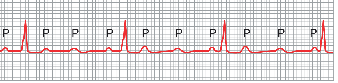
Figure 13.7: Complete (third-degree) AV block - multiple P waves (labeled "P") are seen dissociated from the rare QRS complexes, Lead II.
Stokes-Adams Syndrome (Ventricular Escape):
- Occurs when complete block comes and goes intermittently (borderline ischemia of the conduction system)
- When AV conduction ceases, the ventricles undergo overdrive suppression and do not begin beating immediately - there is a delay of 5 to 30 seconds
- During this pause, cerebral blood flow stops -> the patient faints (the brain cannot tolerate more than 4-7 seconds without blood)
- Eventually a Purkinje focus (usually distal AV node or AV bundle) escapes and begins discharging at 15-40 beats/min - "ventricular escape"
- These periodic syncopal episodes are called Stokes-Adams syndrome
- If the ventricular standstill is prolonged, it may be life-threatening
- Treatment: artificial pacemaker (implanted, battery-operated, electrodes usually in right ventricle)
Summary Table
| Type | ECG Finding | Rate | Treatment |
|---|---|---|---|
| 1st degree | P-R > 0.20 s | Normal | Treat cause |
| 2nd degree Mobitz I | Progressive P-R lengthening, dropped beat | Near normal | Usually none |
| 2nd degree Mobitz II | Fixed dropped beats, fixed P-R | Reduced | Pacemaker often needed |
| 3rd degree (complete) | P-QRS dissociation | Ventricular rate <40 | Pacemaker |
- Guyton and Hall Textbook of Medical Physiology (ISBN 9780443111013), Chapter 13, pp. 166-168
This is a shared conversation. Sign in to Orris to start your own chat.